Magnetic resonance imaging device

a magnetic resonance imaging and magnetic field technology, applied in the direction of measuring devices, magnetic measurements, instruments, etc., can solve the problem of very long total imaging time in parallel imaging, and achieve the effect of accurately calculating the sensitivity distribution of each receiving coil

Inactive Publication Date: 2007-05-29
FUJIFILM HEALTHCARE CORP
View PDF16 Cites 7 Cited by
  • Summary
  • Abstract
  • Description
  • Claims
  • Application Information

AI Technical Summary

Benefits of technology

[0014]In this manner, to remove the component of aliasing artifact in the image data acquired by executing the second pulse sequence in which the phase encoding steps in the measurement space, i.e. the k space are thinned out, the sensitivity distribution data on each slice position of image data are necessary. These sensitivity distribution data are calculated from the sensitivity image data acquired by executing the first pulse sequence for acquiring measured data of central portion (low-frequency portion) in the k space, whereby the time for acquiring the sensitivity distribution of receiving coil can be shortened.
[0015]In the MRI apparatus according to the present invention, the slice number of the sensitivity image data acquired in the first pulse sequence is n in number, which is smaller than m in number of examination image data including the aliasing artifact component acquired in the second pulse sequence. When sensitivity distribution data on a slice position do not exist in the image data for m slices, they are generated by performing interpolation using the above acquired sensitivity distribution data. Therefore, because the repetition number of the first pulse sequence for acquiring the sensitivity distribution of receiving coil can be reduced, the total time of repeatedly executing the first pulse sequence is shortened, and the total imaging time in the parallel imaging including the first pulse sequence and the second pulse sequence can be thus shortened.
[0017]Further, in the MRI apparatus according to the present invention, the plurality of receiving coils may include a first receiving coil and a second receiving coil. In this case, it is preferable that the sensitivity distribution of the first receiving coil is more uniform than that of the second receiving coil. Thus, by using as a reference an image having a uniform sensitivity distribution obtained by the first receiving coil, the sensitivity distribution of the second receiving coil can be accurately calculated.
[0018]Further, in a structure where the first receiving coil does not have a uniform sensitivity distribution, an image having a more uniform sensitivity distribution than that generated on the basis of the measured data acquired by a single receiving coil is generated by combining the measured data obtained by using a plurality of receiving coils. By using as a reference this image having a uniform sensitivity distribution, the sensitivity distribution of each receiving coil can be accurately calculated.

Problems solved by technology

Therefore, there is a problem that the total imaging time in the parallel imaging becomes very long.

Method used


Image

  • Magnetic resonance imaging device
  • Magnetic resonance imaging device
  • Magnetic resonance imaging device

Examples


first embodiment

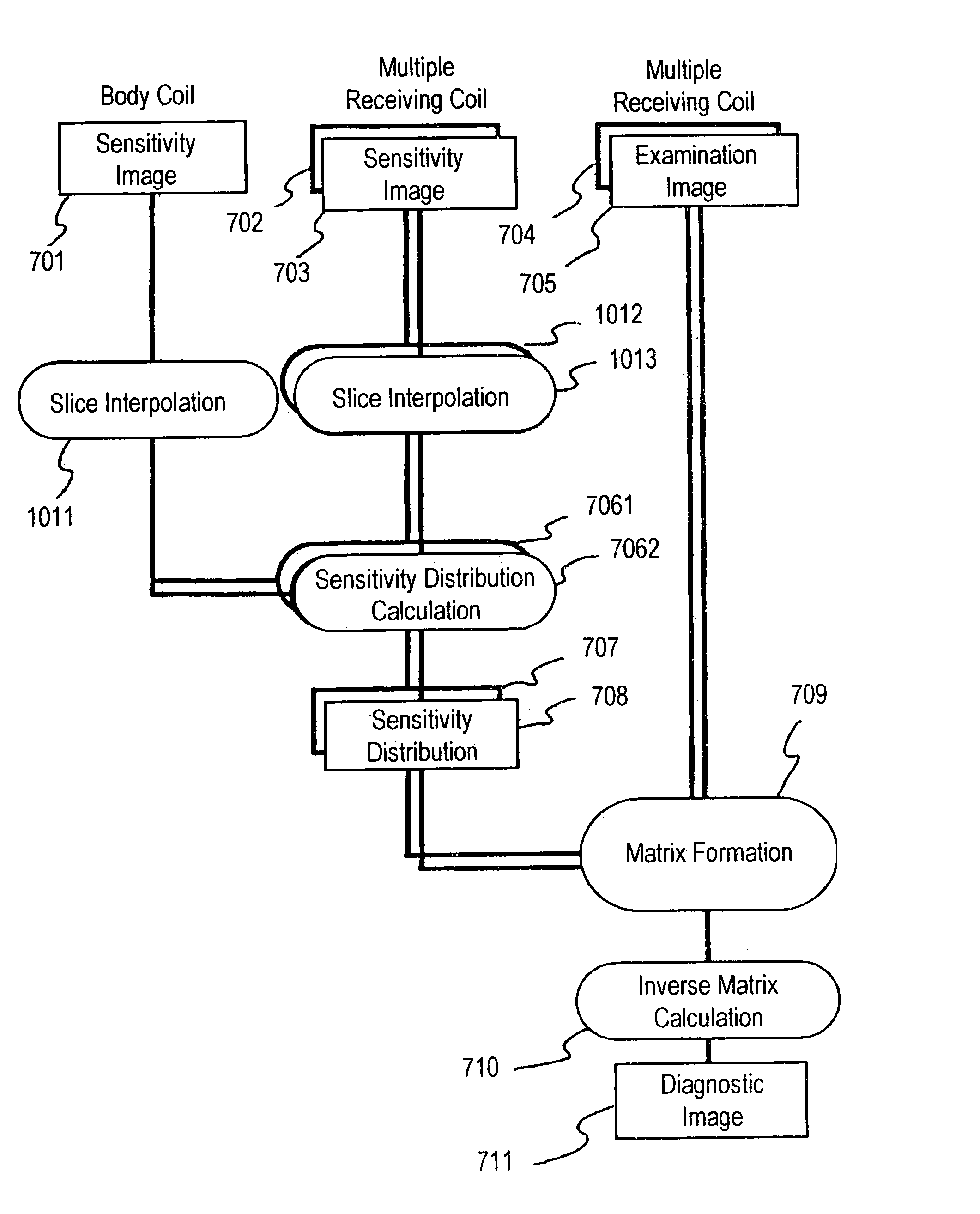
[0030]Hereinafter, an MRI apparatus and an MR imaging method according to the present invention will be described with reference to the drawings. First, a first embodiment of the MRI apparatus according to the present invention will be described with reference to FIG. 1 to FIG. 7.

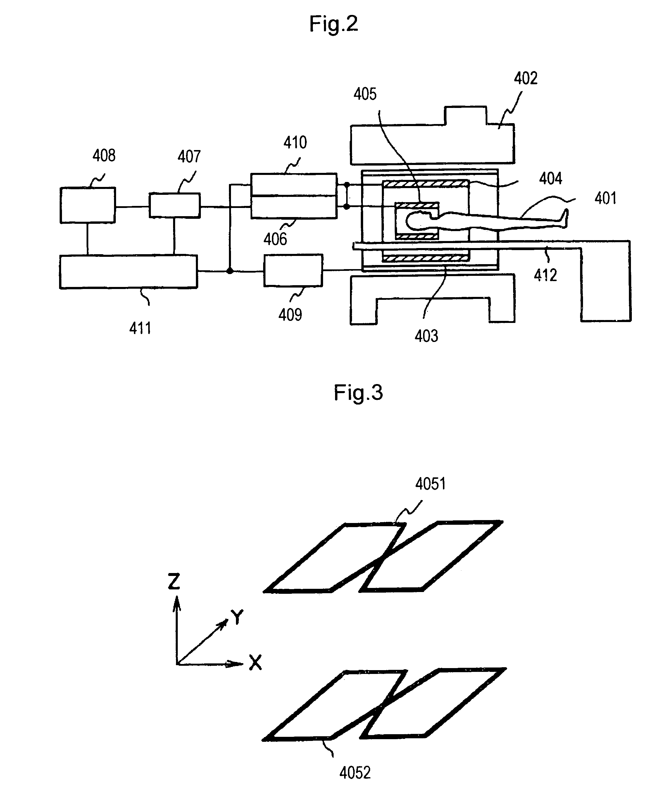
[0031]As shown in FIG. 2, the MRI apparatus according to this embodiment includes magnet 402 for generating a uniform static magnetic field having a predetermined strength to a space (hereinafter referred to as “imaging space”) having a predetermined size for accommodating object 401 to be examined, gradient magnetic field coil 403 for generating gradient magnetic fields to the imaging space, body coil 404 for generating a radio-frequency magnetic field to the imaging space and detecting an NMR signal generated by object 401, and RF probe 405 for detecting the NMR signal generated by object 401. Here and hereinafter, “RF” is an abbreviation of “Radio Frequency.” Although FIG. 2 shows an MRI apparatus of hor...

second embodiment

[0078]Second embodiment of MRI apparatus according to the present invention will be described with reference to FIG. 8 and FIG. 9. Because the same functions as in the first embodiment are given the same reference marks, only the difference with the first embodiment will be hereinafter described.

[0079]The difference between this embodiment and the first embodiment is, as shown in FIG. 8, that slice interpolation processing 801 is performed after sensitivity distribution calculation processing 706 is performed using sensitivity image 701 of body coil 404 and sensitivity images 702 and 703 of receiving coils 7051 and 4052 of multiple receiving coils 301 acquired in the pre-measurement pulse sequence for the sensitivity image data acquisition. Therefore, in the signal processing unit according to this embodiment, sensitivity distribution data 7061 and 7062 of receiving coils 4051 and 4052 are calculated from sensitivity image 701 of body coil 404 and sensitivity images 702 and 703 of r...

third embodiment

[0087]Next, the third embodiment of MRI apparatus according to the present invention will be described with reference to FIG. 10 and FIG. 11. Meanwhile, in this embodiment, too, the same function as in First embodiment is given the same reference mark and the description thereof will be omitted. The structure different from First embodiment and the characteristic part will be described hereinafter.

[0088]The difference between this embodiment and First embodiment lies in that the multiple receiving coils include four receiving coils. That is, as shown in FIG. 10, multiple receiving coils 301A forming an RF probe in the MRI apparatus according to this embodiment include four figure-of-eight shaped receiving coils 4051 to 4054, which are arranged opposite on the X-Y plane and on the T-Z plane at a predetermined distance.

[0089]Further, as shown in FIG. 11, four receiving coils 4051 to 4054 of multiple RF receiving coils 301A according to this embodiment are respectively connected to pre...


PUM


Abstract

A first pulse sequence is executed on an imaging portion of an object to be examined using a multiple receiving coils including a plurality of receiving coils to obtain sensitivity images 701 to 703, each of n in number, which is smaller than the number of examination image. When those sensitivity images are calculated, NMR signals are measured only in a low-frequency region of a k space. Next, a second pulse sequence is executed while phase encoding steps are thinned out to acquire examination images 704 and 705, each of m in number (m>n), of the object with each receiving coil. When sensitivity distributions 707 and 708 are generated on the basis of sensitivity images 701 to 703, if a sensitivity distribution on a slice corresponding to examination images 704 and 705 is not yet measured, it is calculated with a slice interpolation processing on the basis of sensitivity distributions 701 to 703, and an aliasing artifact in examination images 704 and 705 are removed using sensitivity distributions 707 and 708 with a matrix calculation.

Description

TECHNICAL FIELD[0001]The present invention relates to a magnetic resonance imaging apparatus (hereinafter referred to as “MRI apparatus”) for imaging a tissue structure inside an object to be examined by utilizing a magnetic resonance phenomenon, more particularly to an MRI apparatus suitable for parallel imaging for rapidly imaging the object using a plurality of receiving coils.BACKGROUND OF THE INVENTION[0002]In an MRI apparatus, a pulse sequence is sequentially executed while a phase encoding amount given to a nuclear magnetic resonance signal (hereinafter referred to as “NMR signal”) is varied, and an echo signal necessary for reconstructing one image is thus acquired. Therefore, imaging time depends on time of repetition (TR) and the repetition number of times of pulse sequence. When the rapid imaging is performed, a multi-echo pulse sequence in which plural echo signals can be measured by executing the pulse sequence one time and a pulse sequence in which the TR is shortened ...

Claims


Application Information

Patent Timeline
Patent Type & Authority Patents(United States)
IPC IPC(8): G01V3/00G01R33/561
CPCG01R33/5611
Inventor TAKIZAWA, MASAHIROTAKAHASHI, TETSUHIKO
Owner FUJIFILM HEALTHCARE CORP
Why Eureka
Try Eureka