
Intra-cavitary ultrasound medical system and method

a technology of intra-cavitary ultrasound and medical system, applied in the field of ultrasonography, can solve the problems of difficult to keep water within the body cavity without the use of balloons or sheaths, and medical problems

Active Publication Date: 2007-07-24
CILAG GMBH INT
View PDF54 Cites 143 Cited by
  • Summary
  • Abstract
  • Description
  • Claims
  • Application Information

AI Technical Summary

Benefits of technology

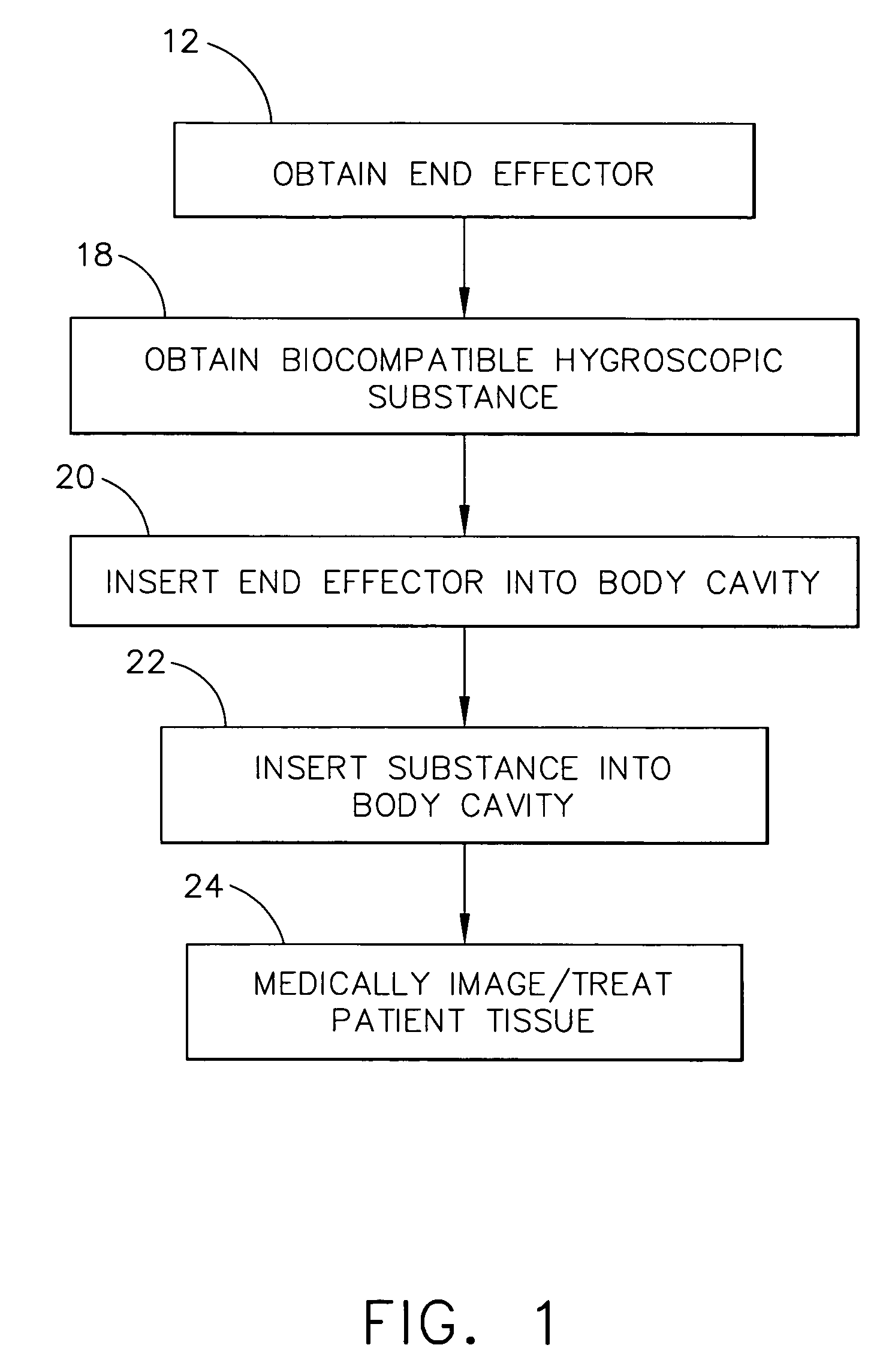
The invention is a method and system for using ultrasound in a patient's body cavity for medical purposes. The system includes an end effector with a medical ultrasound transducer assembly and a biocompatible hygroscopic substance. The substance has a non-expanded anhydrous state and an expanded and fluidly-loculated hydrated state. The end effector is insertable into the body cavity of the patient, and the substance is inserted in its anhydrous state. The medical transducer assembly is adapted to medically image and / or treat patient tissue with ultrasound. The use of the hygroscopic substance provides better acoustic coupling between the ultrasound transducer assembly and the wall of the body cavity, allowing for better imaging and treatment without the need for additional balloons or sheaths. The invention has applications in conventional endoscopic surgical instrumentation and robotic-assisted surgery.

Problems solved by technology

Balloons and sheaths do not contact all of the nearby surrounding patient tissue, especially when the body cavity has sharp body contours, and it is difficult to keep water within the body cavity without the use of a balloon or sheath.
However, having micropellets in an artery can pose potential medical problems, and ligating and fulgurating an artery are relatively invasive procedures.

Method used


Image

  • Intra-cavitary ultrasound medical system and method
  • Intra-cavitary ultrasound medical system and method
  • Intra-cavitary ultrasound medical system and method

Examples


Embodiment Construction

[0017]Before explaining the present invention in detail, it should be noted that the invention is not limited in its application or use to the details of construction and arrangement of parts illustrated in the accompanying drawings and description. The illustrative embodiments of the invention may be implemented or incorporated in other embodiments, variations and modifications, and may be practiced or carried out in various ways. Furthermore, unless otherwise indicated, the terms and expressions employed herein have been chosen for the purpose of describing the illustrative embodiments of the present invention for the convenience of the reader and are not for the purpose of limiting the invention.

[0018]It is understood that any one or more of the following-described embodiments, examples, etc. can be combined with any one or more of the other following-described embodiments, examples, etc.

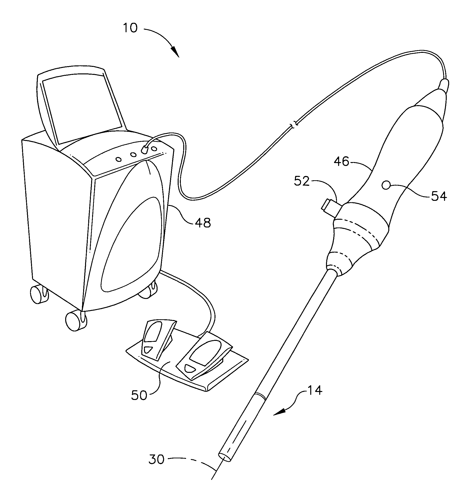
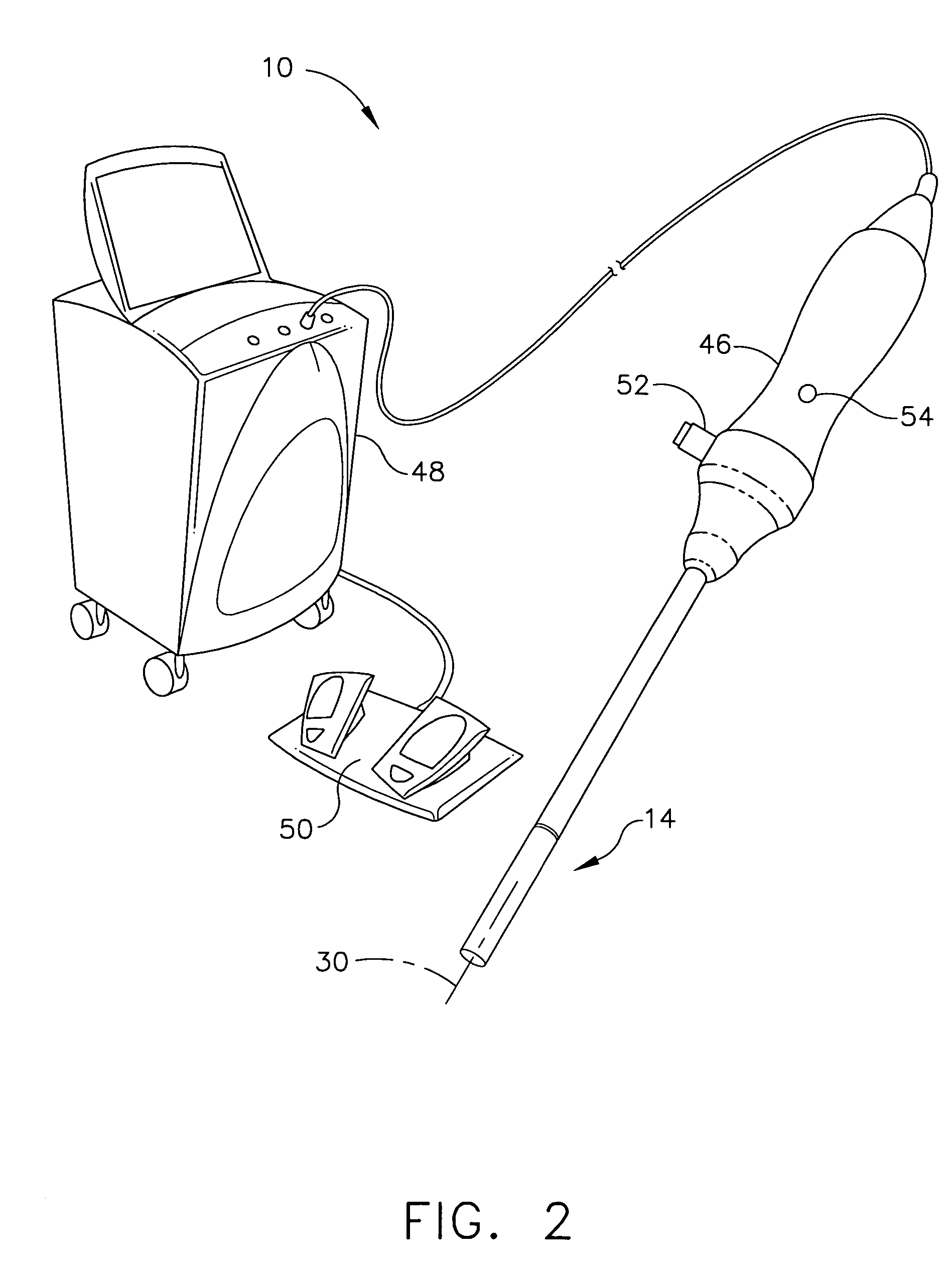
[0019]Referring now to the Figures, in which like numerals indicate like elements, FIG. 1 ill...


PUM


Abstract

A method for medically employing ultrasound within a body cavity of a patient. An end effector is obtained having a medical ultrasound transducer assembly. A biocompatible hygroscopic substance is obtained having a non-expanded anhydrous state and having an expanded and fluidly-loculated hydrated state. The end effector, including the transducer assembly, and the substance in substantially its anhydrous state are inserted into a body cavity (such as endoscopically inserted into a uterus) of a patient. The transducer assembly is used to medically image and / or medically treat patient tissue (such as stopping blood flow to, and / or ablating, a uterine fibroid). A system for medically employing ultrasound includes the end effector and the substance. In another system, the end effector includes the substance. The substance in its hydrated state expands inside the body cavity providing acoustic coupling between the wall of the body cavity and the transducer assembly.

Description

FIELD OF THE INVENTION[0001]The present invention is related generally to ultrasound, and more particularly to a medical system and method employing ultrasound within a body cavity of a patient.BACKGROUND OF THE INVENTION[0002]Known ultrasound medical methods include using ultrasound imaging (at low power) of patients to identify patient tissue for medical treatment and include using ultrasound (at high power) to ablate identified patient tissue by heating the tissue. Known ultrasound imaging includes Doppler ultrasound imaging to detect blood flow, and a proposed known use of ultrasound includes using an ultrasound transducer outside the body to stop internal bleeding (by sealing ruptured blood vessels) of a patient brought to an emergency room of a hospital.[0003]In one known ultrasound uterine medical treatment method, an ultrasound transducer is endoscopically inserted into the vagina but outside the uterus of a female patient and is used to ablate a portion of a uterine fibroid...

Claims


Application Information

Patent Timeline
Patent Type & Authority Patents(United States)
IPC IPC(8): A61B8/00
CPCA61B8/12A61B17/2202A61B17/42A61B8/445A61B2017/2253A61B2017/4216A61B8/4483
Inventor MAKIN, INDER RAJ S.AVIDOR, YOAVSLAYTON, MICHAEL H.BARTHE, PETER G.
Owner CILAG GMBH INT
Learn More