Single-tailed suturing method

a single-tailed, suturing technology, applied in the field of suturing technique and the formation of suturing loops, can solve the problems of difficult knot knot development at best, limited surgeon's work space, and inability to perform surgery

Inactive Publication Date: 2007-10-23
ARTHROCARE
View PDF20 Cites 308 Cited by
  • Summary
  • Abstract
  • Description
  • Claims
  • Application Information

AI Technical Summary

Problems solved by technology

In some cases, the development of a knot in a surgical procedure may require dexterity beyond the capability of the surgeon.
A commonality in these procedures is that the spaces in which the surgeon works are limited, and the tools used for suturing make tying knots difficult at best.
In endoscopic procedures, either the knots need to be tied externally to the body and inserted into the body and to the operative site using some kind of knot pushing device, or they need to be tied inside the body using long, clumsy instruments.
Because of their size, the needles used in these procedures are generally not able to be held in the jaws of the needle driver while being introduced through the operative trocar.
This is a difficult and time-consuming aspect of this current endoscopic technique for suturing.
This process is a time consuming, and sometimes frustrating exercise in hand to eye coordination, which is complicated by the fact that the surgeon is viewing the three dimensional space inside the body cavity through a two dimensional video monitor.
The major disadvantage of these kinds of staplers is that they leave metal in the body.
For some tissues this is not a problem, however in some procedures, metal staples left within the tissues can be a major hindrance to the healing process.
While endoscopy has certainly found favor with many physicians as an alternative operative modality, the advanced skill set and operative time necessary to become an efficient and practiced endoscopist have proven to be a challenge for a large portion of the surgical community.
The cost pressures brought about by large scale patient management (the continued rise and success of health maintenance organizations or HMO's) have also caused the surgical community to cast a critical eye on the overall costs and long-term outcomes of some of the procedures that have been tried via a endoscopic approach.
While the laparascopic cholecystectomy (gall bladder removal) has certainly proven its worth in the past 8–10 years, many other procedures have not shown similar cost effectiveness and positive long-term outcomes.
It is an increasingly common problem for tendons and other soft, connective tissues to tear or to detach from associated bone.
One such type of tear or detachment is a “rotator cuff” tear, wherein the supraspinatus tendon separates from the humerus, causing pain and loss of ability to elevate and externally rotate the arm.
Because of this maneuver, the deltoid requires postoperative protection, thus retarding rehabilitation and possibly resulting in residual weakness.
Although the above described surgical technique is the current standard of care for rotator cuff repair, it is associated with a great deal of patient discomfort and a lengthy recovery time, ranging from at least four months to one year or more.
It is the above described manipulation of the deltoid muscle together with the large skin incision that causes the majority of patient discomfort and an increased recovery time.
However, as will now be described, there are cases where the knots themselves are a hindrance to the healing of the wound.
In cases where joint re-constructions are undertaken by orthopedic surgeons, oftentimes the space available within joint is quite limited.
The knots in the tendon can be bulky and create a painful impingement of the tendon on the bone.
So it may be seen that none of the currently extant approaches to the placement and securing of sutures in, for example, rotator cuff surgery have fulfilled all of the surgeon's requirements.

Method used


Image

  • Single-tailed suturing method
  • Single-tailed suturing method
  • Single-tailed suturing method

Examples


Embodiment Construction

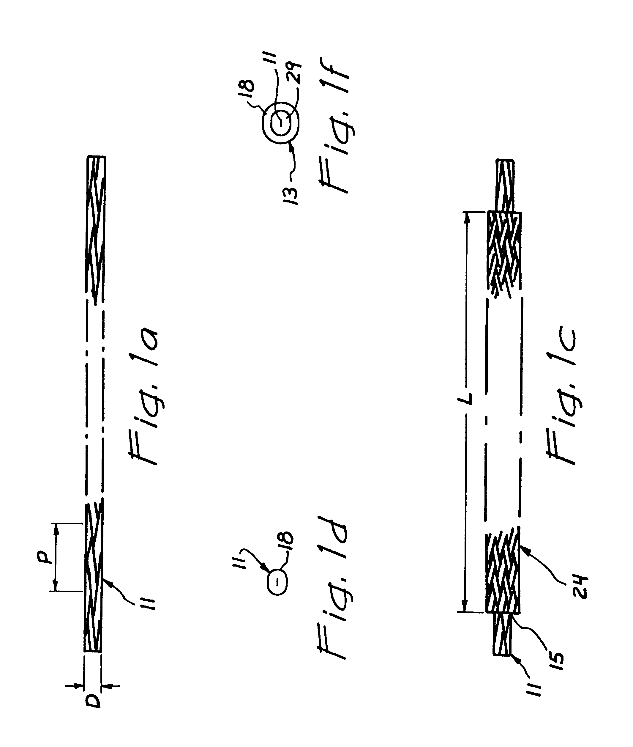
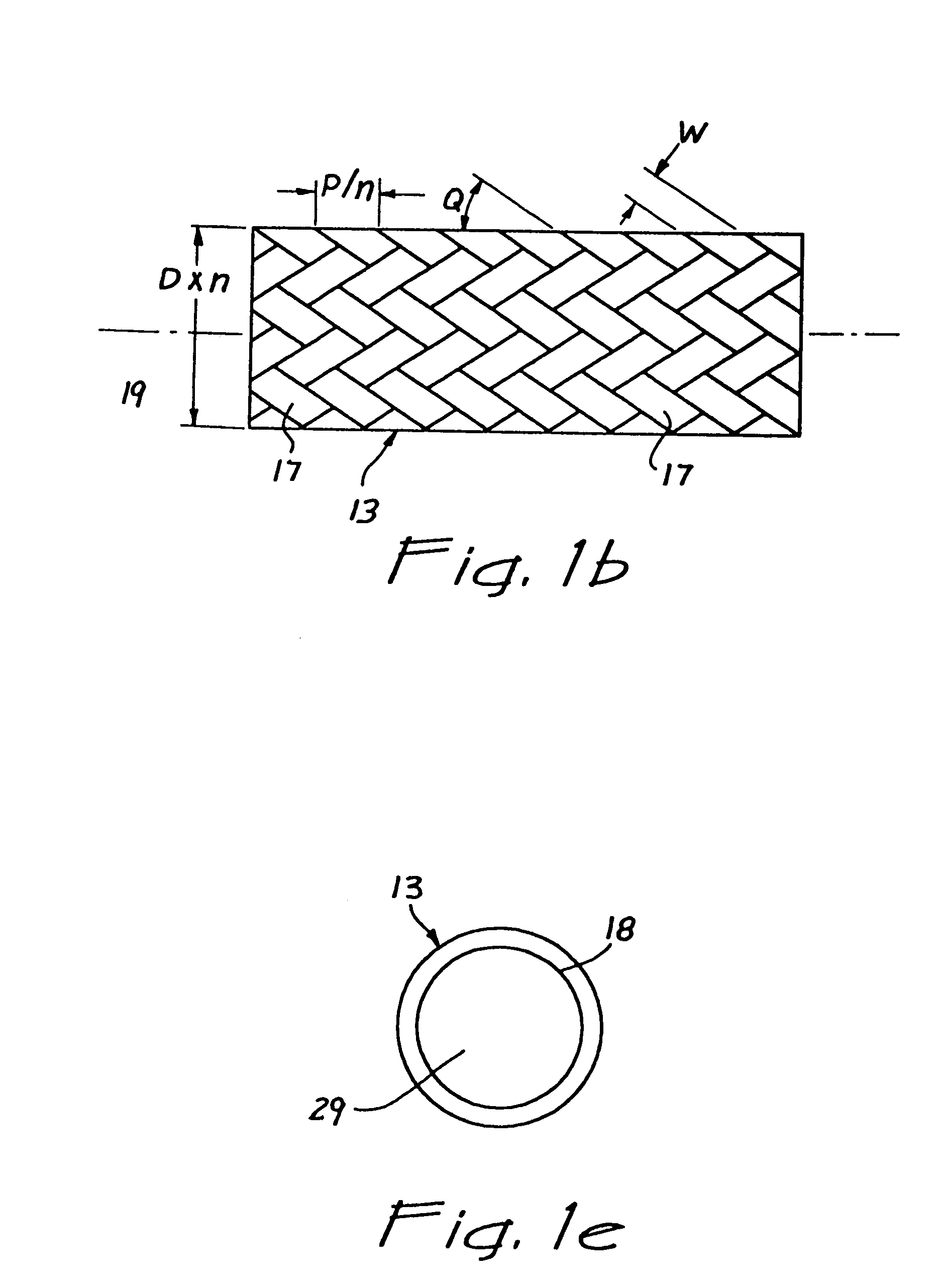
[0047]Referring now more particularly to the drawings, FIG. 1a shows a tensioned suture 11 of a braided construction, in tension. The tension on the suture preferably sets characteristics of the suture so that it is of diameter D and pitch P. FIG. 1b shows the tensioned suture 11 loaded with axial compression to form a compressed suture 13, the suture braid being designed so that its pitch and diameter are affected by the axial compression on the suture by a factor “n” as shown. The factor “n” is of such a value that it makes possible the passage of the tensioned suture 11, having the diameter D, through the center of the compressed suture 13. The factor “n” is also of such a value that the interior of compressed suture 13 further provides for the passage of any instrument that is required for the manipulation of the suture.

[0048]In a preferred configuration, the factor “n” ranges in value from a minimum of about 1.5 to a maximum of about 15.0 in order to achieve acceptable performa...


PUM


Abstract

A suture loop is formed in a hollow braided suture by feeding one end of a length of suture through a part in the braid of the suture and into the inner lumen formed by the hollow braid. The braided configuration of the suture allows it to be expanded in diameter by pushing and reduced in diameter by pulling. Said end of suture is passed continuously through said inner lumen forming a loop of suture with a single tail. The loop may be tightened by pulling on said first end of the suture while pushing on said outer hollow braid. The loop may be locked by extending or pulling on said outer hollow braid to reduce its diameter and lock it down around said first end of the suture.

Description

BACKGROUND OF THE INVENTION[0001]This invention relates generally to the creation of a sliding and locking loop of cord, and more particularly to a surgical technique of suturing and the formation of a suture loop that may be tightened and locked.[0002]Suturing is a necessary aspect of virtually any surgical procedure. Numerous techniques of tying sutures have been developed by surgeons over the years to address various applications of sutures. For example, a surgeon's knot, in which an overhand knot is modified to include two wraps of the suture ends around each other, was developed to minimize the amount of slippage in the suture as the second or locking throw of a ligation or approximation of tissue was accomplished. Another knot called a Roeder knot was developed to allow surgeons to place a loop of suture around a vessel for ligation in an endoscopic environment. The Roeder knot is basically a pre-tied slip knot that may be cinched and locked around a vessel or other structure....

Claims


Application Information

Patent Timeline
Patent Type & Authority Patents(United States)
IPC IPC(8): A61B17/04A61B17/06
CPCA61B17/0469A61B17/0482A61B17/06A61B17/0485A61B17/0487A61B2017/06028A61B17/06166A61B2017/0454A61B2017/0496A61B2017/06185
Inventor FOERSTER, SETH A.
Owner ARTHROCARE
Why Eureka
Try Eureka