Method and apparatus for volumetric cardiac computed tomography imaging

a computed tomography and volumetric cardiac technology, applied in the field of medical imaging arts, can solve the problems of time-consuming reconstruction of this plethora of data, disconnected arrangement, and little guidance to users, and achieve the effect of reducing image reconstruction processing and streamlining a cardiac ct imaging session

Inactive Publication Date: 2007-10-30
KONINKLIJKE PHILIPS ELECTRONICS NV
View PDF15 Cites 21 Cited by
  • Summary
  • Abstract
  • Description
  • Claims
  • Application Information

AI Technical Summary

Benefits of technology

[0017]One advantage of the present invention resides in a comprehensive cardiac CT imaging workflow that provides substantial guidance to the user in performing cardiac CT imaging with a specific diagnostic objective.
[0018]Another advantage of the present invention resides in reduced image reconstruction processing for a diagnostic objective which utilizes a sub-set of the complete cardiac CT imaging data set.
[0019...

Problems solved by technology

Modern multi-slice and helical CT scanners produce large quantities of data.
Reconstruction of this plethora of data is time consuming.
Typically, the operator is given little guidance on optimizing the survey, monitoring, and diagnostic imaging steps.
The disconnected nature of the a...

Method used


Image

  • Method and apparatus for volumetric cardiac computed tomography imaging
  • Method and apparatus for volumetric cardiac computed tomography imaging
  • Method and apparatus for volumetric cardiac computed tomography imaging

Examples


Embodiment Construction

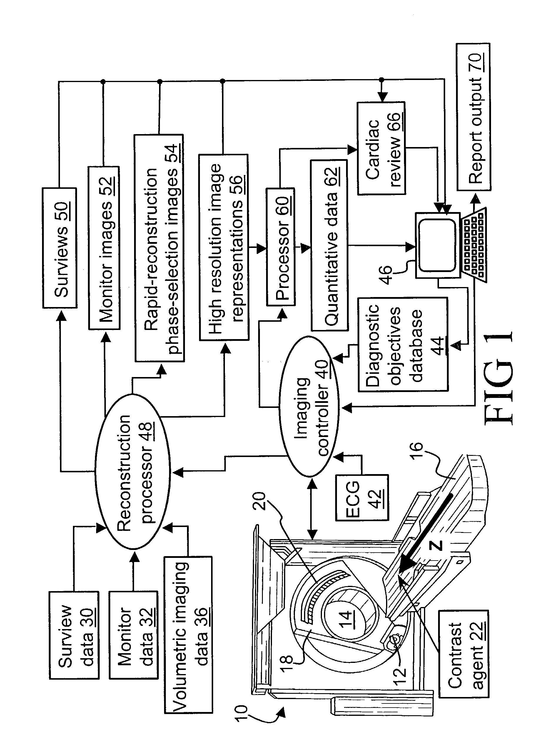
[0034]With reference to FIG. 1, a computed tomography (CT) imaging scanner 10 includes an x-ray source 12 that produces a fan-shaped, cone-shaped, wedge-shaped, or otherwise-shaped x-ray beam directed into an examination region 14 which contains a patient arranged on a patient support 16 with the patient's heart substantially centered within the examination region 14. The patient support 16 is linearly movable in a Z-direction while the x-ray source 12 is mounted on a rotating gantry 18 that rotates around the Z-axis.

[0035]In a helical CT imaging mode, the rotating gantry 18 rotates simultaneously with linear advancement of the patient support 16 to produce a generally helical trajectory of the x-ray source 12 about the examination region 14. For helical CT imaging, the x-ray source 12 preferably produces a cone-shaped x-ray beam that diverges in the Z-direction.

[0036]In a multi-slice imaging mode, the rotating gantry 18 rotates while the patient support 16 remains stationary to pro...


PUM


Abstract

In a diagnostic cardiac imaging session of a patient's heart using a computed tomography imaging scanner (10) and a cardiac cycle monitor (42), a diagnostic objective (100) is received. Survey imaging (104) of the heart is performed to determine optimized imaging parameter values for the received diagnostic objective (100). Monitor imaging (108) of a limited portion of the heart is performed during influx of a contrast agent (22) using a low patient x-ray exposure condition to detect a trigger condition. Volume imaging (110) of the heart responsive to detection of the trigger condition is performed using the optimized imaging parameter values to obtain volumetric imaging data. Cardiac cycle data is recorded during at least a portion of the survey imaging (104), the monitor imaging (108), and the volume imaging (110). High resolution reconstructing (130) of at least some volumetric imaging data is performed to produce high resolution image representations (132).

Description

BACKGROUND OF THE INVENTION[0001]The present invention relates to the medical imaging arts. It particularly relates to helical or multi-slice volumetric cardiac computed tomography (CT) imaging, and will be described with particular reference thereto. However, the invention will also find application in the computed tomographic imaging of other dynamically moving organs, in angiographic imaging, and in medical imaging of dynamically moving organs using other medical imaging techniques such as magnetic resonance imaging (MRI) and nuclear medical imaging.[0002]Cardiac volumetric computed tomography (CT) imaging typically employs an x-ray source that generates a fan-beam, wedge-beam, or cone-beam of x-rays that traverse an examination region within which a patient's heart is disposed. The cardiac tissue, coronary arteries, and blood therewithin interacts with and absorbs a portion of the traversing x-rays. Typically, a contrast agent is administered to the patient to improve blood cont...

Claims


Application Information

Patent Timeline
IPC IPC(8): A61B6/00A61B5/352A61B6/03
CPCA61B5/02014A61B5/0456A61B6/032A61B6/507A61B6/503A61B6/504A61B6/481A61B6/027A61B5/352
Inventor JOHNSON, PETER C.CHANDRA, SHALABHSHRETER, URISTEINBERG, AMMONHEUSCHER, DOMINIC J.
Owner KONINKLIJKE PHILIPS ELECTRONICS NV
Why Eureka
Try Eureka