Method for producing a digital subtraction angiography and apparatus

a digital subtraction angiography and digital subtraction technology, applied in the field of digital subtraction angiography and equipment, can solve the problems of difficult detection of the artery(ies) supplying the avm and the vein(s) draining the avm from the many arteries represented, and the risk of severe brain hemorrhage,

Active Publication Date: 2021-03-25
SIEMENS HEATHCARE GMBH
View PDF0 Cites 0 Cited by
  • Summary
  • Abstract
  • Description
  • Claims
  • Application Information

AI Technical Summary

Benefits of technology

[0017]According to a further embodiment, the at least two intensity classes are differentiated through the use of at least one intensity threshold value. When differentiating into two intensity classes, for example, precisely one threshold value is therefore selected or is already preset. All pixels or voxels with an intensity value lying below the threshold value belong to the first intensity c

Problems solved by technology

In AVMs in the brain, there is a great risk of a rupture with severe brain hemorrhaging due to the high pressure in the vessels, for example.
By known DSA methods, although it is pos

Method used


Image

  • Method for producing a digital subtraction angiography and apparatus
  • Method for producing a digital subtraction angiography and apparatus
  • Method for producing a digital subtraction angiography and apparatus

Examples


Embodiment Construction

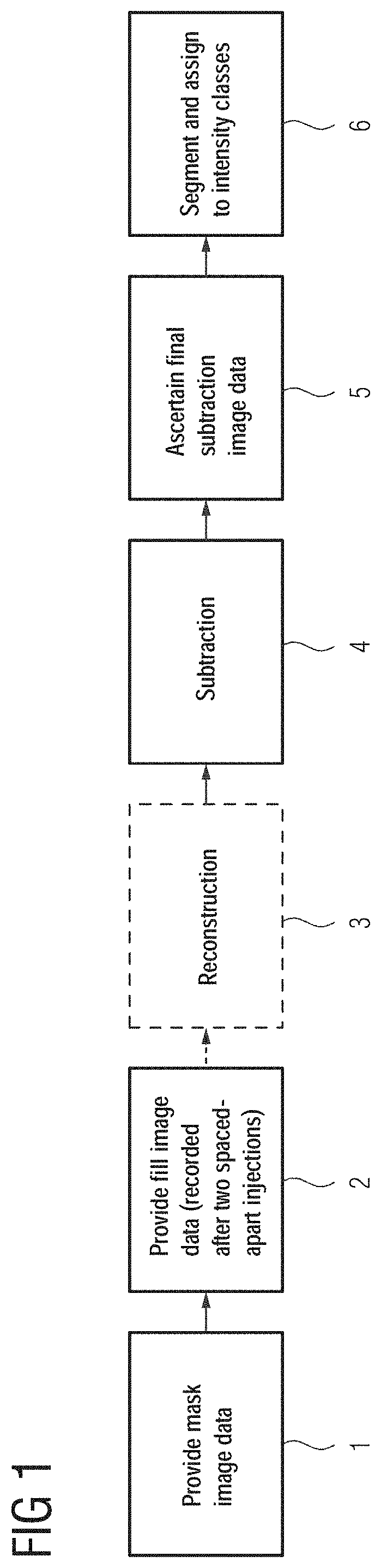
[0024]In FIG. 1, a sequence is shown of certain acts of a method for producing a digital subtraction angiography of a hollow organ system of a patient. The hollow organ system may, for example, involve a cerebral vascular system or also a vascular system in the heart or liver or another organ of a patient. By way of the method, the desired vascular system may be represented comprehensively and particularly highly perfused vessels, (e.g., AVM-draining veins), may be identified.

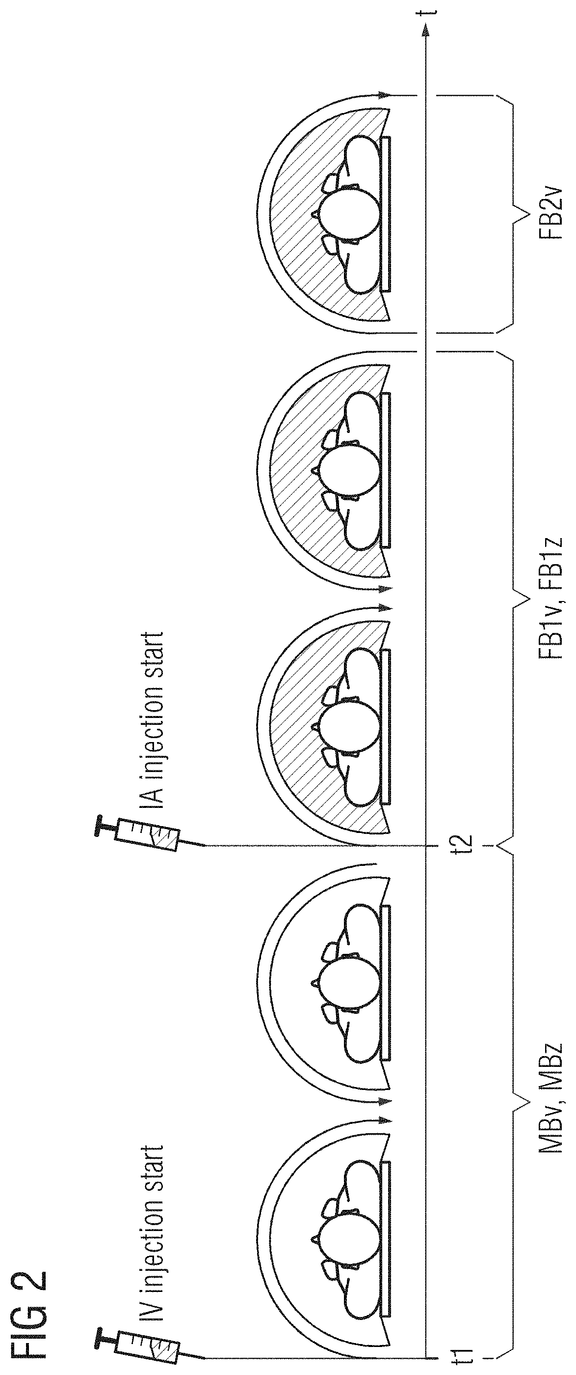
[0025]In act 1, mask image data of the hollow organ system recorded by an X-ray device is provided and, in act 2, at least first fill image data is provided which has been recorded by the same X-ray device during an at least partial filling of the hollow organ system with a contrast agent, starting from a first intravenous and a second intraarterial contrast agent injection following in time. Depending on the application, the image data may involve 2D, 3D, or 4D image data. An exemplary 4D DSA acquisition proto...


PUM


Abstract

For a particularly comprehensive identification of hollow organ systems, a method is provided for producing a digital subtraction angiography of a hollow organ system of a patient. The method includes: providing mask image data recorded by an X-ray device; providing at least first fill image data recorded by the X-ray device, which has been recorded during an at least partial filling of the hollow organ system with a contrast agent; starting from a first intravenous and a second intraarterial contrast agent injection following in time; ascertaining at least first subtraction image data by subtracting the mask image data from the at least first fill image data; ascertaining final subtraction image data from the at least first subtraction image data; and segmenting the final subtraction image data and assigning the pixels or voxels of the final subtraction image data to at least two different intensity classes based on their respective intensity value.

Description

[0001]The present patent document claims the benefit of U.S. Provisional Patent Application No. 62 / 904,897, filed Sep. 24, 2019, and German Patent Application No. 10 2019 215 904.4, filed Oct. 16, 2019, which are hereby incorporated by reference.TECHNICAL FIELD[0002]The disclosure relates to a method for producing a digital subtraction angiography of a hollow organ system of a patient in accordance as well as an apparatus for performing a method of this kind.BACKGROUND[0003]Digital subtraction angiography (DSA) may serve to examine blood vessels or hollow organs. In one example, two-dimensional variants, temporally successive (two-dimensional) projection images of a vascular system of a patient to be examined are produced by an X-ray angiography system (e.g., by a C-arm system), during which time a contrast agent is injected. This results in one projection image without contrast agent, also known as the mask image, and further projection images with a distribution of contrast agent ...

Claims


Application Information

Patent Timeline
IPC IPC(8): G06T7/00G06T7/11G06T11/00A61B6/00
CPCG06T7/0014G06T7/11G06T11/001G06T2207/30101A61B6/504G06T2207/10121A61B6/481A61B6/52A61B6/54A61B6/4441A61B6/5235G06T5/50G06T2207/20221G06T11/008G06T2211/404
Inventor KOWARSCHIK, MARKUSSCHAFER, SEBASTIAN
Owner SIEMENS HEATHCARE GMBH
Why Eureka
Try Eureka