Handheld optical diagnostic device having image system array

a technology of image system array and optical diagnostic device, which is applied in the field of clinical chemistry, can solve the problems of inability to carry, inability to accurately measure, and the aggregation of samples from multiple patients, and achieves the effect of improving accuracy and accuracy

Inactive Publication Date: 2007-12-25
SIEMENS HEALTHCARE DIAGNOSTICS INC
View PDF5 Cites 13 Cited by
  • Summary
  • Abstract
  • Description
  • Claims
  • Application Information

AI Technical Summary

Problems solved by technology

Color comparisons made with the naked eye can lead to imprecise measurement.
A common feature of these instruments is that their relative size and complexity, particularly with respect to those utilizing automated test pad transport systems, render them relatively unportable.
Unfortunately, such aggregation of samples from multiple patients presents opportunities for error due to mislabeling of the samples and / or the test results.
Moreover, in many instances, the time required for transporting the samples to and from the processing center, and for testing and recording the results, may be problematic.

Method used


Image

  • Handheld optical diagnostic device having image system array
  • Handheld optical diagnostic device having image system array
  • Handheld optical diagnostic device having image system array

Examples


Embodiment Construction

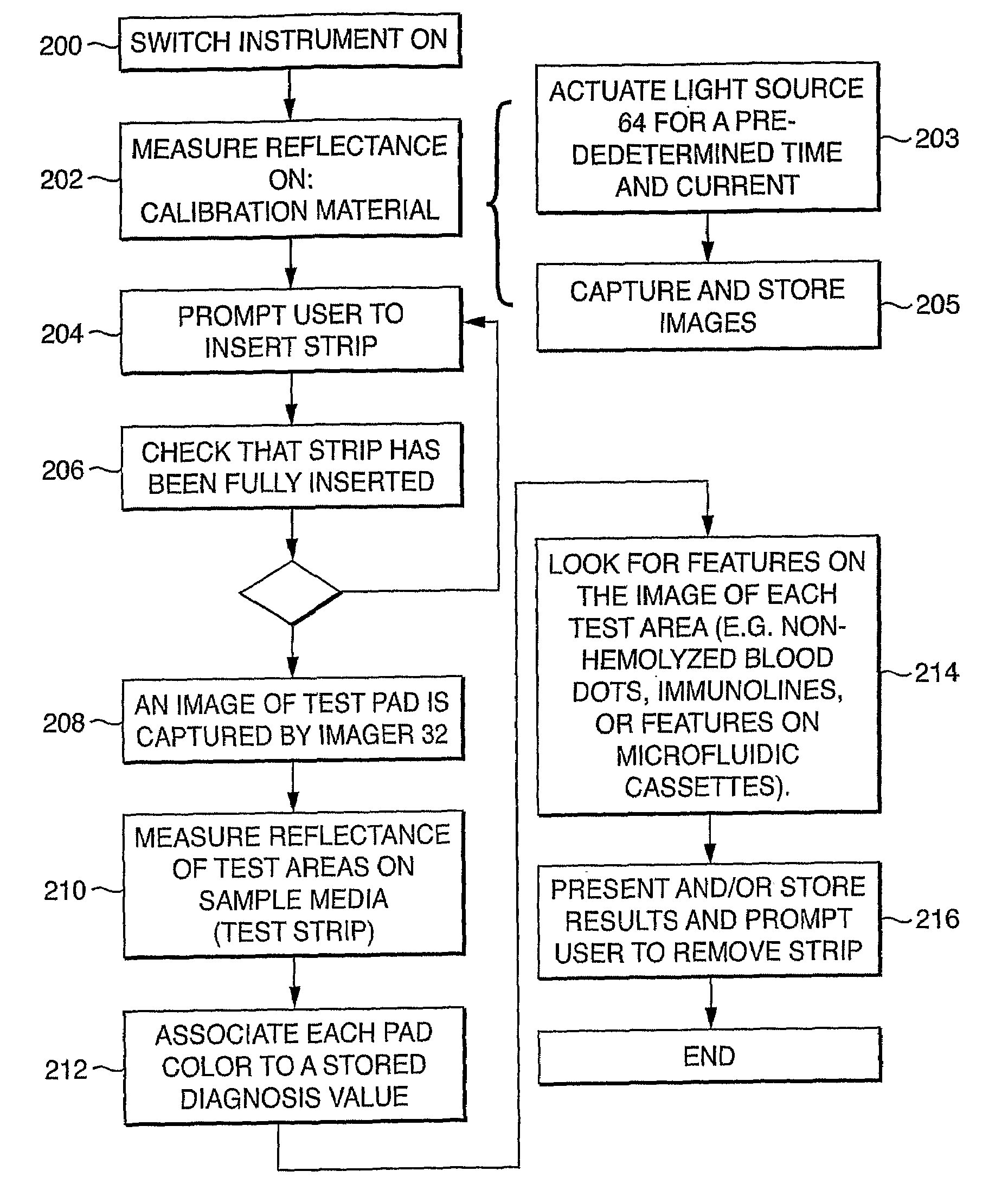
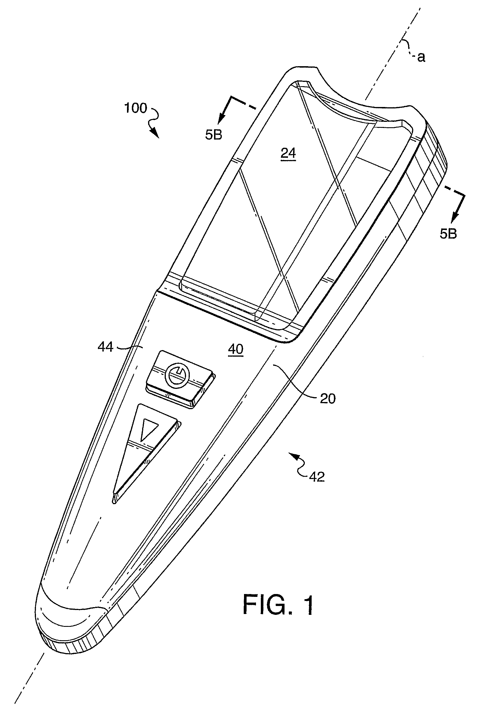
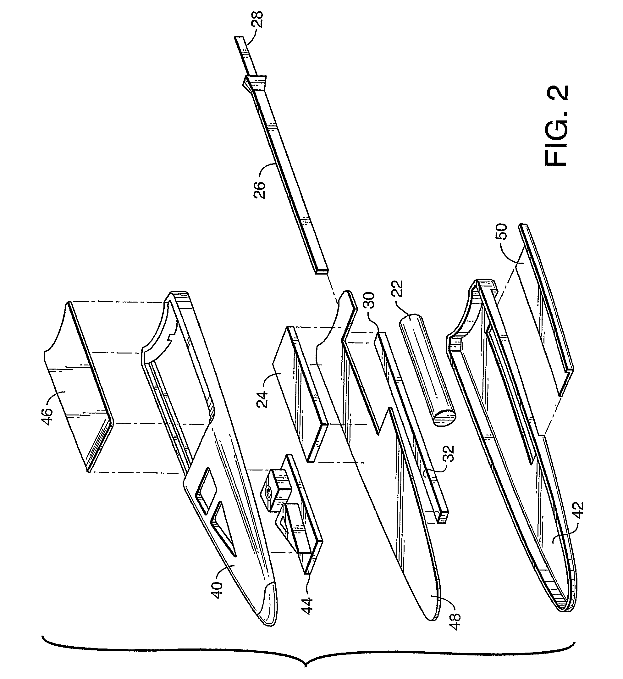
[0028]In the following detailed description, reference is made to the accompanying drawings that form a part hereof, and in which is shown by way of illustration, specific embodiments in which the invention may be practiced. These embodiments are described in sufficient detail to enable those skilled in the art to practice the invention, and it is to be understood that other embodiments may be utilized. It is also to be understood that structural, procedural and system changes may be made without departing from the spirit and scope of the present invention. The following detailed description is, therefore, not to be taken in a limiting sense, and the scope of the present invention is defined by the appended claims and their equivalents. For clarity of exposition, like features shown in the accompanying drawings are indicated with like reference numerals and similar features as shown in alternate embodiments in the drawings are indicated with similar reference numerals.

[0029]An overv...


PUM

PropertyMeasurementUnit
voltageaaaaaaaaaa
reflectanceaaaaaaaaaa
percent reflectanceaaaaaaaaaa

Abstract

A self-contained, optical hand-held diagnostic device is provided with a body having a pocket-sized form factor sized and shaped for engagement by a user's hand. The body includes an integral power supply and an integral display. A channel receives reagent sample media in an indexed fit. The sample media has a plurality of spaced test areas which change color according to an amount of a constituent or property in the sample. Imagers are located within the body so that each of the imagers is superposed with one of the test areas when the sample media is indexed within the channel, to capture an image thereof. A processor is coupled with the imagers to analyze the captured images. The processor also derives a diagnosis value from the analysis, and generates an output corresponding thereto. The display is configured to receive and display the output.

Description

BACKGROUND[0001]1. Technical Field[0002]The present invention generally relates to the field of clinical chemistry. More particularly, the present invention relates to a diagnostic imaging system that analyzes the color change associated with one or more test areas on sample media following contact thereof with a liquid specimen, such as urine or blood.[0003]2. Background Information[0004]Throughout this application, various patents are referred to by an identifying citation. The disclosures of the patents referenced in this application are hereby incorporated by reference into the present disclosure.[0005]Reagent test strips are widely used in the field of clinical chemistry. A test strip usually has one or more test areas, and each test area is capable of undergoing a color change in response to contact with a liquid specimen. The liquid specimen usually contains one or more constituents or properties of interest. The presence and concentrations of these constituents or properties...

Claims


Application Information

Patent Timeline
Patent Type & Authority Patents(United States)
IPC IPC(8): G06K9/00G01N21/27G01N21/47G01N21/86
CPCG01N21/253G01N21/276G01N21/8483G01N2201/0222G01N2201/0221
Inventor ROMAN, JUAN FELIX
Owner SIEMENS HEALTHCARE DIAGNOSTICS INC
Why Eureka
Try Eureka