Computed tomography scanning

a computed tomography and computed tomography technology, applied in image data processing, diagnostics, radiation therapy, etc., can solve the problems of not being able to select slices from the resulting data, the technique used in conventional ct scanners cannot be used directly, and the motion cannot be measured in the reconstruction volume data, so as to remove significant uncertainties and normal amplitude and pattern of breathing

Inactive Publication Date: 2008-04-08
ELEKTA AB
View PDF14 Cites 151 Cited by
  • Summary
  • Abstract
  • Description
  • Claims
  • Application Information

AI Technical Summary

Benefits of technology

[0008]The techniques used in conventional CT scanners cannot be used directly in a cone beam system as the data is acquired in 2D projection images, and therefore slices cannot be selected from the resulting data. However, respiration correlation techniques could be applied to the acquired projection images rather than the reconstructed CT volume. To achieve this, we propose monitoring the phase of the patients breathing while acquiring projection images continuously. On completion of the acquisition, projection images that have comparable breathing phases can be selected from the complete set, and these are used to reconstruct the volume data using similar techniques to those of conventional CT. An advantage is that any phase can be selected and therefore the effect of breathing can be studied.
[0010]It is known in the field of convention CT scanning to be advantageous to prompt the patient visually and audibly in order to ensure a regular amplitude and pattern of breathing. Techniques such as these could usefully be applied in the present invention. Furthermore this feature in the projection images can be used to control delivery of therapeutic radiation dependent on the patient's breathing cycle, to ensure that the tumour is in the correct position when the radiation is delivered. This will provide a direct measure of the patient's breathing phase, a significant improvement as compared to current methods that use external markers affixed to the patient. The use of a 3D volume data set generated using the same patient position and contemporaneous with the treatment will remove significant uncertainties.

Problems solved by technology

We have however found that there are other artifacts in the reconstructed volume data of cone beam CT systems, which we have traced to patient breathing movements.
In addition, the motion is not measurable in the reconstructed volume data.
This can be a particular problem in cone beam systems due to the long time required for acquisition, typically 1-2 minutes.
The techniques used in conventional CT scanners cannot be used directly in a cone beam system as the data is acquired in 2D projection images, and therefore slices cannot be selected from the resulting data.

Method used


Image

  • Computed tomography scanning
  • Computed tomography scanning
  • Computed tomography scanning

Examples


Embodiment Construction

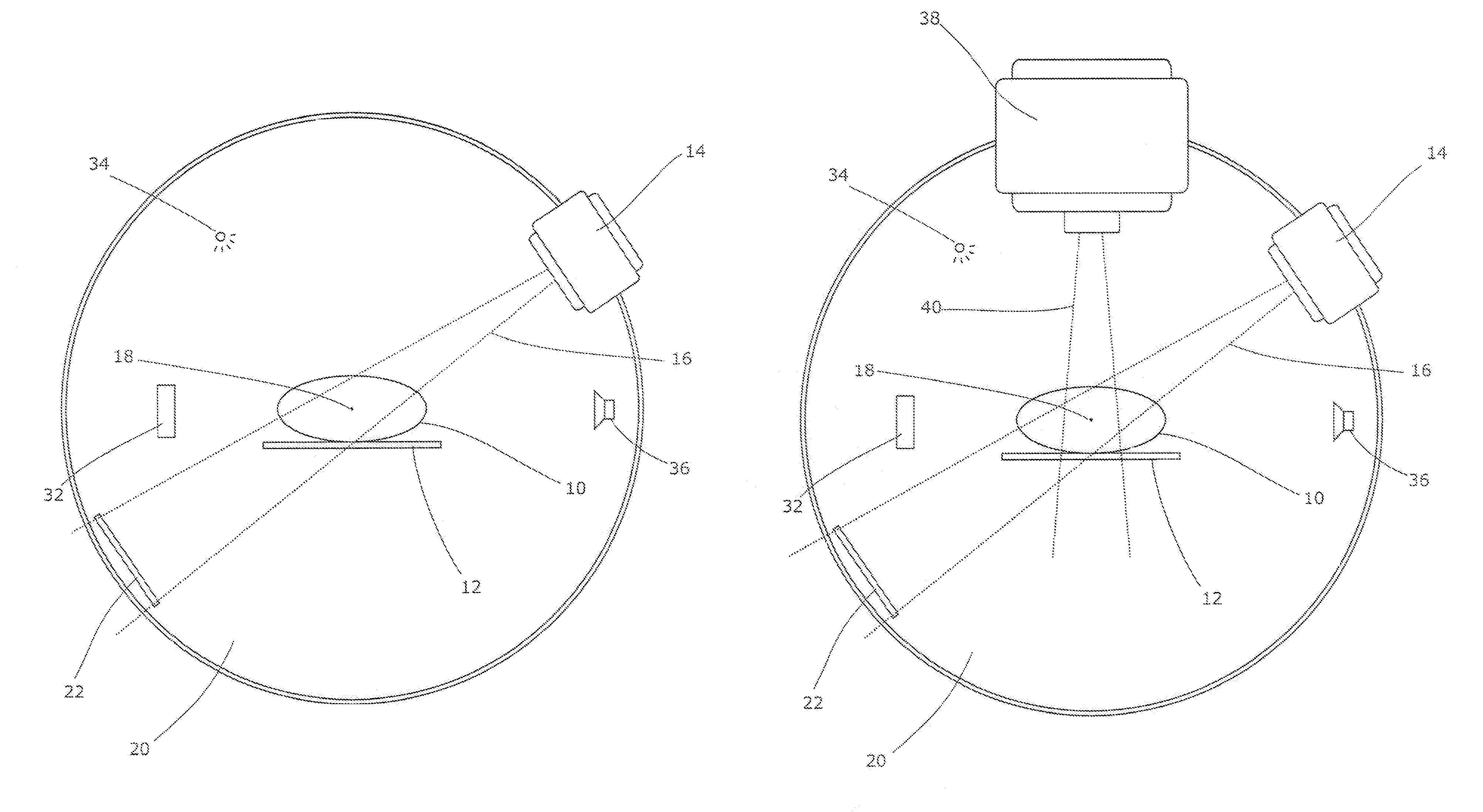
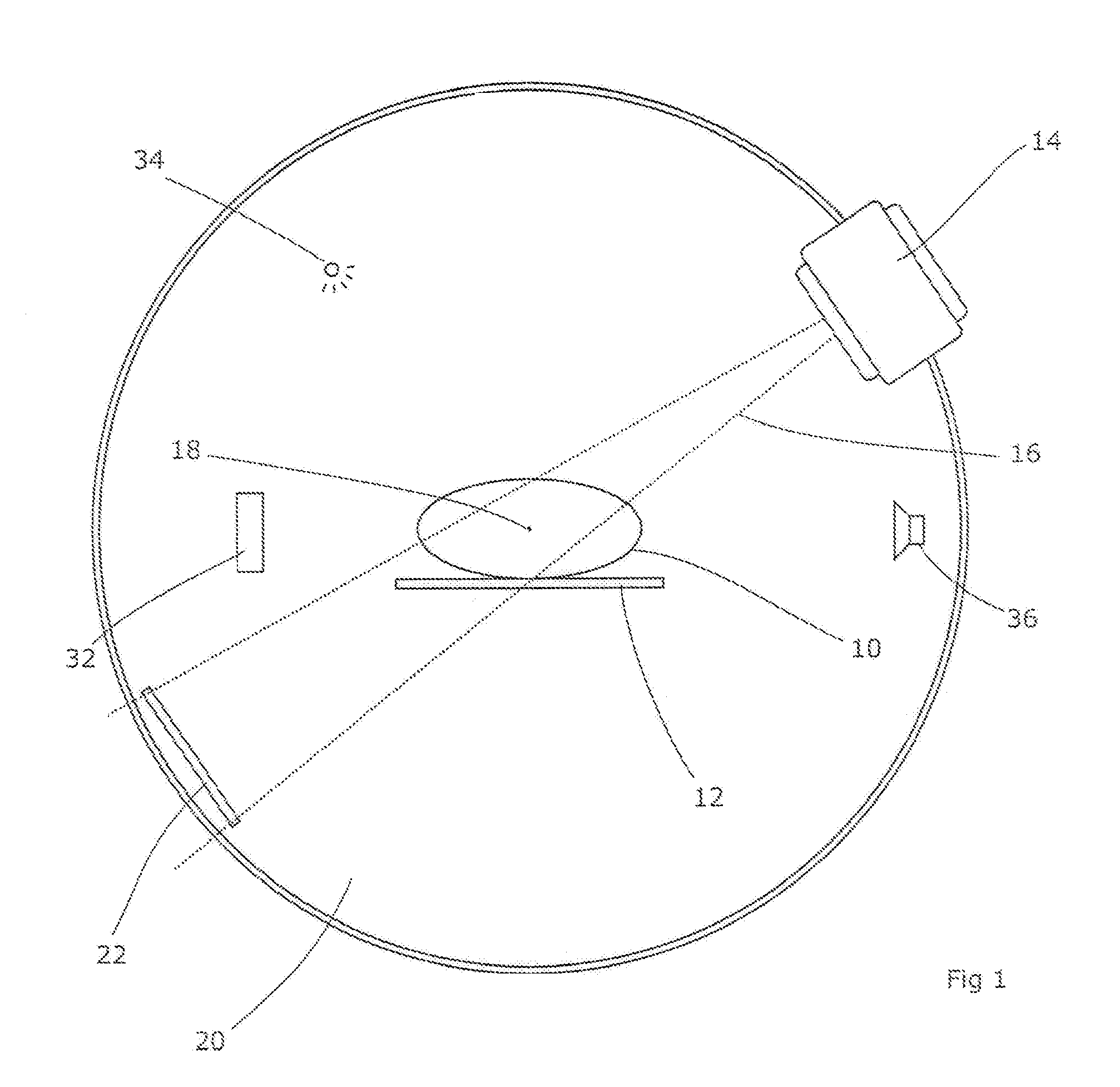
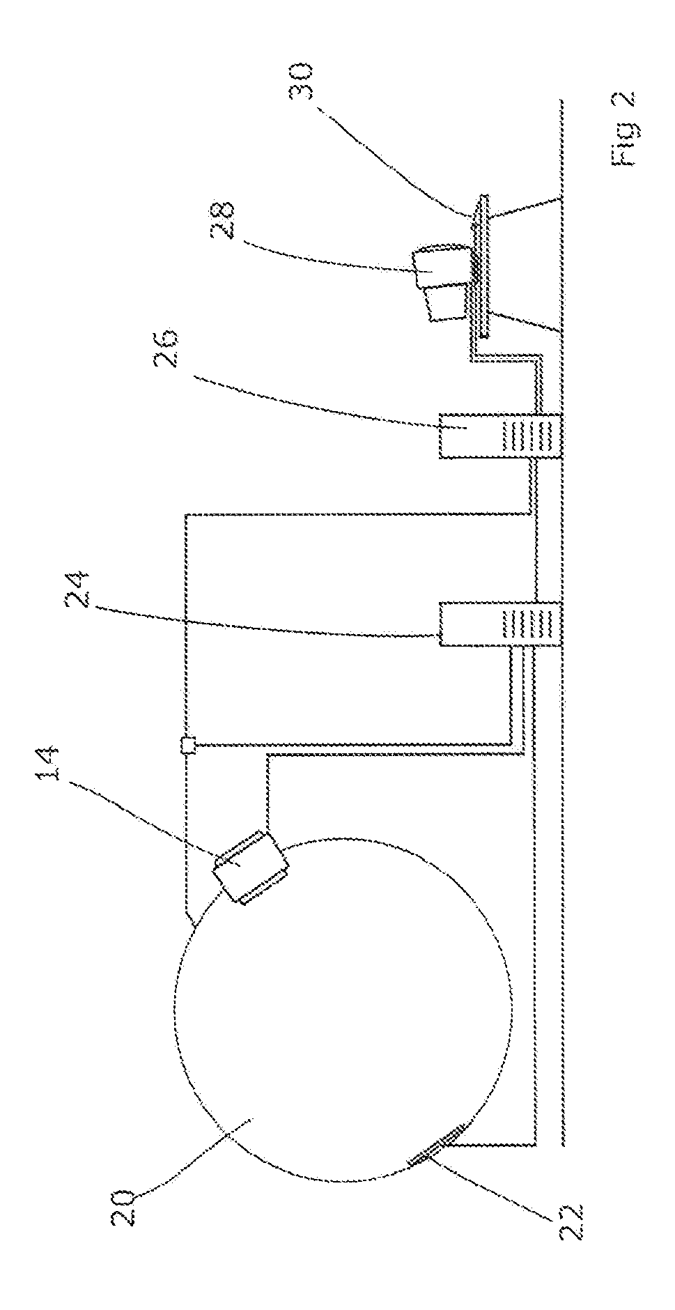
[0016]FIG. 1 shows a cone beam CT scanner. A patient 10 is supported on a couch 12 which may be of any suitable design. Couches typically allow the elevation and longitudinal position of the patient to be adjusted and this may be provided for as desired. An x-ray source 14 is arranged to project a wide beam 16 of radiation generally directed towards the isocentre 18 of the patient. The source 14 is rotatable around the isocentre 18 on a rotational support 20. The support can, for example, be in the form of a ring or annulus around the patient 10 and couch 12 in which the source is mounted, or it can be a C-arm, or any suitable support allowing the source to rotate, or any combination thereof. A two-dimensional flat-panel detector 22 is also mounted on the support 20, opposite the source 14 and arranged to rotate in synchronism therewith. If the support includes a C-arm then this can be achieved by mounting the detector on the opposite arm.

[0017]Thus, radiation emitted by the source ...


PUM


Abstract

Artifacts in the reconstructed volume data of cone beam CT systems can be removed by the application of respiration correlation techniques to the acquired projection images. To achieve this, the phase of the patients breathing is monitored while acquiring projection images continuously. On completion of the acquisition, projection images that have comparable breathing phases can be selected from the complete set, and these are used to reconstruct the volume data using similar techniques to those of conventional CT. Any phase can be selected and therefore the effect of breathing can be studied. It is also possible to use a feature in the projection images such as the patient's diaphragm to determine the breathing phase. This feature in the projection images can be used to control delivery of therapeutic radiation dependent on the patient's breathing cycle, to ensure that the tumor is in the correct position when the radiation is delivered.

Description

[0001]The present application is a continuation application of U.S. patent application Ser. No. 10 / 760,627, filed Jan. 20, 2004, now abandoned which in turn claimed priority from United Kingdom Patent Application 0327675.5, filed Nov. 28, 2003, and from United Kingdom Patent Application 0301278.8, filed Jan. 21, 2003; each of which is hereby incorporated by reference.FIELD OF THE INVENTION [0002]The present invention relates to scanning by computed tomography (CT).BACKGROUND ART [0003]CT scanning is a process for imaging the internal structure of a patient. In conventional CT scanning, a beam of x-rays is projected through the patient and its attenuation is measured. At the same time, the apparatus is rotated about an axis passing longitudinally through the patient. Thus, data is acquired as to the attenuation of the beam in each direction in the plane in which rotation takes place. From this data, the internal structure of the patient on that plane can be computed. The patient or a...

Claims


Application Information

Patent Timeline
Patent Type & Authority Patents(United States)
IPC IPC(8): A61B6/00A61N5/10G06K9/00G06T5/00G06T7/00G06T7/20G06T11/00H05G1/10
CPCG06T7/0016G06T7/20G06T11/005G06T5/003G06T5/009A61N5/1064G06T2207/10081G06T2207/20068G06T2207/30061G06T2207/30096G06T2211/412G06T2207/20192G06T5/92G06T5/73
Inventor BROWN, KEVIN JOHNJAFFRAY, DAVIDSIEWERDSEN, JEFFREY H.VAN HERK, MARCELSONKE, JAN-JAKOB
Owner ELEKTA AB
Why Eureka
Try Eureka