Ultrasonic device and method for tissue coagulation

a tissue coagulation and ultrasonic technology, applied in the field of surgical instruments, can solve the problems of diffuse bleeding, coagulated tissue to be re-opened and bleed, and undesirable thermal trauma to adjacent tissues, so as to improve coagulation, improve coagulation, and coagulate tissue.

Active Publication Date: 2008-04-22
SOLTA MEDICAL
View PDF58 Cites 198 Cited by
  • Summary
  • Abstract
  • Description
  • Claims
  • Application Information

AI Technical Summary

Benefits of technology

[0011]The present invention provides a novel, improved ultrasonic surgical device for and method for coagulating tissue. The device of the present invention has a surgical handle with an ultrasonic transducer mounted therein for generating ultrasonic vibrations. An ultrasonic applicator is attached to the ultrasonic transducer for transmitting longitudinal ultrasonic vibrations and extends distally from the surgical handle. The ultrasonic applicator is generally and substantially round in cross-section at the distal end and has a diameter between approximately 2 millimeters and 6 millimeters. A clamp with a jaw surface is supported on an elongated support member that is releasably attached to the surgical handle that generally surrounds the ultrasonic applicator along its length. The clamp and jaw surface are designed so that the clamp cannot be completely closed against the vibrating ultrasonic applicator, but is stopped at a predefined clearance, i.e., distance between the jaw surface and the vibrating ultrasonic applicator. This predefined clearance provides a zone for controlled tissue flow as the vibrating ultrasonic member heats the tissue. The shape and thickness of the predefined clearance determine the quality and final shape of the coagulated tissue. The predefined clearance may be varied between between about 0.075 to about 1.9 mm, and preferably between about 0.075 and about 0.75 mm, depending on the type and structure of the targeted tissue to be coagulated. The surgical device can include means for adjusting the clearance within this range. Thus, the vibrating ultrasonic applicator is not a vibrating “blade” and is not used for the cutting of tissues, but only for improved coagulation. The zone of controlled tissue flow also contributes to improved coagulation by creating an improved coagulation effect and by avoiding simultaneous cutting during coagulation. The thickness and shape of the tissue flow is carefully controlled.
[0012]If a cutting capability is desired with the surgical device, a separate non-ultrasonic cutting element may be provided that can be advanced and retracted to accomplish the cutting function as a separate step. The cutting element may be advanced after the coagulation has been completed, and the jaw is still closed to the maximum permitted extent. Preferably, the cutting element may be a surgical blade with a sharpened leading edge that cuts the coagulated tissue as it is advanced. Other forms of mechanical cutting tools can be employed. The surgeon can wait to advance the cutting tool until sufficient time has elapsed for the tissue to have been coagulated and “cooled” to minimize bleeding during the cutting process.

Problems solved by technology

Sutures are safe, reliable, and commonly used on larger vessels or structures, but are difficult to use on small vessels or structures or in situations involving diffuse bleeding.
It is effective on the smaller vessels and structures but may cause undesirable thermal trauma to adjacent tissues due to stray electrical conduction in a wet surgical environment.
Bipolar instruments may suffer from tissue adherence to the electrodes, causing the coagulated tissue to be re-opened and bleed again as the probes are removed.
This “end-on” design blocks tissue access to the clamped region between the ultrasonic member and clamp mechanism from the axial direction, requiring that tissue be accessed laterally, and thereby severely limits application of the device for surgical application because tissue cannot be accessed in a scissor-like fashion.
None of the patents discloses a device for limiting the closure of the clamp mechanism relative to the vibrating ultrasonic member for the purpose of creating a predetermined clearance there between so that an improved coagulation effect is achieved.
Holding the clamp against the ultrasonic member in the prior art devices will inevitably result in the cutting of the tissue.
The surgeon has no way of knowing how far the process has occurred from the intended coagulation to undesirable cutting.
It is impossible to reliably separate these two processes in the prior art devices.
Deficiencies in the performance of prior art coagulation devices have been noted in the literature.
While the authors personally concluded that the devices “can be considered safe,” the devices were not uniformly successful.
The LCS device was successful in all of the “small vessel” tests but had two complete failures in the twelve tests of medium-sized vessels and two additional instances where the medium-sized vessel commenced bleeding before the defined pressure limit was reached.
This is an unacceptable failure rate of 33%.
If the predefined clearance is greater than about 1.9 mm, it has been found that insufficient tissue flow is achieved and complete coagulation may not occur.

Method used


Image

  • Ultrasonic device and method for tissue coagulation
  • Ultrasonic device and method for tissue coagulation
  • Ultrasonic device and method for tissue coagulation

Examples


Embodiment Construction

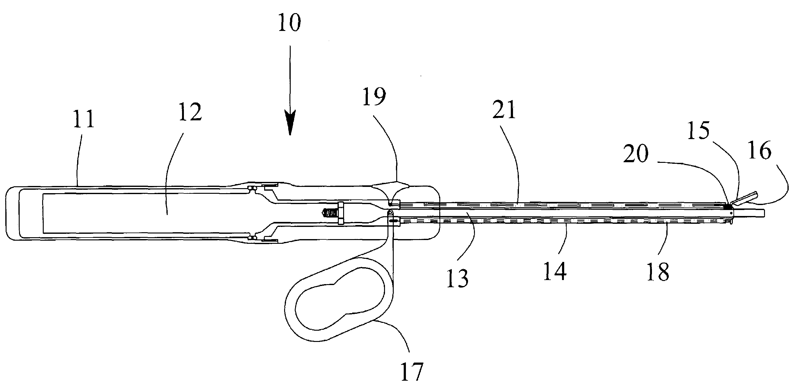
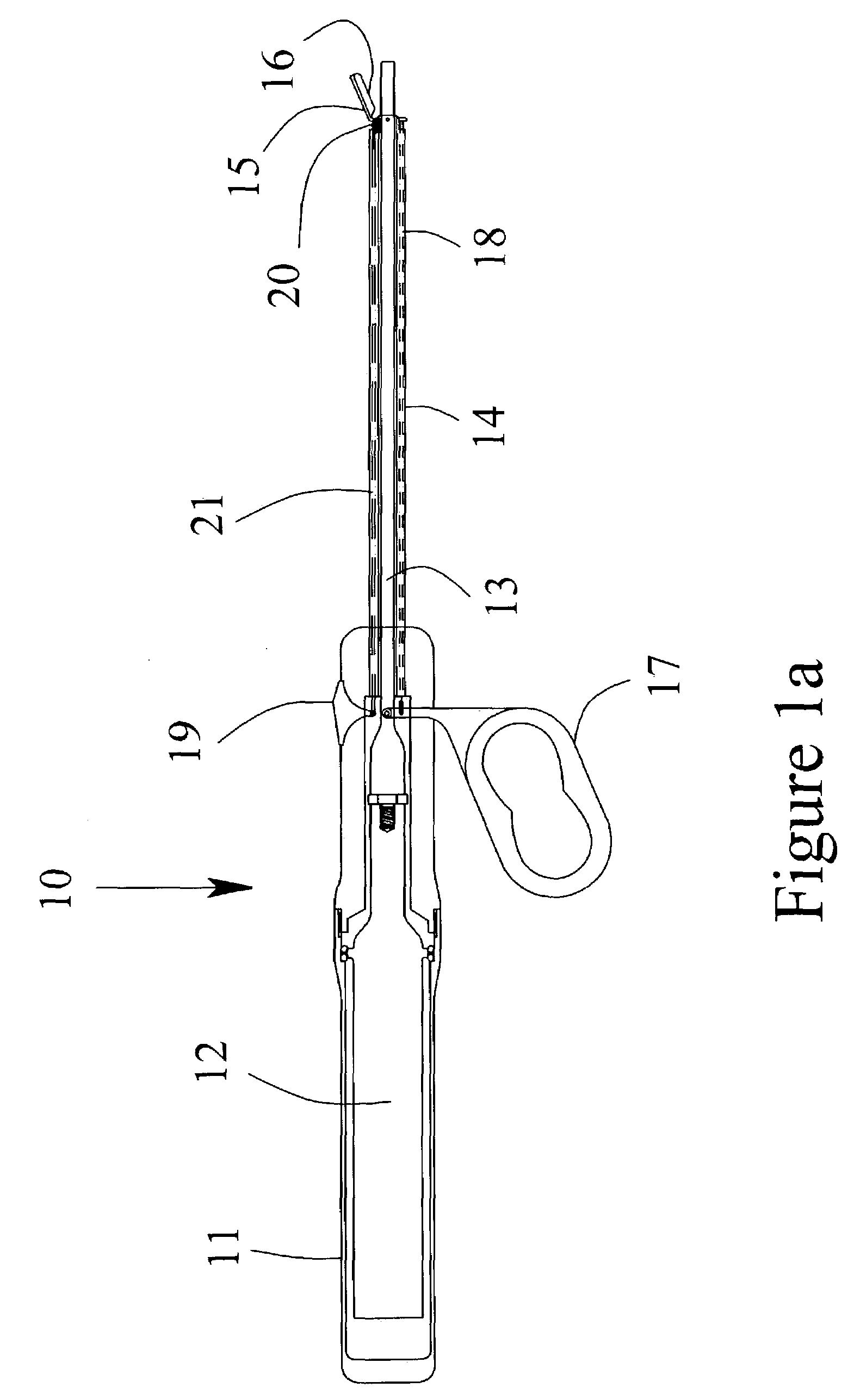
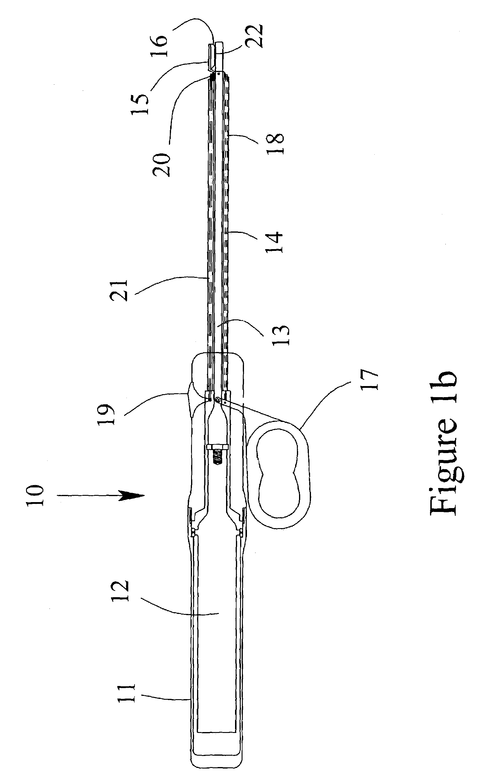
[0026]Referring to the drawings, FIG. 1a is a schematic representation of one preferred embodiment of the invention. FIG. 1 illustrates a partial cut-away view of the present invention including an ultrasonic surgical instrument, generally designated 10. The instrument has a surgical handle 11 to be held and manipulated by the surgeon. The surgical handle 11 may be fabricated from either machined or molded plastic components. An ultrasonic transducer 12 is mounted within the surgical handle 11 for generating ultrasonic vibrations. The ultrasonic vibrations may be generated using any common and well-known means such as the use of PZT crystals held in compression.

[0027]An ultrasonic applicator 13 is attached to the ultrasonic transducer 12 and extends distally from the ultrasonic transducer 12. The preferred method of attachment is a threaded joint. The ultrasonic applicator may be fabricated from any suitable metallic material including, for example, titanium alloys, aluminum alloys,...


PUM


Abstract

An ultrasonic surgical device for the coagulation of animal tissue having an ultrasonic applicator and a movable jaw with a jaw surface adjacent the distal portion of the ultrasonic applicator for movement toward the applicator to a closed position at a predefined clearance of between about 0.075 to about 1.9 millimeters from the applicator. The device may also include a mechanical cutting element that can be extended into the clearance to cut the tissue and means to vary the predefined clearance without removing the applicator from the patient. Tissue coagulating and cutting can be maximized and performed separately and can be easily monitored by the surgeon.

Description

[0001]This application claims the benefit of priority from U.S. provisional patent application Ser. No. 60 / 386,119, filed Jun. 4, 2002.I. FIELD OF THE INVENTION[0002]The present invention relates generally to surgical instruments, and more particularly, to an ultrasonic surgical device for use in coagulation of tissues of a patient.II. BACKGROUND OF THE INVENTION[0003]Hemostasis of bleeding or potentially bleeding tissues is of premium importance in open or laparoscopic surgery. Several methods are currently used to coagulate tissues to achieve the desired hemostasis. Sutures are safe, reliable, and commonly used on larger vessels or structures, but are difficult to use on small vessels or structures or in situations involving diffuse bleeding. Monopolar electrosurgery works by electrically heating and burning the tissue to achieve coagulation. It is effective on the smaller vessels and structures but may cause undesirable thermal trauma to adjacent tissues due to stray electrical c...

Claims


Application Information

Patent Timeline
Patent Type & Authority Patents(United States)
IPC IPC(8): A61B18/04A61B17/00A61B17/32A61B18/00A61B18/18A61NA61N7/02
CPCA61B17/320092A61N7/02A61B2017/00504A61B2017/320095A61B2017/320094A61B2017/320093A61B18/18
Inventor CIMINO, WILLIAM C.
Owner SOLTA MEDICAL
Why Eureka
Try Eureka