Method for computer-aided detection of three-dimensional lesions
What is AI technical title?
AI technical title is built by PatSnap AI team. It summarizes the technical point description of the patent document.
a three-dimensional, computer-aided technology, applied in the field of systems and methods for computer-aided detection of three-dimensionally extended organ lesions, can solve the problems of increasing the perceptual error, prone to perceptual errors, and time-consuming interrogation of an entire ctc examination, so as to maximize sensitivity, reduce false positives, and reduce false positives
Active Publication Date: 2008-05-27
UNIVERSITY OF CHICAGO
View PDF11 Cites 64 Cited by
Summary
Abstract
Description
Claims
Application Information
AI Technical Summary
This helps you quickly interpret patents by identifying the three key elements:
Problems solved by technology
Method used
Benefits of technology
Benefits of technology
[0084]An aspect of the present invention is the computation of novel three-dimensionally derived geometric features for the detection of lesions. These geometric features are employed specifically for differentiating lesions from folds and from the target organ wall, and thus they can be effective in maximizing sensitivity.
[0085]Another aspect of the present invention is the calculation of a novel three-dimensionally derived volumetric feature, directional gradient concentration, which characterizes the internal structures of lesions and folds, and is useful for reducing false positives.
[0086]Another aspect of the present invention is the combined, instead of independent, use of three-dimensionally derived volumetric features by a linear or a nonlinear classifier to reduce false positives.
Problems solved by technology
Currently, however, interpretation of an entire CTC examination is time-consuming [5, 8, 9].
In addition, the diagnostic performance of CTC currently remains undetermined and prone to perceptual errors.
These factors increase the perceptual error even for experienced radiologists.
The absence of visual cues which normally exist with colonoscopy, such as mucosal color changes, and a large number of images for each patient, also makes image interpretation tedious and susceptible to perceptual error.
Perceptual errors due to the large number of images are becoming more important as there is a tendency to use thinner collimation and reconstruction intervals for production of high-quality images [20].
However, these existing schemes suffered from either a low sensitivity or a high false-positive rate when they were applied to clinical cases.
Most of the previously proposed methods use a surface-generation method for extraction of the inner surface of the colonic wall [28, 30]; this has the risk of losing a part of a polyp, in particular, the internal structure of the polyp.
The colon was reportedly segmented satisfactorily, but a small amount of bowel or stomach was present in a majority of the segmentations, and the unoptimized version of the technique took sixty minutes to compute.
The segmentation was satisfactory in fifteen data sets, but there were problems in six datasets.
Method used
Image
Click on the blue label to locate the original text in one second.
Reading with bidirectional positioning of images and text.
Examples
Embodiment Construction
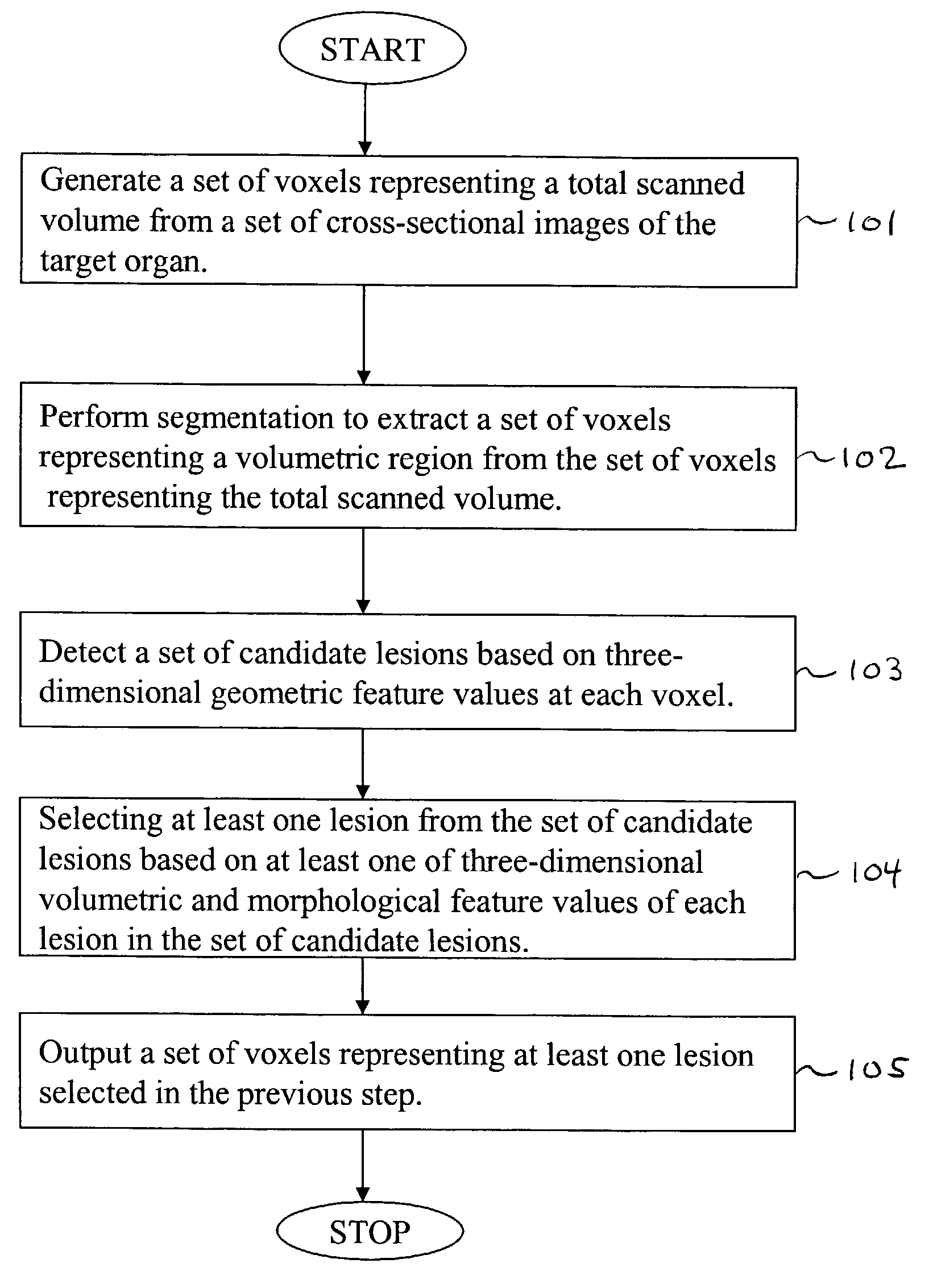
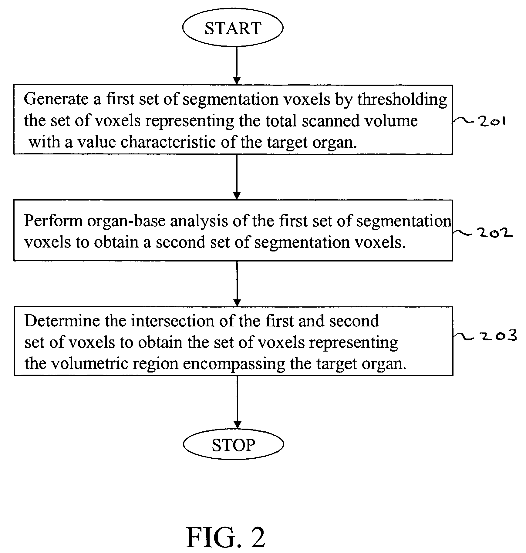
[0117]Referring now to the drawings, wherein like reference numerals designate identical or corresponding parts throughout the several views, FIG. 1 is a flowchart illustrating steps in the computer-aided detection of three-dimensionally extended lesions within a volumetric region encompassing a target organ, according to the present invention.
[0118]First, in step 101, a set of voxels representing a total scanned volume is generated from a set of cross-sectional images of the target organ. Note that in this context, we mean a “set” to include at least one member.
[0119]Next, in step 102, segmentation is performed to extract a set of voxels representing the thick volumetric region from the set of voxels representing the total scanned volume.
[0120]In step 103, a set of candidate lesions is detected based on three-dimensionally derived geometric feature values of each voxel in the set of voxels representing the thick volumetric region.
[0121]In step 104, at least one three-dimensionally ...
PUM
Abstract
A method, system, and computer program product for identifying at least one three-dimensionally extended lesion within a volumetric region encompassing an inner surface, an outer surface, and intervening tissue of a target organ. The method includes: (1) generating a set of voxels representing a total scanned volume from a set of cross-sectional images of the target organ; (2) performing segmentation to extract a set of voxels representing the volumetric region from the set of voxels representing the total scanned volume; (3) detecting a set of candidate lesions based on geometric feature values of each voxel in the set of voxels representing the volumetric region; and (4) selecting the at least one three-dimensionally extended lesion from the set of candidate lesions based on at least one of volumetric, morphologic, and texture feature values of each lesion in the set of candidate lesions.
Description
CROSS-REFERENCE TO CO-PENDING APPLICATIONS[0001]The present application is related to and claims priority to U.S. Provisional Application Ser. No. 60 / 329,322, filed Oct. 16, 2001. The contents of that application are incorporated herein by reference.BACKGROUND OF THE INVENTION[0002]1. Field of the Invention[0003]The present invention relates generally to systems and methods for the computer-aided detection of three-dimensionally extended organ lesions.[0004]The present invention also generally relates to automated techniques for the detection of abnormal anatomic regions, for example, as disclosed, in particular, in one or more of U.S. Pat. Nos. 4,907,156; 5,133,020; 5,832,103; and 6,138,045; all of which are incorporated herein by reference.[0005]The present invention also generally relates to computerized techniques for automated analysis of digital images, for example, as disclosed in one or more of U.S. Pat. Nos. 4,839,807; 4,841,555; 4,851,984; 4,875,165; 4,918,534; 5,072,384; ...
Claims
Application Information
Patent Timeline
Application Date:The date an application was filed.
Publication Date:The date a patent or application was officially published.
First Publication Date:The earliest publication date of a patent with the same application number.
Issue Date:Publication date of the patent grant document.
PCT Entry Date:The Entry date of PCT National Phase.
Estimated Expiry Date:The statutory expiry date of a patent right according to the Patent Law, and it is the longest term of protection that the patent right can achieve without the termination of the patent right due to other reasons(Term extension factor has been taken into account ).
Invalid Date:Actual expiry date is based on effective date or publication date of legal transaction data of invalid patent.

