

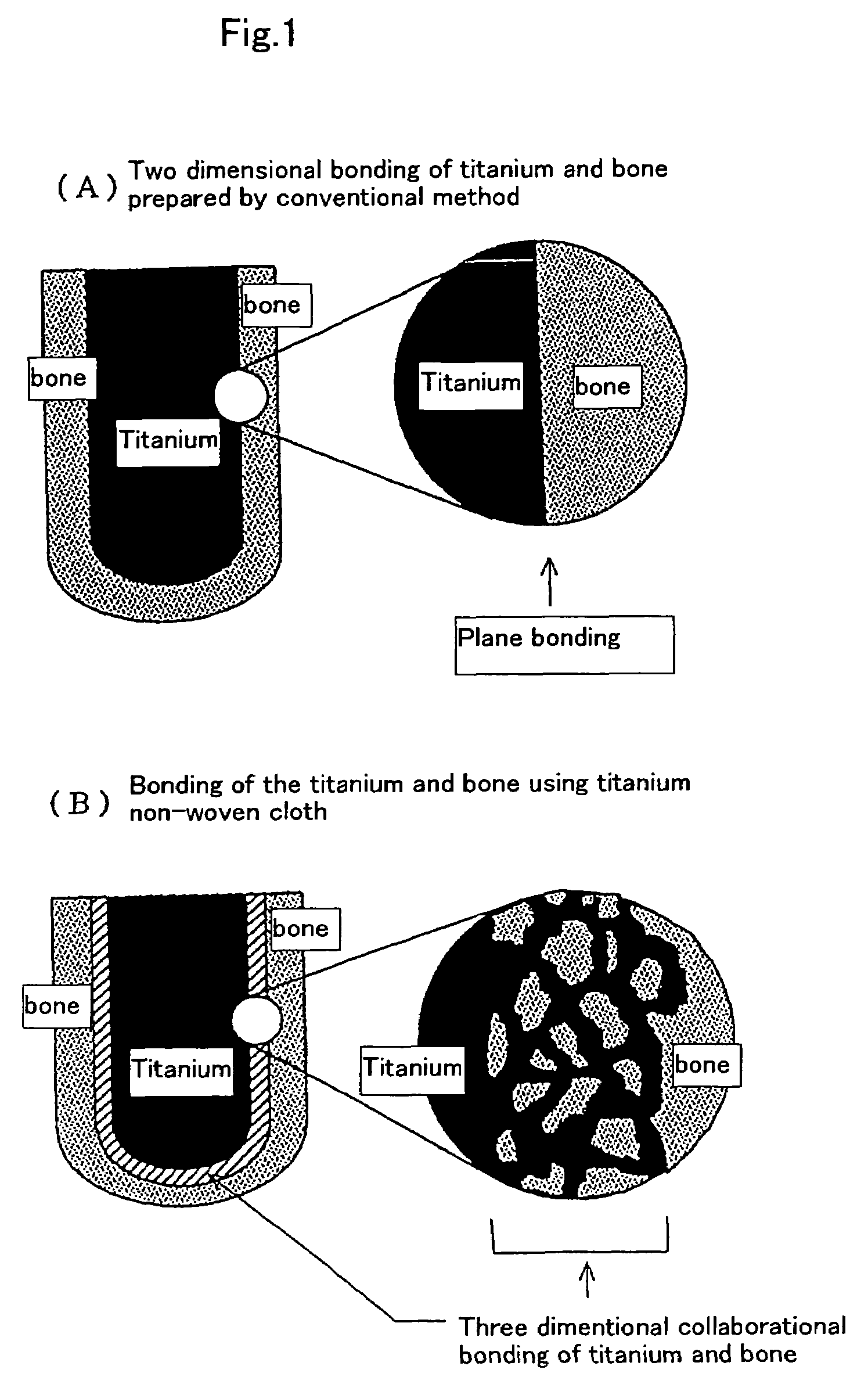
Medical implant having a layer of titanium or titanium alloy fibers
a technology of titanium alloy fibers and medical implants, which is applied in the direction of prosthesis, impression caps, on/in inorganic carriers, etc., can solve the problems of insufficient biological and mechanical insufficient satisfaction of medical materials composed of titanium or titanium group alloys, and insufficient bonding of metal materials with bone tissue, etc., to achieve excellent action and effect, improve the effect of health and welfar
- Summary
- Abstract
- Description
- Claims
- Application Information
AI Technical Summary
Benefits of technology
Problems solved by technology
Method used
Image

Examples
example 1
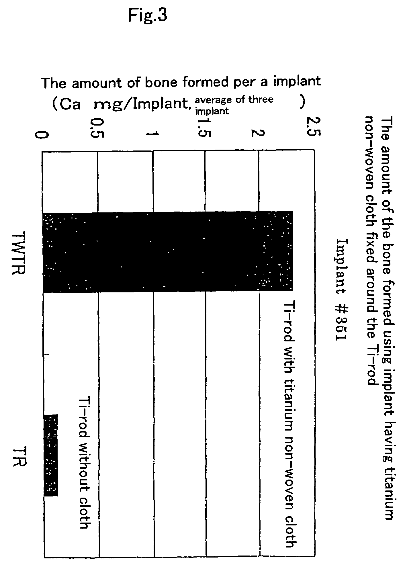
Heterotopic Bone Forming Experiment Under the Skin of Rat
[0059]I. Preparation of a Specimen for Experiment: Following Examples ① and ② are Prepared:
[0060]① Preparing a non-woven cloth of 85% void fraction and 0.9 g / mL density composed of titanium metal fiber having 8 μm-80 μm diameter and 20 or more aspect ratio (product of Bekinit Co., Ltd.). This non-woven cloth is wounded firmly to a titanium rod by a voluntarily thickness, and a composite consisting of titanium non-woven cloth and a titanium rod of 1.5 mm diameter is prepared. Said composite is filled in a sintering syringe made of ceramics and sintered at 1000° C. for 5 hours in high vacuum condition. Consequently, at many contacting points of fibers themselves and contacting points with titanium rod surface, fibers are fused. And thus the rigid composite which does not sink or does not cause the transformation of shape by adding forth on the surface is prepared.
① A titanium metal rod having 1.5 mm diameter.
[0061]II. Method for...
example 2
Experiment to Confirm the Effect of Hydroxyl Apatite Coating Treatment to the Formation of a Heterotopic Bone
[0066]Experimental Method:
[0067]After titanium non-woven cloth is put on a titanium rod having 1.5 mm diameter without sintering in vacuum following two composites are prepared. That is, apatite coated composite ③ prepared by a liquid dipping method which is an apatite coating treatment disclosed in Example 4 mentioned later, and non apatite coated composite ④ are prepared. These composites are implanted under the skin of rat for 4 weeks and the difference of bone tissue formation between these two composites is compared.
[0068]Experimental Results:
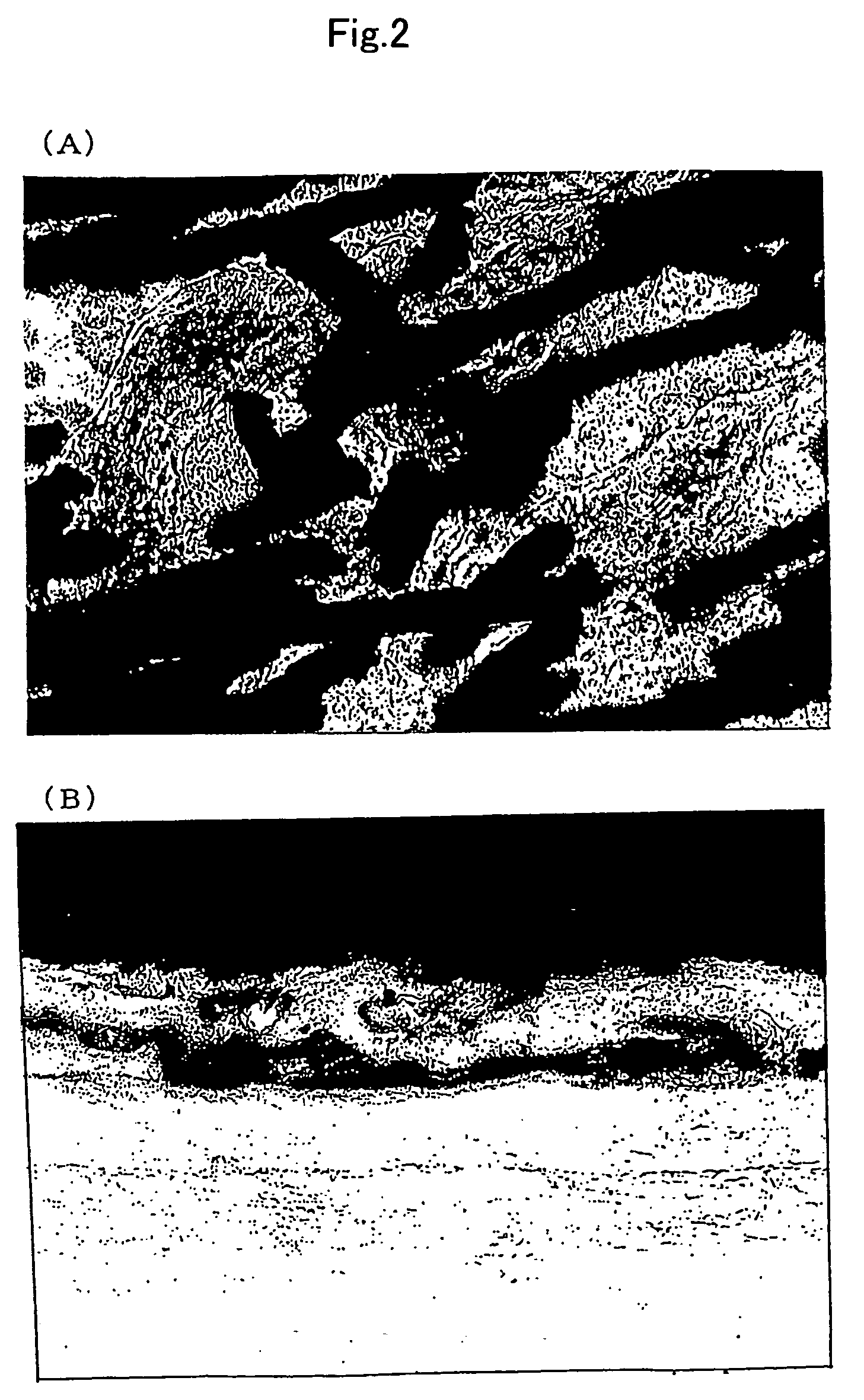
[0069]Results are shown in FIG. 4. In the apatite coated composite ③, vigorous bone formation is observed at the titanium non-woven cloth part. Bone is clearly distinguished as the red colored areas or as thicker and fused contour in gray or black FIG. 4 (A). However, since the titanium non-woven cloth is not treated by sintering in...
example 3
Orthotopic Bone Forming Experiment in Head Bone of Rabbit
[0073]I. Experimental Method
Experiments are carried out according to the procedures 1, 2 and 3 mentioned below.
[0074]1. A rabbit of 2.5 kg weight is anesthetized by nenbutal intravenous anesthesia, and perioste of head bone is partially incised and a hole of 3 mm diameter and 3 mm thickness which passes through the calvarial bone is dug at the parietal area by a diamond round disk for dental use.[0075]2. A titanium rod equipped with titanium non-woven cloth (cut off to cylindrical shape of 3 mm diameter and 3 mm thickness) is inserted into the hole and perioste and dermis is closured.[0076]3. The rabbit is killed after 4 weeks and the bone at the parietal area is removed and embeded by resin, then a ground specimen of 20 μm thickness is prepared. The specimen is dyed by hematoxylin-eosin dying method.
[0077]II. Experimental Results:
[0078]A tissue section specimen for microscope observation obtained in above item 3 is inspected ...
PUM
| Property | Measurement | Unit |
|---|---|---|
| diameter | aaaaa | aaaaa |
| aspect ratio | aaaaa | aaaaa |
| diameter | aaaaa | aaaaa |
Abstract
Description
Claims
Application Information