
De-immunized streptokinase

a streptokinase and deimmunization technology, applied in the field of substantially nonimmunogenic proteins, can solve the problems of reduced antibody effectiveness, limited use of rodent monoclonal antibodies for therapeutic and in vivo diagnostic applications in man, and no method has provided any means of avoiding or eliminating immunogenic epitopes at framework:cdr junctions

Inactive Publication Date: 2008-12-16
MERCK PATENT GMBH
View PDF2 Cites 7 Cited by
  • Summary
  • Abstract
  • Description
  • Claims
  • Application Information

AI Technical Summary

Benefits of technology

[0009](c) modifying the amino acid sequence to eliminate at least one of the T-cell epitopes identified in step (b) thereby to eliminate or reduce the immunogenicity of the protein or part thereof when exposed to the immune system of the given species.
[0043]The invention has particular and widespread application in the field of therapeutic molecules including monoclonal antibodies whereby rodent or other non-human antibodies can be de-immunized for applications in humans and whereby previously humanized or chimeric antibodies with B- or T-cell epitopes could be converted into a less immunogenic form for use in humans. It will also be understood that even antibodies derived from human immunoglobulin genes such as antibodies derived from bacteriophage-display libraries (Marks et al., J Mol. Biol., 222: 581-597 (1991)), transgenic mice with human immunoglobulin genes (Bruggermann et al., Proc. Nat'l. Acad. Sci. USA, 86: 6709-6713 (1989)) and natural human monoclonal antibodies can carry B- and T-cell epitopes especially as somatic mutations are introduced into framework sequences in immunoglobulin genes during the maturation of antibodies. Therefore, de-immunization may be required in order to prepare such antibodies for use in humans. Finally, it will be understood that CDRs from any naturally derived antibodies have been subjected to selection by somatic mutation of V region genes and thus might have T-cell epitopes capable of triggering immune responses in humans. The de-immunization method might be applicable without severe loss of antibody binding affinity (depending on the contribution of particular CDRs to antigen binding).

Problems solved by technology

The use of rodent, especially mouse, monoclonal antibodies for therapeutic and in vivo diagnostic applications in man was found to be limited by immune responses made by patients to the rodent antibody.
The development of so-called “HAMA” (human anti-mouse antibody) responses in patients was shown to limit the ability of antibodies to reach their antigenic targets resulting in a reduced effectiveness of the antibodies.
However, the means for achieving this objective has been the introduction into the rodent antibody of as much human sequence as possible and it has been assumed that such a general introduction of human sequence will render the antibodies non-immunogenic.
It is known that certain short peptide sequences (“epitopes”) can be immunogenic in humans and none of the methods for chimeric or humanized antibodies have considered how to eliminate or avoid such epitopes in the resultant antibody.
Furthermore, most of the methods (e.g. EP-B-0451216) have advocated the introduction of non-human amino acids into human V region frameworks without considering the possible creation of immunogenic epitopes, and none of the methods has provided any means for avoiding or eliminating immunogenic epitopes at framework:CDR junctions and, where practical, within CDRs themselves.
Thus, of the methods devised with the objective of creating substantially non-immunogenic antibodies, none can be considered as actually achieving the creation of such substantially non-immunogenic antibodies.

Method used


Image

  • De-immunized streptokinase
  • De-immunized streptokinase
  • De-immunized streptokinase

Examples


example 2

[0097]In this example, a range of antibodies were tested in mice to compare immune responses. As a source of antibody to elicit an immune response in mice, the humanized VH fragment from Example 1 was deliberately altered to insert two murine MHC class II epitopes as shown in FIG. 8. This was undertaken by SOE PCR (Higuchi et al., Nucleic Acids Research, 16: 7351 (1988)) using primers as detailed in FIG. 9. Using methods as in Example 1, for the murine de-immunized version the MHC class II epitopes were removed from the altered humanized VH fragment and this was also veneered to substitute exposed residues from the murine 340 sequence. The resultant sequence is shown in FIG. 10 and the synthetic oligonucleotides used shown in FIG. 11.

[0098]The murine de-immunized VH fragment from above and the humanized and murine VH fragments from Example 1 were joined either to human or murine C region fragments of isotope IgG2. For human, a 7.2 kb HindIII-BamHI genomic fragment from IgG2 C region...

example 3

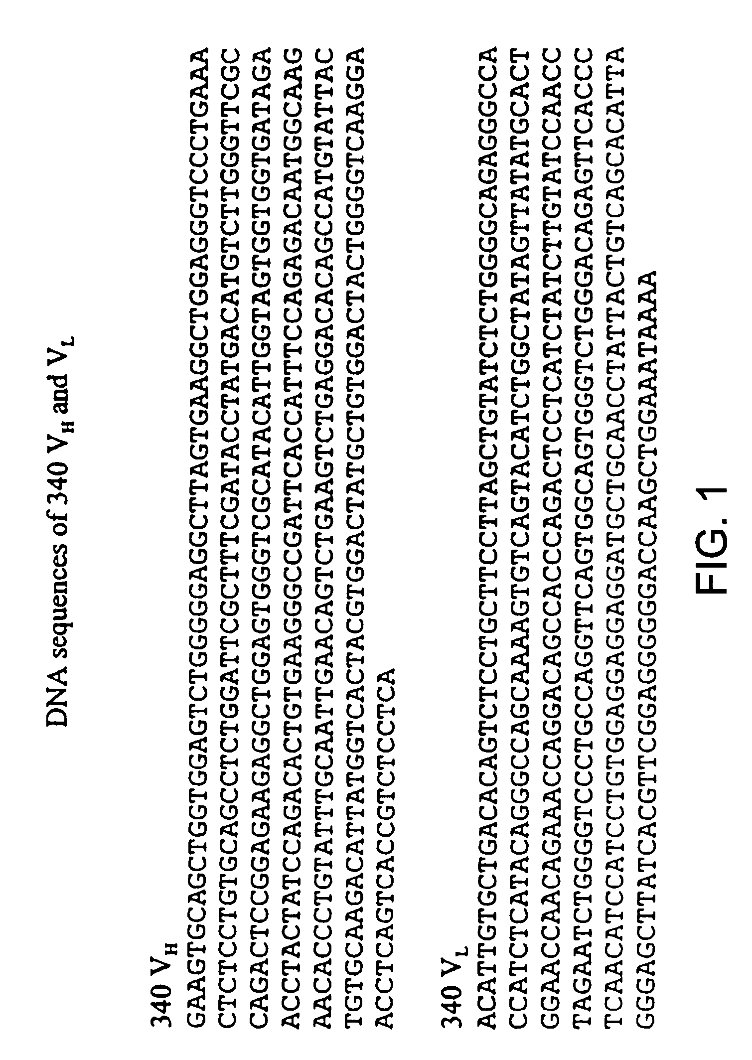
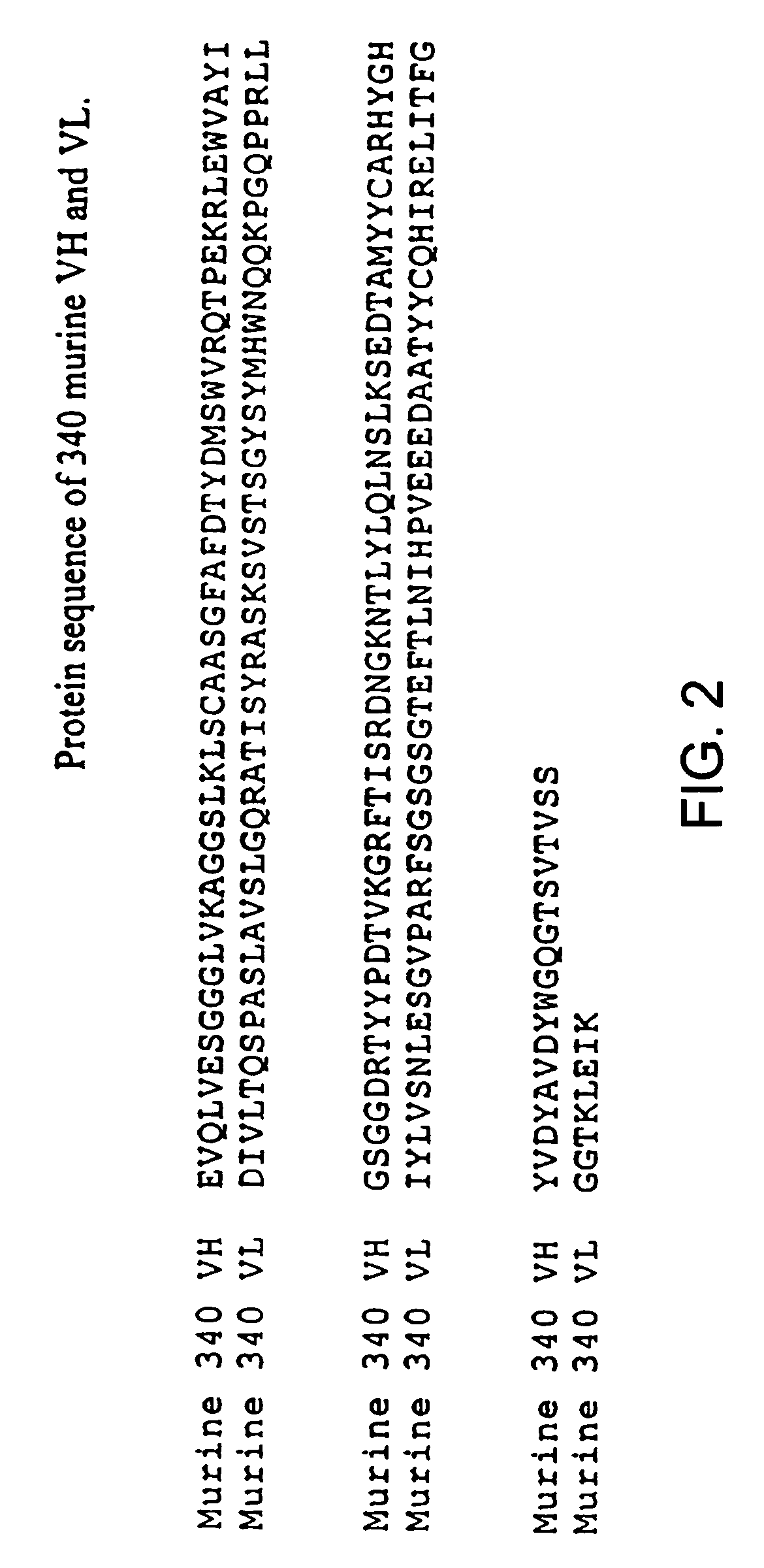
[0101]mRNA was isolated from 5×10⁶ hybridoma 708 cells (Durrant et al., Int. J. Cancer, 50: 811 (1992) using TRIZOL™ reagent (Life Technologies, Paisley, UK) according to the manufacturers' instructions. The MRNA was converted to cDNA / mRNA hybrid using READY-TO-GO™ T-primed First Strand Kit (Pharmacia Biotech, St. Albans, UK). Variable region heavy (VH) and light (VL) chain cDNAs were amplified using the primer sets using the method of Jones and Bendig (Bio / Technology, 9: 188 (1991)). PCR products were cloned into pBLUESCRIPT II SK (Stratagene, Cambridge, UK) or pCRTM3 (Invitrogen, The Netherlands) and six individual clones each of VH and VL were sequenced ion both directions using the Applied Biosystems automated sequencer model 373A (Applied Biosystems, Warrington, UK). Resultant VH and VL sequences are shown in FIG. 13 and the corresponding protein sequences in FIG. 14.

[0102]A de-immunized antibody was generated by analysis of the sequence of FIG. 14. To remove B-cell epitopes, t...

example 4

[0103]A set of vaccine molecules were constructed based on the 708 antibody. As before, the various VH and VL molecules were assembled from long synthetic oligonucleotides using the method of PCR recombination (Daugherty et al, ibid.). Cloning, sequencing, addition of human IgG1 and κ constant regions and expression in NS0 cells was as for the 340 antibody (Example 1).

[0104]The first antibody vaccine (“Vaccine 1”) comprised the 708 heavy and light chains from which all potential human T-cell epitopes have been removed from both antibody chains, using the method described in Example 1, including epitopes found in the CDRs, apart from the region encompassing CDRs 2 and 3 and framework 3 of the heavy chain which contains the desired human epitopes. The antibody chains were not “veneered” to remove B-cell epitopes. The resultant protein sequences are shown in FIG. 17. The oligonucleotides for assembly of 708 Vaccine 1 VH and VK are shown in FIG. 18. The primary PCR used oligonucleotides...


PUM

PropertyMeasurementUnit
molecular weightaaaaaaaaaa
pharmaceutical compositionaaaaaaaaaa
structuresaaaaaaaaaa

Abstract

A target protein is rendered less immunogenic to a given species by (a) determining at least part of the amino acid sequence of the target protein; (b) identifying in the amino acid sequence one or more potential epitopes for T-cells (“T-cell epitopes”) of the given species; and (c) modifying the amino acid sequence to eliminate at least one of the T-cell epitopes identified in step (b) to reduce the immunogenicity of the protein when exposed to the immune system of the given species.

Description

CROSS-REFERENCE TO RELATED APPLICATIONS[0001]This application is a division of U.S. application Ser. No. 10 / 300,215, filed on Nov. 20, 2002, now U.S. Pat. No. 7,125,689, which is a continuation of U.S. application Ser. No. 09 / 438,136 filed on Nov. 10, 1999, now abandoned, which is a continuation of International Application Serial No. PCT / GB98 / 01473, filed on May 21, 1998, which designates the United States and claims the benefit of U.S. Provisional Application Ser. No. 60 / 067,235, filed Dec. 2, 1997, the disclosures of which are incorporated herein by reference.FIELD OF THE INVENTION[0002]The present invention relates to the production of substantially non-immunogenic proteins, especially antibodies, and their uses. The invention uses a combination of recombinant DNA and monoclonal antibody technology for the generation of novel therapeutic and in vivo diagnostic agents for particular use in man.BACKGROUND OF THE INVENTION[0003]The use of rodent, especially mouse, monoclonal antibo...

Claims


Application Information

Patent Timeline
Patent Type & Authority Patents(United States)
IPC IPC(8): C12N9/70A61K39/395A61K38/43C07K14/00C12P21/06C12N9/00A61K38/00G01N33/563A61K38/45C07K14/315C07K16/46G01N33/68
CPCC07K14/3153C07K16/461G01N33/6878A61K38/00C07K2319/00
Inventor CARR, FRANCIS JOSEPHADAIR, FIONA SUZANNEHAMILTON, ANITA ANNECARTER, GRAHAM
Owner MERCK PATENT GMBH
Learn More