System and method for filtering noise from a medical image

a medical image and filtering technology, applied in the field of image processing, can solve the problems of not providing a method for choosing convergence criteria, not taking into account inhomogeneous data sampling, etc., and achieve the effect of increasing or decreasing the amount of noise removed

Active Publication Date: 2009-03-10
SIEMENS MEDICAL SOLUTIONS USA INC
View PDF5 Cites 41 Cited by
  • Summary
  • Abstract
  • Description
  • Claims
  • Application Information

AI Technical Summary

Benefits of technology

[0022]The Dirichlet boundary conditions are applied with an increased or decreased sampling density to respectively increase or decrease the amount of noise removed from the medical image.

Problems solved by technology

Medical images are frequently noisy, due both to varying image data and artifacts resulting from an acquisition system such as an ultrasound, positron emission tomography (PET) or computed tomography (CT) scanner.
A drawback of the technique proposed by Perona and Malik is that it does not it does not take into account inhomogeneous data sampling or enable the adjustment of tuning constants.
In addition, it does not provide a method for choosing convergence criteria (e.g., the stopping parameter).

Method used


Image

  • System and method for filtering noise from a medical image
  • System and method for filtering noise from a medical image
  • System and method for filtering noise from a medical image

Examples


Embodiment Construction

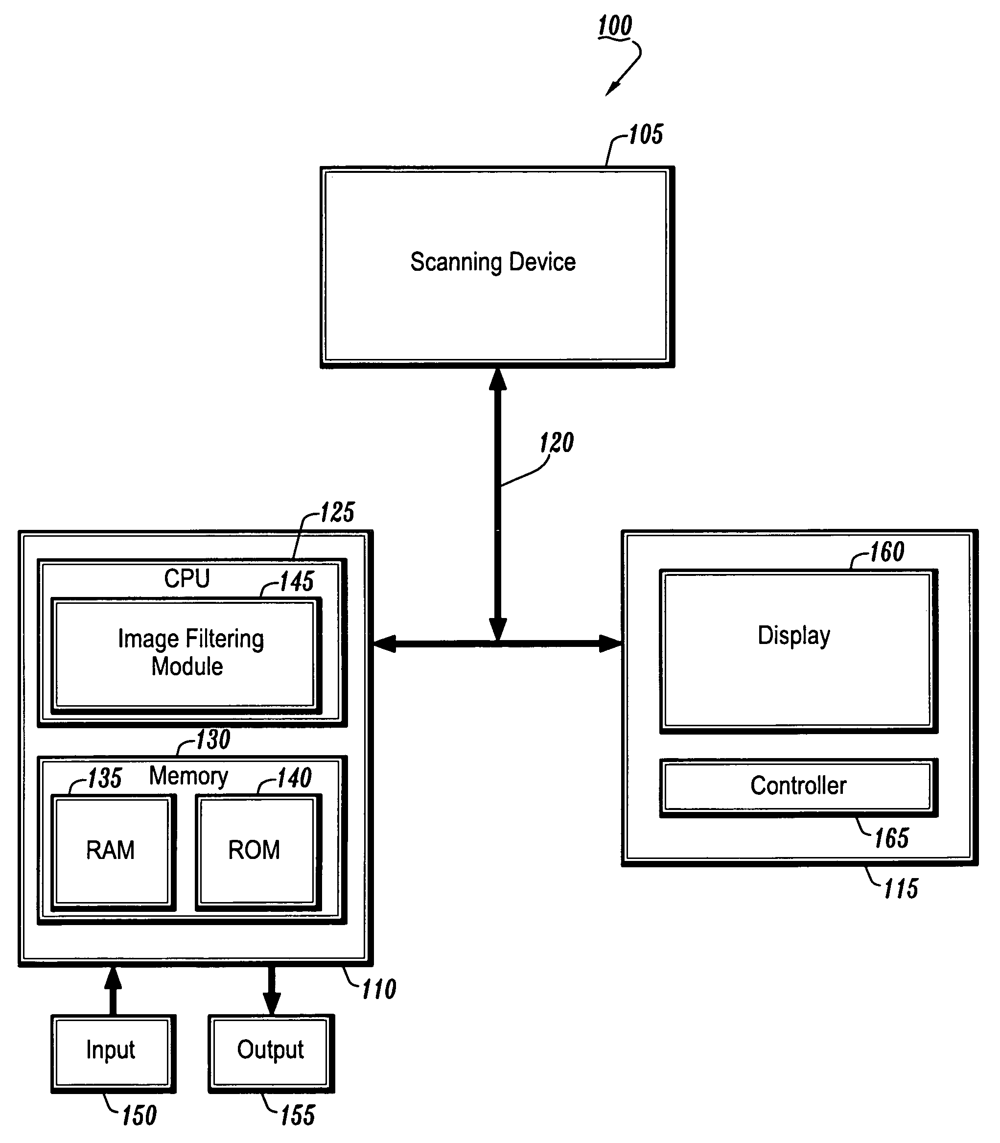
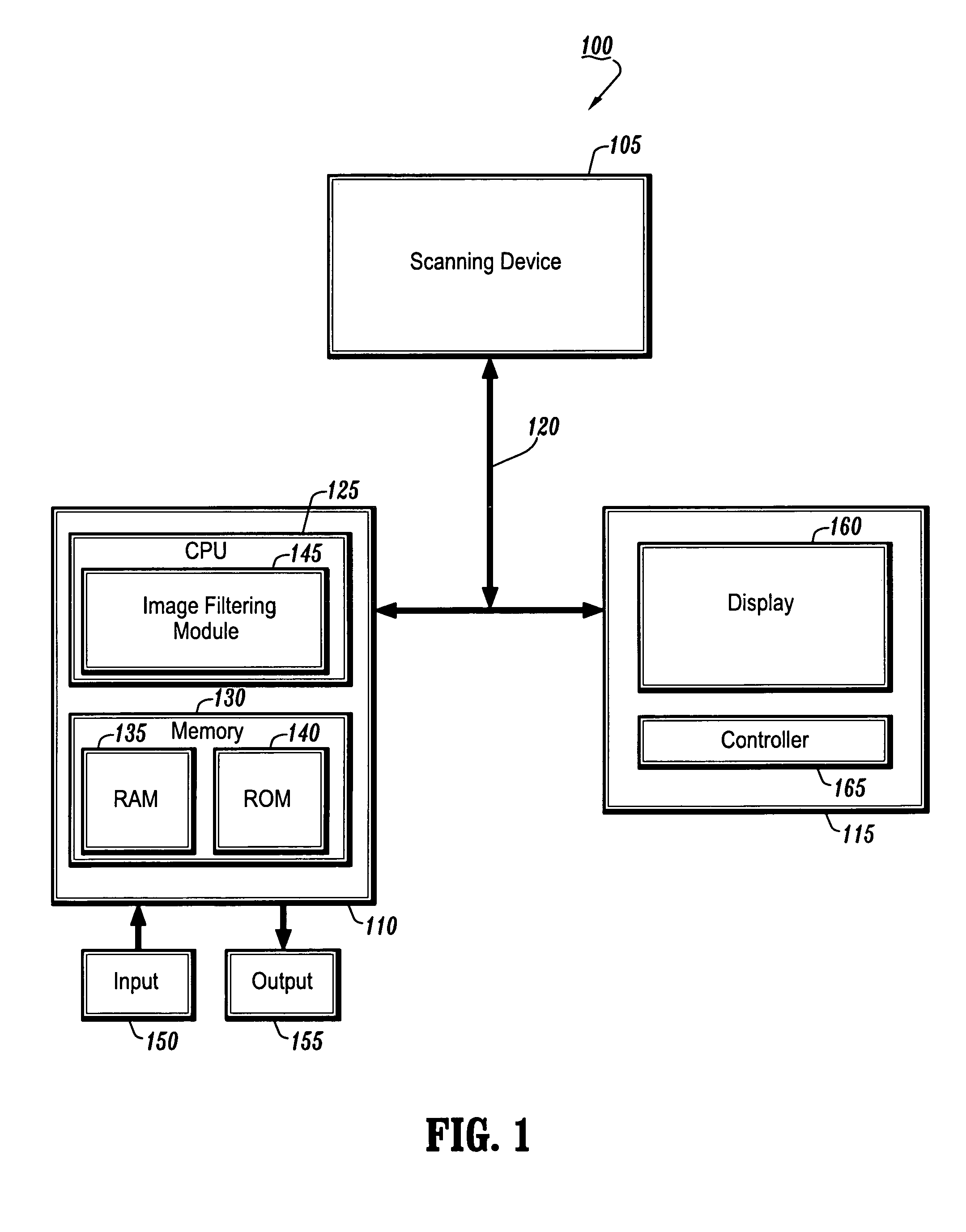
[0030]FIG. 1 is a block diagram of a system 100 for filtering noise from a medical image according to an exemplary embodiment of the present invention.

[0031]As shown in FIG. 1, the system 100 includes, inter alia, an acquisition device 105, a personal computer (PC) 110 and an operator's console 115 connected over, for example, an Ethernet network 120. The acquisition device 105 may be a magnetic resonance (MR) imaging device, a CT imaging device, a helical CT device, a PET device, a two-dimensional (2D) or three-dimensional (3D) fluoroscopic imaging device, a 2D, 3D, or four-dimensional (4D) ultrasound imaging device, or an x-ray device.

[0032]The acquisition device 105 may also be a hybrid-imaging device capable of CT, MR, PET or other imaging techniques. The acquisition device 105 may further be a flatbed scanner that takes in an optical image and digitizes it into an electronic image represented as binary data to create a computerized version of a photo or illustration.

[0033]The P...


PUM


Abstract

A system and method for filtering noise from a medical image are provided. A method for filtering noise from an image comprises: generating a weighted graph representing the image; selecting a plurality of nodes from the image to retain grayscale values of the plurality of nodes; and determining filtered grayscale values of the plurality of nodes.

Description

CROSS-REFERENCE TO RELATED APPLICATION[0001]This application claims the benefit of U.S. Provisional Application No. 60 / 624,368, filed Nov. 2, 2004, a copy of which is herein incorporated by reference.BACKGROUND OF THE INVENTION[0002]1. Technical Field[0003]The present invention relates to image processing, and more particularly, to a system and method for filtering noise from a medical image.[0004]2. Discussion of the Related Art[0005]Medical images are frequently noisy, due both to varying image data and artifacts resulting from an acquisition system such as an ultrasound, positron emission tomography (PET) or computed tomography (CT) scanner. Recently, nonlinear diffusion methods have proven useful for reducing image noise in many fields ranging from medical applications and image-sequence analysis to computer aided quality control and post-processing of noisy data.[0006]One such nonlinear diffusion method presented by Perona and Malik, “Scale-Space and Edge Detection Using Anisot...

Claims


Application Information

Patent Timeline
Patent Type & Authority Patents(United States)
IPC IPC(8): G06K9/00G06K9/32
CPCG06T5/002G06T2207/30004G06T2207/20072
Inventor GRADY, LEO
Owner SIEMENS MEDICAL SOLUTIONS USA INC
Why Eureka
Try Eureka