Sensor guided epicardial lead

a technology of epicardial leads and sensors, which is applied in the direction of epicardial electrodes, internal electrodes, therapy, etc., can solve the problems of affecting the cardiac output of patients, affecting the cardiac function of patients, and not being good candidates for transvenous lead implantation

Inactive Publication Date: 2009-04-07
CARDIAC PACEMAKERS INC
View PDF20 Cites 118 Cited by
  • Summary
  • Abstract
  • Description
  • Claims
  • Application Information

AI Technical Summary

Problems solved by technology

Without the synchronization afforded by the normally functioning specialized conduction pathways, the heart's pumping efficiency is greatly diminished.
Patients who exhibit pathology of these conduction pathways may suffer compromised cardiac output, such as that associated with congestive heart failure.
While transvenous lead delivery is appropriate for many patients that experience adverse synchronization conditions, there are a significant number of patients who could benefit from cardiac resynchronization therapy or other cardiac stimulation therapies, but are not good candidates for transvenous lead implantations.
Many of these patients are considered poor candidates for transvenous lead implantation for various reasons, including inability to locate the coronary sinus, presence of coronary sinus stenosis, inability to catheterize a desired branch vein, instability of the transvenous lead, or an unacceptably high pacing threshold, for example.
For procedures utilizing epicardial lead placement, many challenges sometimes make lead placement difficult or impossible.
If the target implantation site is on the posterior or lateral wall of the left ventricle, it may be difficult to deploy an endoscope to allow visualization of the myocardial surface.
Without visualization, lead placement may be difficult or impossible.
Without visualization, there is a risk that the lead may be inserted into fat or a coronary artery or blood vessel.

Method used


Image

  • Sensor guided epicardial lead
  • Sensor guided epicardial lead
  • Sensor guided epicardial lead

Examples


Embodiment Construction

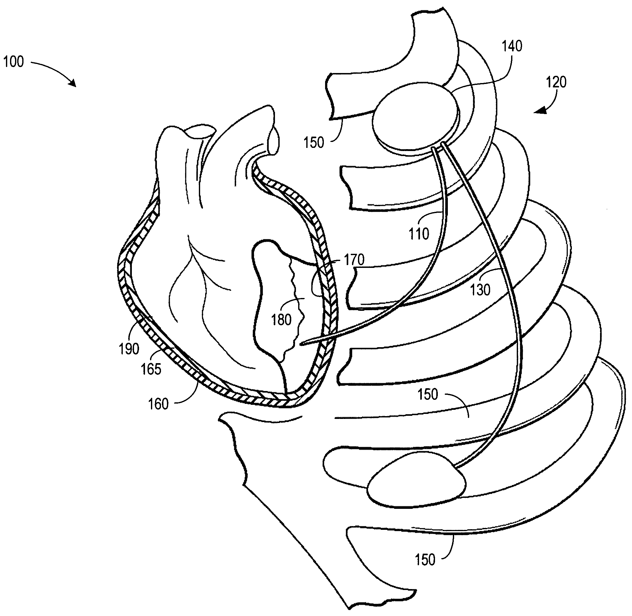
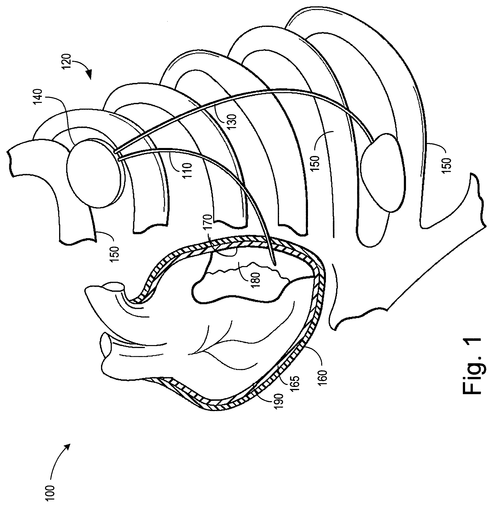
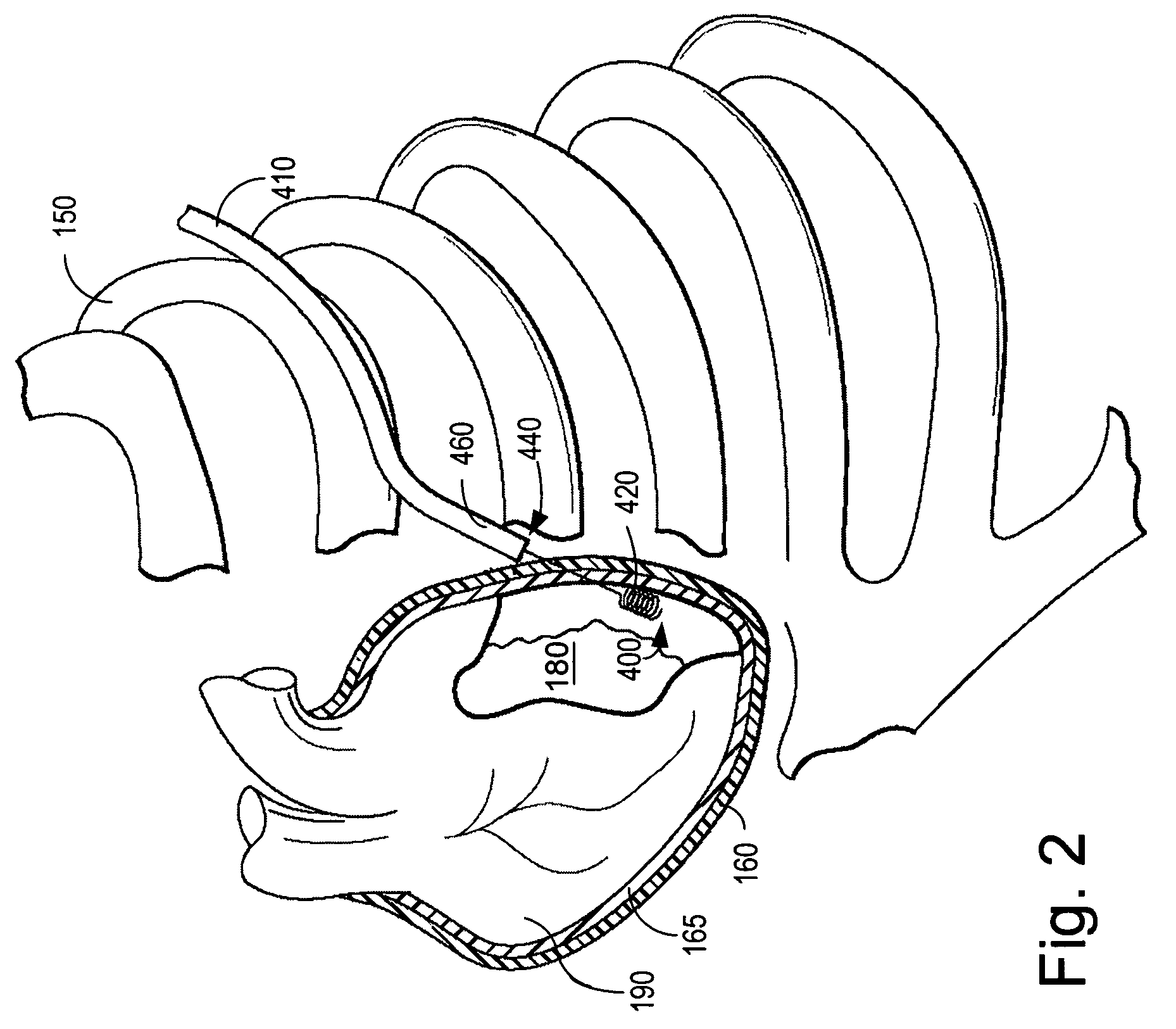
[0032]In the following description of the illustrated embodiments, references are made to the accompanying drawings, which form a part hereof, and in which is shown by way of illustration various embodiments in which the invention may be practiced. It is to be understood that other embodiments may be utilized, and structural and functional changes may be made without departing from the scope of the present invention.

[0033]Methods and devices employing an implantable cardiac lead in accordance with the present invention may incorporate one or more of the features, structures, methods, or combinations thereof described herein below. For example, devices and / or leads having sensing arrangements to provide feedback to the clinician during implantation may be implemented to include one or more of the features and / or processes described below. It is intended that such a device or method need not include all of the features and functions described herein, but may be implemented to include ...


PUM


Abstract

Implantable cardiac monitoring and stimulation methods and devices with epicardial leads having sensor feedback. A fixed or extendable / retractable sensor may be displaceable within the lead's lumen and configured to sense the presence of an anatomical feature or physiological parameter of cardiac tissue in proximity with the lead body's distal end. The sensor may include an ultrasonic sensing element, a perfusion sensor, a photoplethysmographic sensor, or a blood oximetry sensor. Methods of determining suitability for implanting a lead involve the steps of accessing an epicardial surface of the heart, and moving the cardiac lead to an implant site at the epicardial surface. A transmitted signal is directed at the implant site. A reflected signal is received, indicative of the presence of a blood vessel at the implant site. A determination may be made to determine whether the implant site is suitable or unsuitable based on the reflected signal.

Description

FIELD OF THE INVENTION[0001]The present invention relates generally to leads for implantable cardiac monitoring and stimulation devices and, more particularly, to epicardial leads having sensor feedback capabilities.BACKGROUND OF THE INVENTION[0002]Rhythmic contractions of a healthy heart are normally initiated by the sinoatrial node that includes specialized cells located in the superior right atrium. The sinoatrial node is the normal pacemaker of the heart, typically initiating 60-100 heartbeats per minute. When the sinoatrial node is pacing the heart normally, the heart is said to be in normal sinus rhythm (NSR).[0003]The heart has specialized conduction pathways in both the atria and the ventricles that enable the rapid conduction of excitation impulses (i.e. depolarizations) from the sinoatrial node throughout the myocardium. These specialized conduction pathways conduct the depolarizations from the sinoatrial node to the atrial myocardium, to the atrio-ventricular node, and to...

Claims


Application Information

Patent Timeline
Patent Type & Authority Patents(United States)
IPC IPC(8): A61N1/05
CPCA61N1/0587
Inventor TOCKMAN, BRUCE A.WESTLUND, RANDY
Owner CARDIAC PACEMAKERS INC
Why Eureka
Try Eureka