Enhanced cardiac radionuclide imaging techniques

a radionuclide imaging and cardiac technology, applied in the field of cardiac radionuclide imaging techniques, can solve the problems of removing unnecessary therapy or additional testing, affecting the actual cause of the patient's problem, and delivering the radioactive substance to the myocardium more poorly than it otherwise,

Active Publication Date: 2009-04-28
MUNN CHARLES S
View PDF4 Cites 0 Cited by
  • Summary
  • Abstract
  • Description
  • Claims
  • Application Information

AI Technical Summary

Problems solved by technology

If a coronary artery supplying blood to the myocardium is obstructed in some fashion, it will deliver the radioactive substance to the myocardium more poorly than it otherwise would.
As a result, an area of the myocardium that has insufficient radioactive substance delivered to it may be visualized as defective with respect to the remainder of the myocardium.
Falsely positive examinations can lead toward unnecessary therapy or additional testing (one or both of which may be dangerous and / or costly) and away from the actual cause of a patient's problem.
In any event, despite the modest gains in decreasing artifactual inferior wall defects achieved in the manner described above, artifactual inferior wall defects remain a serious and important problem in the general clinical practice of radionuclide imaging of the heart.

Method used


Image

  • Enhanced cardiac radionuclide imaging techniques
  • Enhanced cardiac radionuclide imaging techniques

Examples


Embodiment Construction

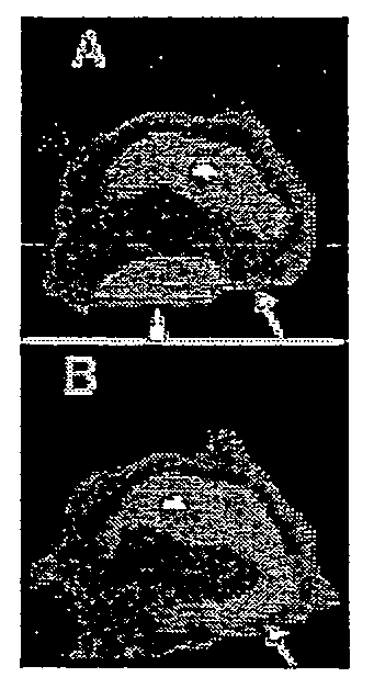
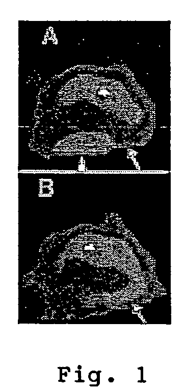
[0020]As noted above, the present invention is based, at least in part, on the unexpected findings that artifactual inferior wall defects of the heart that occur during radionuclide imaging are often attributable to attenuation caused by the fundal wall and possibly nearby cardia wall of the stomach and that such attenuation may be diminished by distending the stomach through inflation with a gas that is not highly attenuating.

[0021]Although not wishing to be limited to any particular theory behind the invention, the present inventor offers the following explanation as to why the invention achieves its desired effect: The stomach is usually just a few millimeters from the heart, on the other side of the diaphragm. It has recently been proposed that liquid in the stomach might sometimes be responsible for inferior defects. In fact, in one such study (Begum et al., “Positional related shifting inferior wall deficits on myocardial perfusion imaging caused by fluid in the gastric fundus...


PUM


Abstract

A cardiac imaging technique comprises (a) introducing a sufficient quantity of a not highly attenuating gas into the stomach of a patient to distend the stomach wall of the patient; and (b) while the stomach wall of the patient is distended, performing radionuclide imaging of the heart and / or of the heart's contents. Preferably, the gas used to distend the stomach wall is introduced into the patient (i) by having the patient ingest a substance that, when contacted with water or with stomach contents, produces the gas, (ii) by having the patient imbibe a liquid which has been mixed or combined with a gas-producing substance, or (iii) by having the patient imbibe a liquid into which the gas has been dissolved.

Description

CROSS-REFERENCE TO RELATED APPLICATIONS[0001]The present application claims the benefit under 35 U.S.C. 119(e) of U.S. Provisional Patent Application Ser. No. 60 / 488,541, filed Jul. 18, 2003, U.S. Provisional Patent Application Ser. No. 60 / 489,738, filed Jul. 23, 2003, and U.S. Provisional Patent Application Ser. No. 60 / 496,352, filed Aug. 19, 2003, all of which are incorporated herein by reference.BACKGROUND OF THE INVENTION[0002]The present invention relates generally to cardiac radionuclide imaging techniques and more particularly to new and improved cardiac radionuclide imaging techniques.[0003]In the practice of human or veterinary medicine, radioactive substances are sometimes used to image various body parts and bodily functions. These radioactive substances may be administered to a patient in a variety of ways, e.g., by being injected, inhaled, ingested, instilled or the like. Images created by capturing or visualizing the radioactivity may be mathematically or otherwise pro...

Claims


Application Information

Patent Timeline
Patent Type & Authority Patents(United States)
IPC IPC(8): A61B6/00
CPCA61B6/00A61B6/037A61B6/503
Inventor MUNN, CHARLES S.
Owner MUNN CHARLES S
Why Eureka
Try Eureka