Biomarker detection in circulating cells

a biomarker and cell technology, applied in the field of circulating cell antigen detection, can solve the problems of false positives often observed using this technique, difficult quantitatively, if not impossible in many situations, etc., and achieves the effects of reducing background, convenient multiplexing capability, and sensitivity

Inactive Publication Date: 2009-05-26
VIROLOGIC INCORPORATED
View PDF135 Cites 41 Cited by
  • Summary
  • Abstract
  • Description
  • Claims
  • Application Information

AI Technical Summary

Benefits of technology

[0007]The present invention provides a rapid and efficient screening method for the characterization of not only tumor cells, but also rare cells, or other biological entities from biological samples, especially blood. The method described herein combines elements of immunomagnetic enrichment with the use of releasable molecular tags that are separated from an assay mixture for detection and quantification. Other means of enrichment such as density gradient centrifugation or panning or alteration of target cell density by appropriate labeling may also be utilized. According to a preferred embodiment, the method of the invention enables assaying whole blood for cancer staging, monitoring and screening. The sensitive nature of the assay facilitates the detection of residual disease, thus making it possible to monitor for cancer recurrence.
[0011]The present invention provides a method of detecting or measuring biomarkers in an enriched population of rare circulating cells. The invention has several advantages over current techniques including, but not limited to, (1) separation and enrichment of a rare cell population from a complex population of biological cells, (2) the detection and / or measurement of molecular tags that are separated from an assay mixture provide greatly reduced background and a significant gain in sensitivity; and (3) the use of molecular tags that are specially designed for ease of separation and detection thereby providing convenient multiplexing capability.

Problems solved by technology

This is being done through the laborious procedure of isolating all of the mRNA from a blood sample and performing reverse transcriptase PCR.
Presently, however, no good correlation exists between the presence of such cells in blood and the ability to predict which patients are in need of vigorous treatment, Gomella, J of Urology.
It is noteworthy that PCR is difficult, if not impossible in many situations, to perform quantitatively, i.e., determine number of tumor cells per unit volume of biological sample.
Additionally false positives are often observed using this technique.
There is an added drawback which is that there is a finite and practical limit to the sensitivity of this technique based on the sample size examined.
As a further consideration, tumor cells are often genetically unstable.

Method used


Image

  • Biomarker detection in circulating cells
  • Biomarker detection in circulating cells
  • Biomarker detection in circulating cells

Examples


example 1

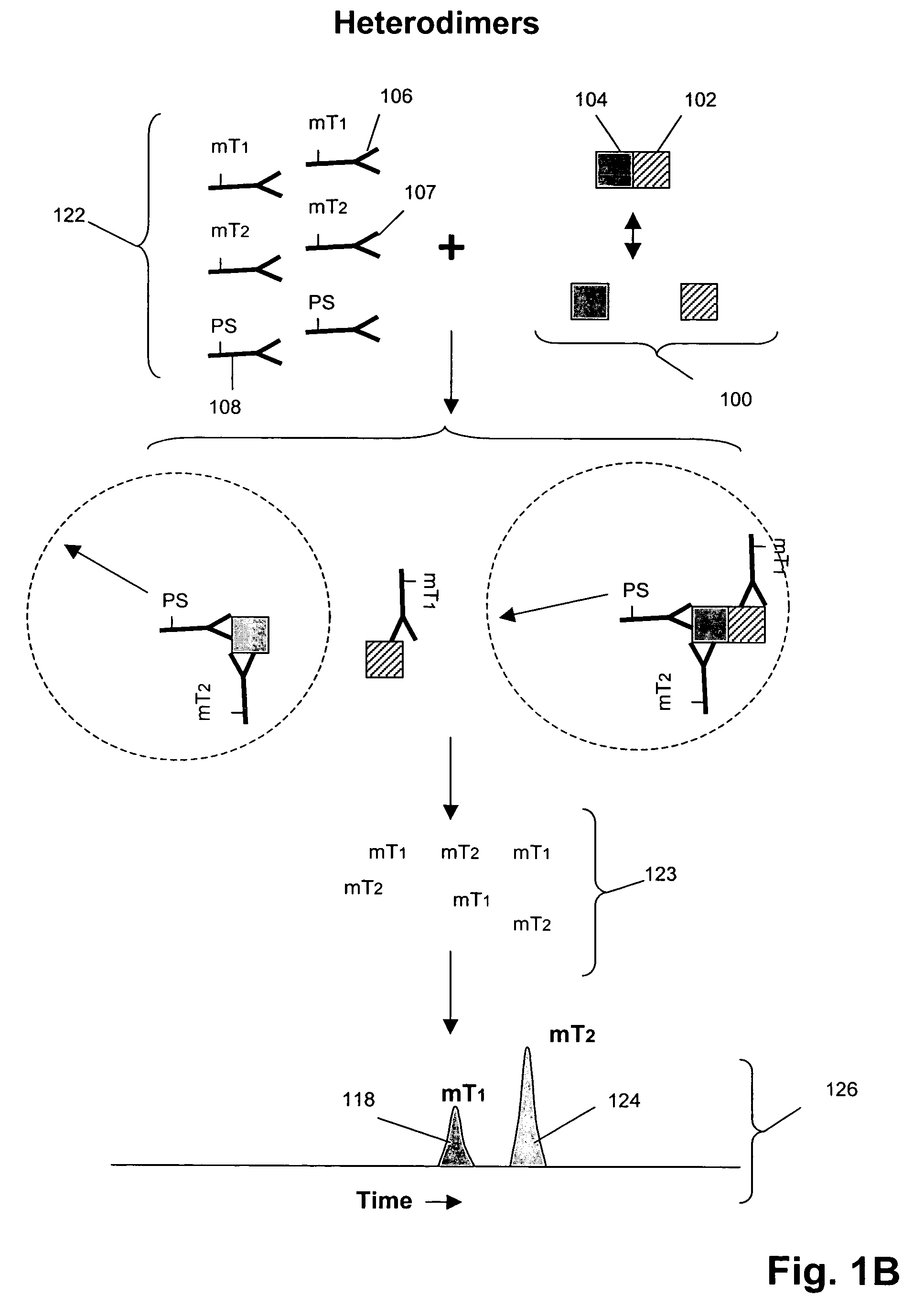
Analysis of Cell Lysates for Her-2 Heterodimerization and Receptor Phosphorylation on Magnetically Isolated Circulating Cells

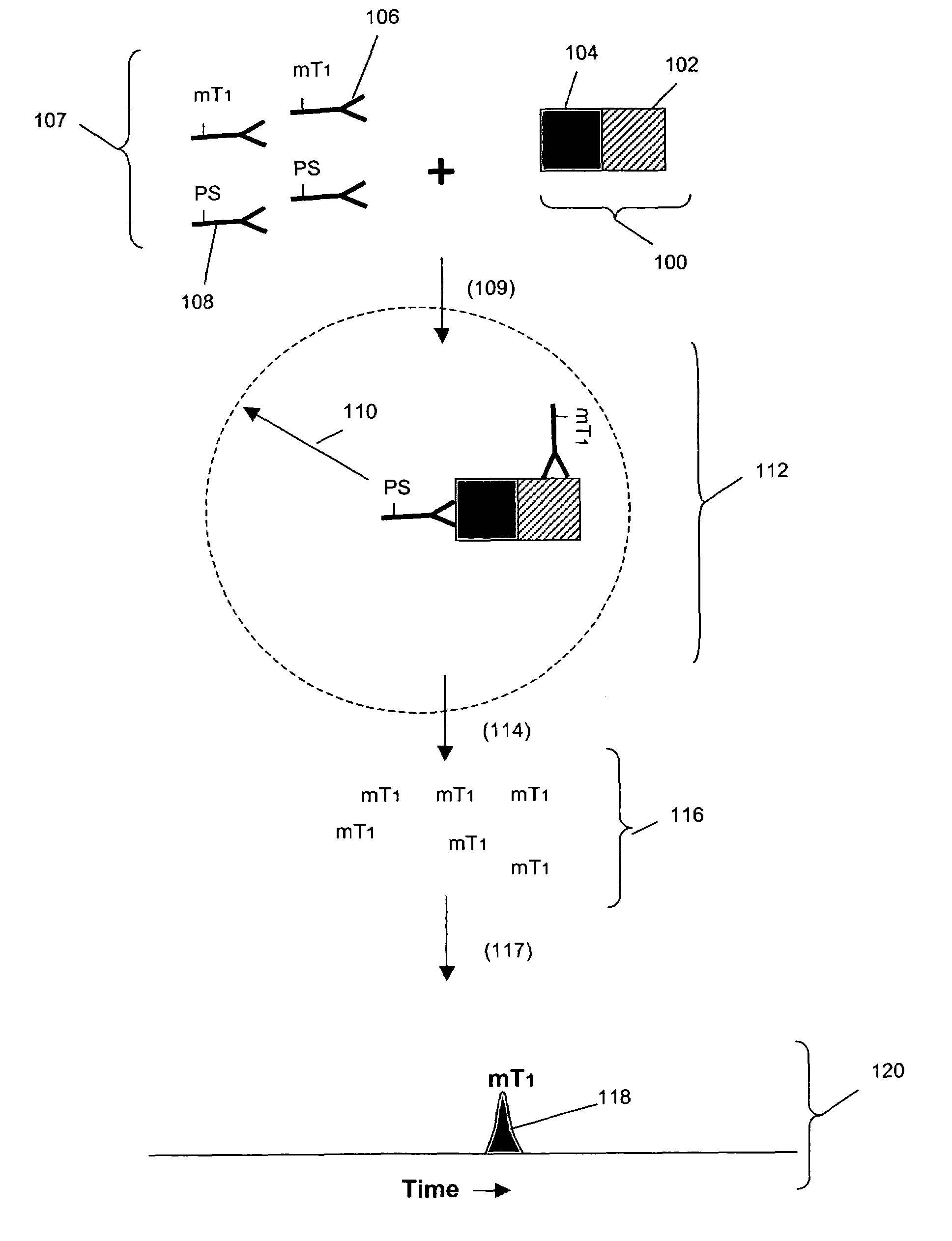
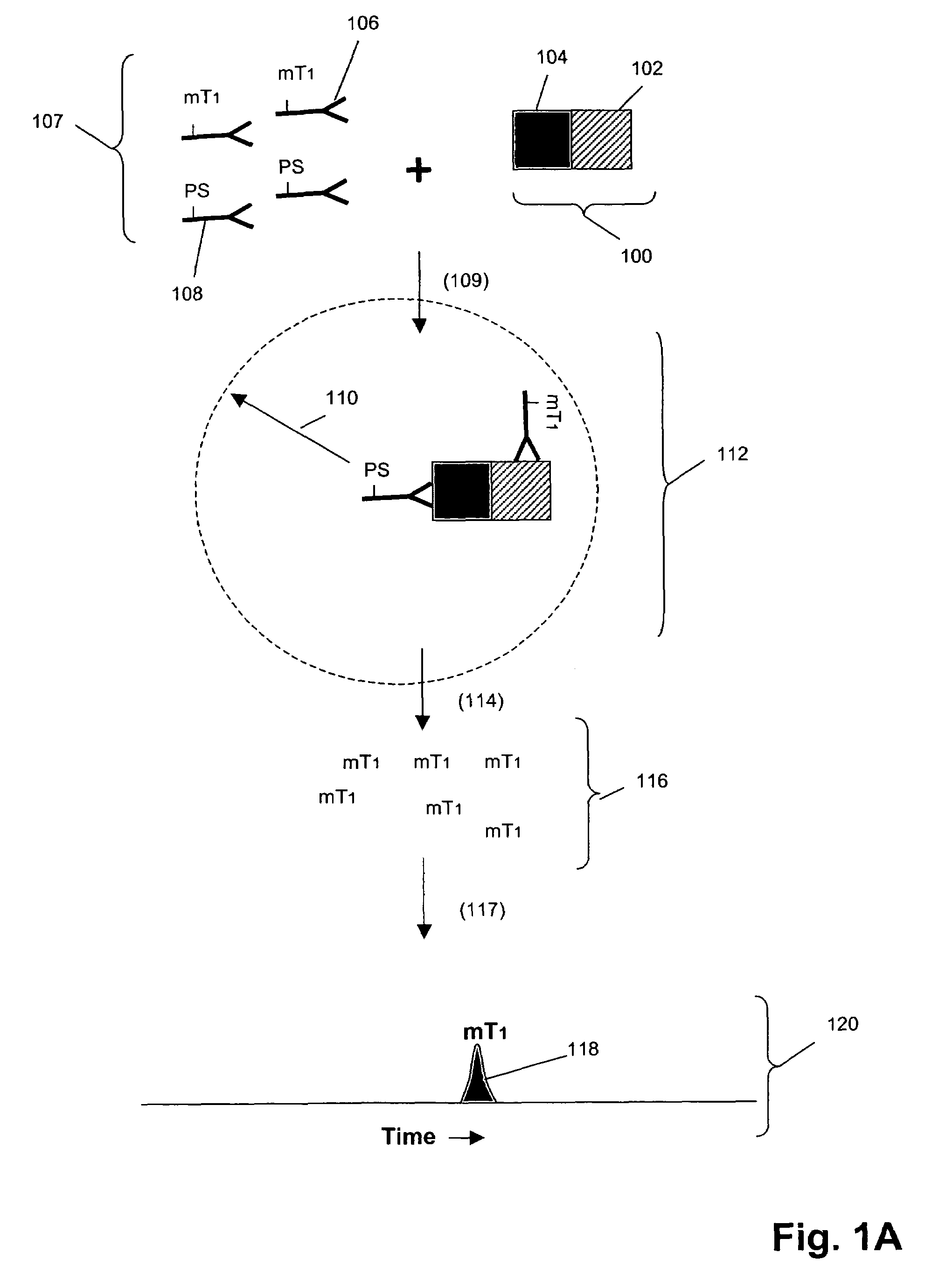
[0102]In this example, Her1-Her2 and Her2-Her3 heterodimers and phosphorylation states are measured in cell lysates from an enriched population of cells from a test blood sample. The test blood sample is made by spiking normal blood with known numbers of the tumor cell line MCF-7 (about 500 cells / mL normal blood). The enriched population is treated with various concentrations of epidermal growth factor (EGF) and heregulin (HRG) then assayed using the binding compounds and a cleaving probe as described below.

Sample Preparation:

[0103]1. 10 mL of test sample blood is incubated with the anti-Her3 conjugated ferrofluid for 15 minutes. The tubes are placed into a separator composed of four opposing magnets for 10 minutes (CellTracks AutoPrep System, Immunicon, Huntingdon Valley, Pa.). After separation, the blood is aspirated and discarded. The tube is taken out of t...


PUM

PropertyMeasurementUnit
molecular weightaaaaaaaaaa
pressureaaaaaaaaaa
pressureaaaaaaaaaa

Abstract

A method is provided for detecting biomarkers in rare circulating cells by forming an enriched population of cells immunomagnetically followed by biomarker detection using binding compounds having releasable molecular tags. Preferably, biomarkers comprising one or more protein-protein complexes are detected in circulating cancer cells metastasized from a solid tumor. In the presence of biomarkers, the molecular tags of the binding compounds are released and separated from the assay mixture for detection and / or quantification.

Description

RELATED APPLICATIONS[0001]This is a continuation-in-part of U.S. patent application Ser. No. 10 / 154,042 filed 21 May 2002 now U.S. Pat. No. 7,255,999 and a continuation-in-part of U.S. patent application Ser. No. 10 / 420,549 filed 18 Apr. 2003, which is a divisional of U.S. patent application Ser. No. 09 / 698,849 filed 27 Oct. 2000 (now U.S. Pat. No. 6,627,400), which is a continuation-in-part of U.S. patent application Ser. No. 09 / 602,586 filed 21 Jun. 2000 (now U.S. Pat. No. 6,514,700), which is a continuation-in-part of U.S. application Ser. No. 09 / 561,579 filed 28 Apr. 2000 (now U.S. Pat. No. 6,682,887), all of which are incorporated by reference.FIELD OF THE INVENTION[0002]The present invention relates generally to a method of detecting antigens of circulating cells, and more particularly, to a method of detecting antigens of circulating cancer cells.BACKGROUND OF THE INVENTION[0003]A biomarker is a characteristic that is objectively measured and evaluated as an indicator of norm...

Claims


Application Information

Patent Timeline
Patent Type & Authority Patents(United States)
IPC IPC(8): G01N33/553G01N33/53G01N33/50G01N33/543G01N33/566G01N33/567G01N33/574G01N33/68
CPCG01N33/5091G01N33/54326G01N33/566G01N33/574G01N33/6845
Inventor KIRAKOSSIAN, HRAIRKLOPACK, THOMASSINGH, SHARAT
Owner VIROLOGIC INCORPORATED
Why Eureka
Try Eureka