Method and device for imaging body lumens

a technology of body lumens and imaging methods, applied in the field of in vivo diagnostics, can solve the problems of bowel wall injury, low detection rate and exposure to x-ray radiation, flexible upper endoscopy,

Inactive Publication Date: 2009-06-30
GIVEN IMAGING LTD
View PDF58 Cites 89 Cited by
  • Summary
  • Abstract
  • Description
  • Claims
  • Application Information

AI Technical Summary

Problems solved by technology

This technique has several drawbacks, namely, low detection rate and exposure to x-ray radiation.
Risks associated with flexible upper endoscopy include injury to the bowel wall, bleeding, and aspiration.
Upper endoscopy is usually performed under conscious sedation, which carries risks as well.
Furthermore, patients typically need to take a day off of normal activities due to the lasting effects of conscious sedation.
Finally, the endoscopy procedure is clearly a cause of discomfort, pain and vomiting in many patients.
Even the physical dimensions of the endoscope can be a cause for fear.
However, in larger or voluminous lumens, such as the stomach or large intestine, the view or sensing capacity of a capsule may not cover the entire surface of the lumen wall.

Method used


Image

  • Method and device for imaging body lumens
  • Method and device for imaging body lumens
  • Method and device for imaging body lumens

Examples


Embodiment Construction

[0023]In the following detailed description, numerous specific details are set forth in order to provide a thorough understanding of the invention. However, it will be understood by those skilled in the art that the present invention may be practiced without these specific details. In other instances, well-known methods, procedures, and components have not been described in detail so as not to obscure the present invention.

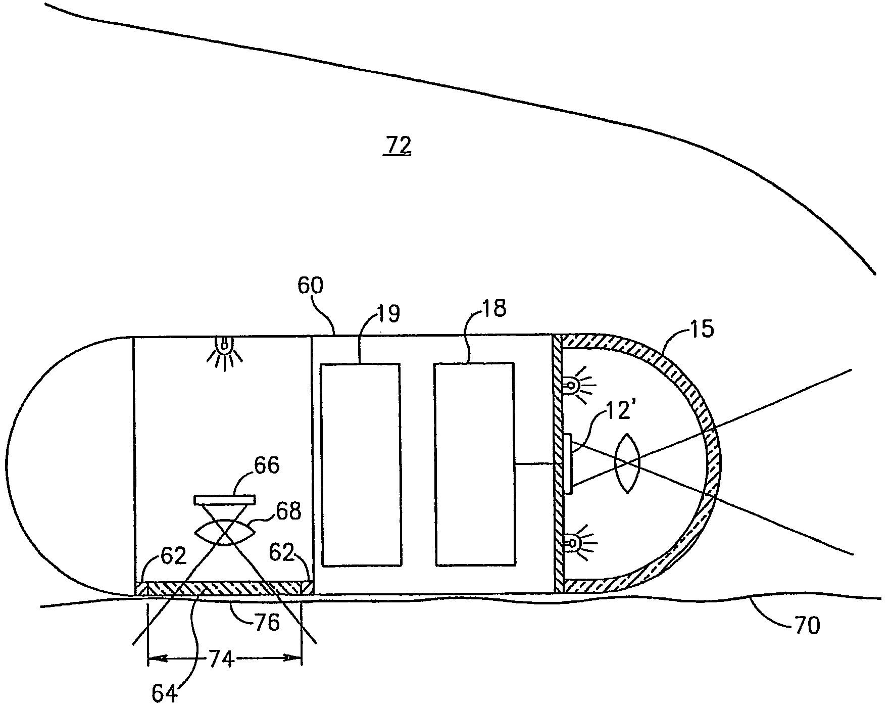
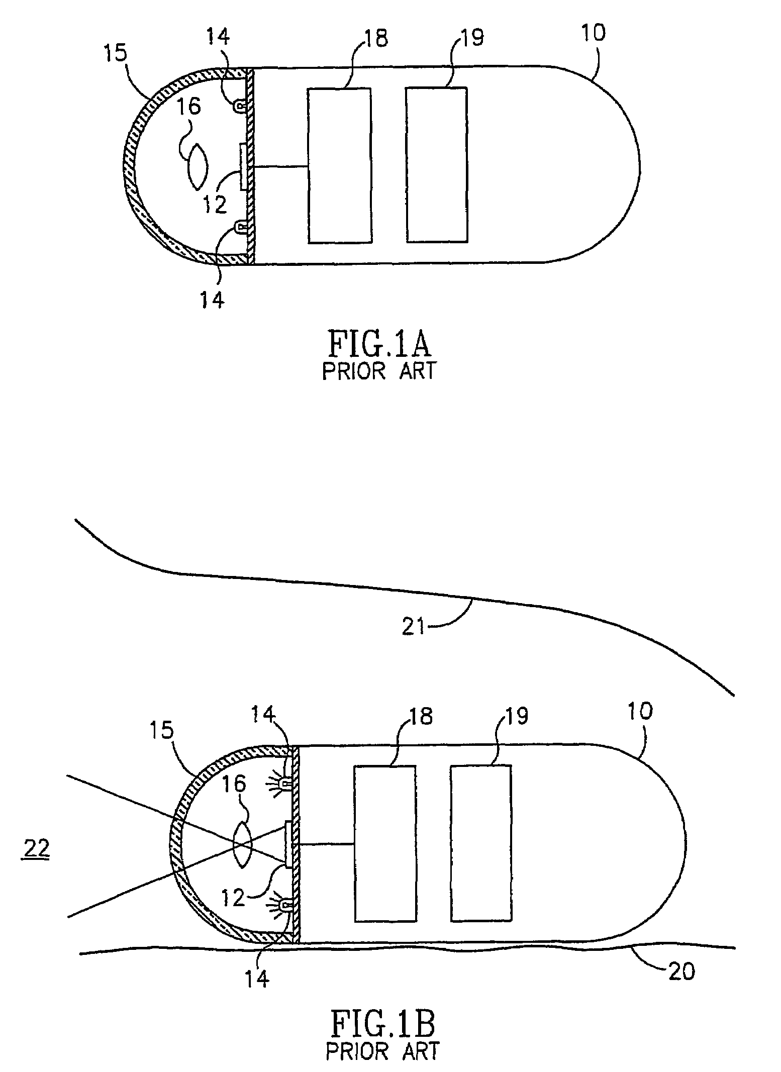
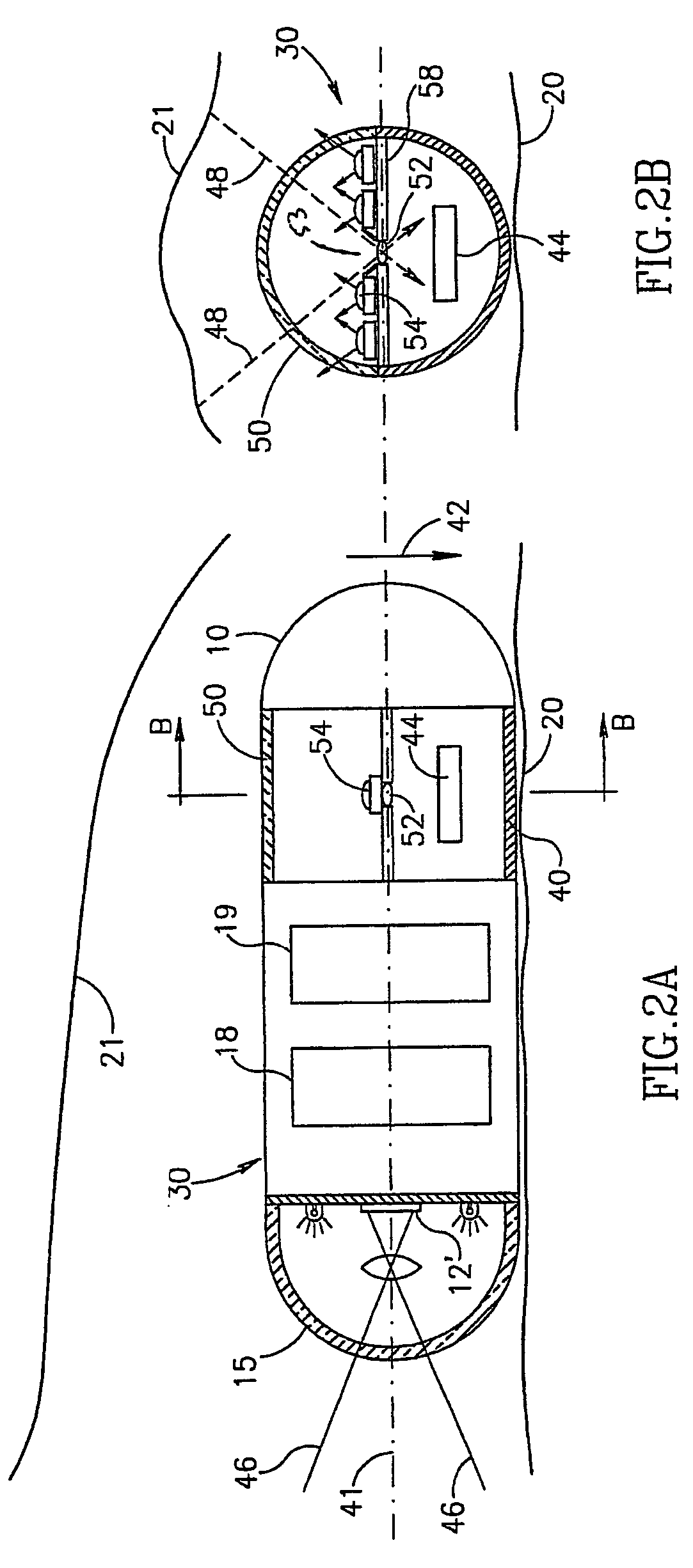
[0024]Reference is now made to FIG. 1A, which illustrates an ingestible device 10, which may be, for example, a capsule, but which may have other shapes or configurations. Device 10 may be, for example, similar to embodiments described in U.S. Pat. No. 5,604,531 to Iddan et al., and / or WO 01 / 65995, entitled “A Device And System For In Vivo Imaging”, published on 13 Sep., 2001, both of which are assigned to the common assignee of the present invention and which are hereby incorporated by reference. However, device 10 may be any sort of in-vivo sensor device and may...


PUM


Abstract

An in-vivo imaging device including a ballast that may for example orient such device in a known orientation relative to the gravitational force on such ballast. Such imaging device may, for example, assume a known orientation when it is free to move, and may capture images from such known orientation. The device may include one or more imagers, which may be oriented at different angles or points of view (e.g., forward and transverse). A method of use may include moving a patient so that the device inside the patient images different fields of view.

Description

CROSS REFERENCE TO RELATED APPLICATIONS[0001]This application claims benefit from U.S. provisional application Ser. No. 60 / 458,438, filed on Mar. 31, 2003, which is incorporated in its entirety by reference herein and U.S. provisional application Ser. No. 60 / 466,729 filed May 1, 2003, which is incorporated in its entirety by reference herein. This application is also a continuation-in-part of prior U.S. patent application Ser. No. 10 / 046,541 filed on Jan. 16, 2002 entitled System and Method for Wide Field Imaging of Body Lumens, which is incorporated in its entirety by reference herein, and which claims benefit of prior provisional application 60 / 261,188 filed on Jan. 16, 2001.FIELD OF THE INVENTION[0002]The present invention relates to the field of in vivo diagnostics. More specifically, the present invention relates to a method for sensing, for example, imaging, body lumens.BACKGROUND OF THE INVENTION[0003]Some pathologies of the gastrointestinal (GI) tract, involve epithelial dam...

Claims


Application Information

Patent Timeline
Patent Type & Authority Patents(United States)
IPC IPC(8): A61B1/06A61B6/00A61B1/04
CPCA61B1/00158A61B1/00179A61B1/00181A61B1/041A61B1/0607
Inventor IDDAN, GAVRIEL J.
Owner GIVEN IMAGING LTD
Why Eureka
Try Eureka