Gross pathology breast map

a breast biopsy and gross pathology technology, applied in the field of pathology, can solve the problems of inability to accurately identify the correct anatomical relationship of the excised specimen margins with the actual surgical cavity, and no standardized method of orienting the excised breast biopsy or lumpectomy (cancer) specimens

Inactive Publication Date: 2009-08-11
XOFT INC +1
View PDF8 Cites 6 Cited by
  • Summary
  • Abstract
  • Description
  • Claims
  • Application Information

AI Technical Summary

Problems solved by technology

However, such technology generally fails to accurately identify the correct anatomical relationship of the excised specimen margins with the actual surgical cavity created by tissue removal.
In particular, within the field of breast surgery, there exists no standardized method of orienting excised breast biopsy or lumpectomy (cancer) specimens whether radiographically or non-radiographically (i.e., by palpation or ultrasound identified abnormalities vis-à-vis the operative site).

Method used


Image

  • Gross pathology breast map
  • Gross pathology breast map
  • Gross pathology breast map

Examples


Embodiment Construction

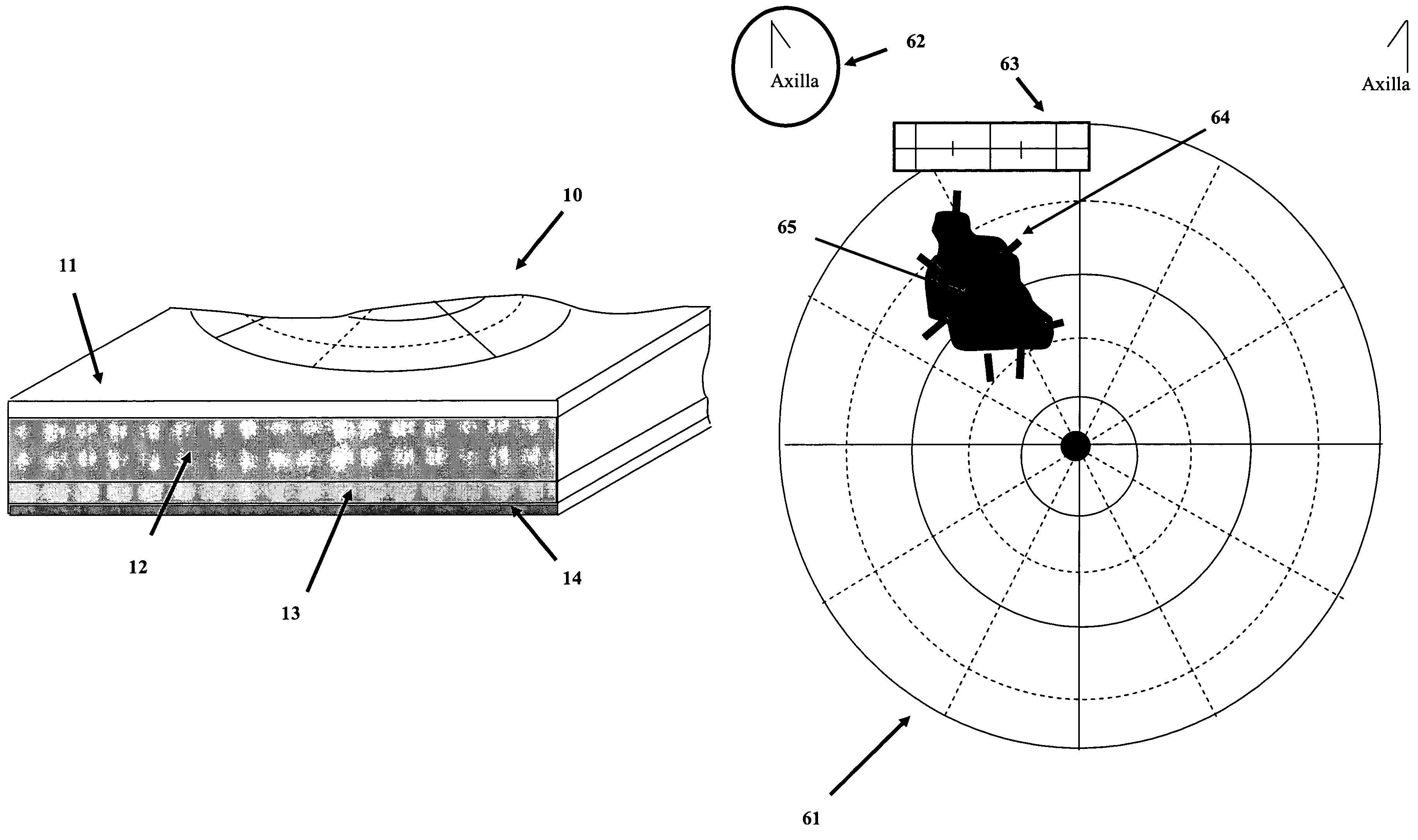
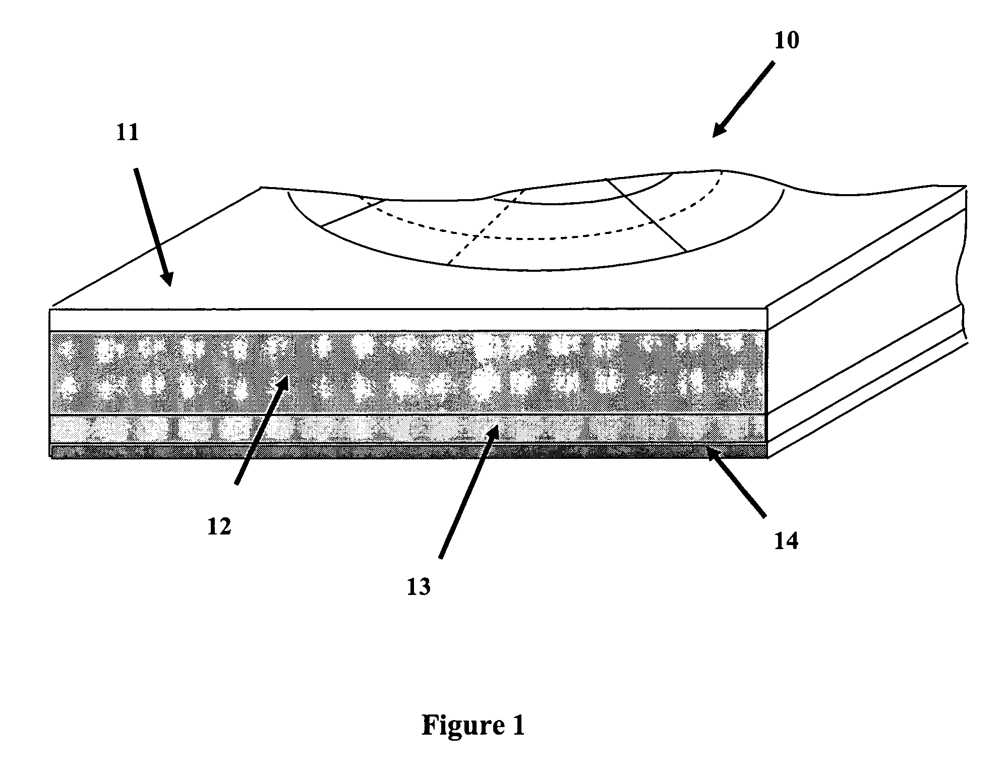
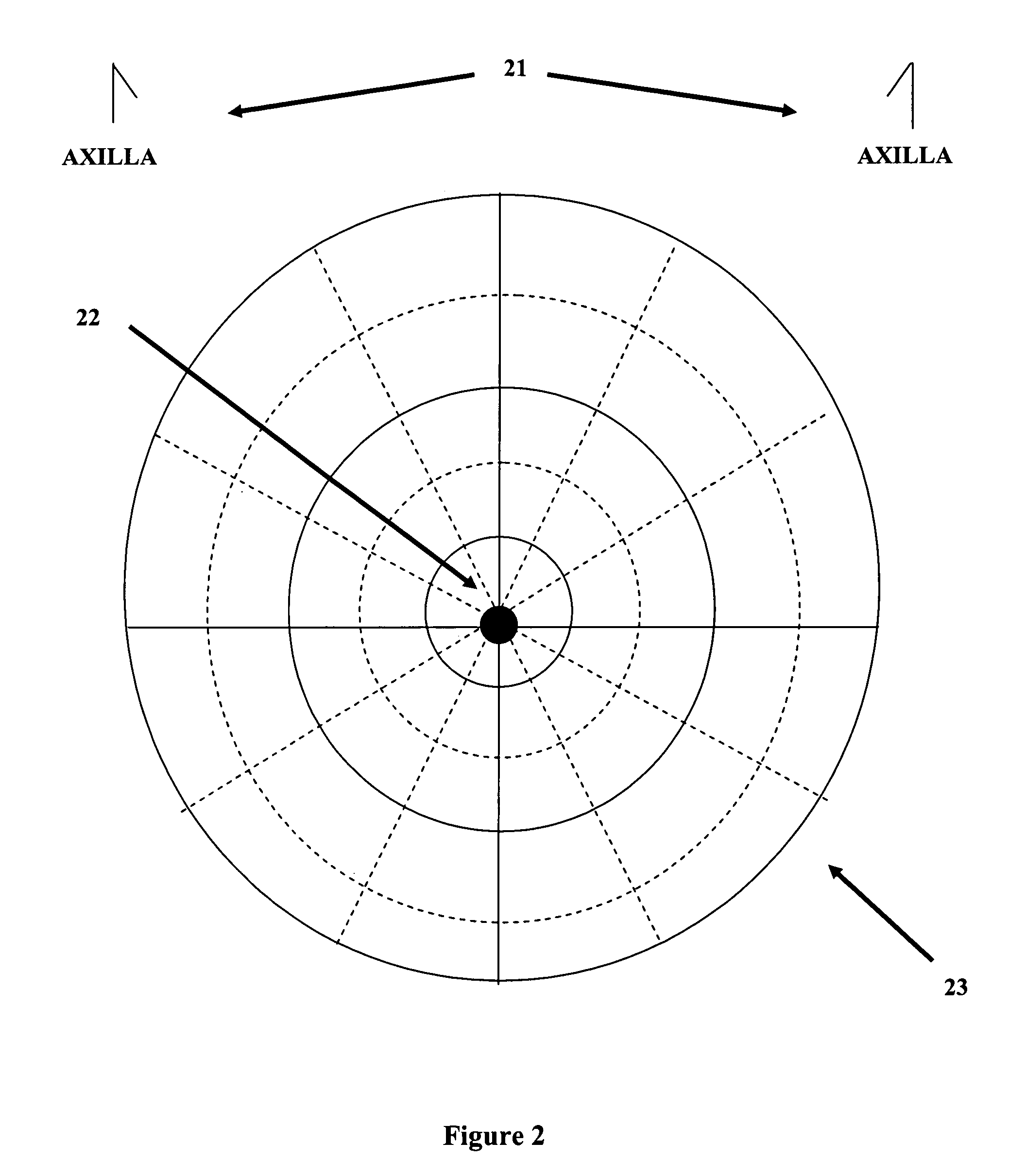
[0024]One embodiment of the present invention allows positioning and orientation of tissue specimens relative to the tissue mass or organ from which excised. This provides reliable and accurate assessment of margin status as well as a reproducible and accurate way to direct, if necessary, a surgical re-excision of an involved margin at the site of surgical excision. In addition, another embodiment of the present invention provides a mapping system wherein an excised tissue specimen can be sutured to, molded into, or otherwise attached thereto. Embodiments of the present invention provide a stable platform for pathologic sectioning. Additional embodiments allow the user to maintain a true, anatomically correct orientation between the operated breast and the tissue specimen.

[0025]In another embodiment, mapping materials for use at an operating table are provided. Such mapping materials include, but are not limited to, a breast map made of a suturable, absorbent material, color-coded s...


PUM


Abstract

A system and method to position and orient an excised breast tissue specimen on a support structure in a duplicate anatomic orientation relative to the operated breast is provided. One embodiment of the system includes an anatomically representative map of the breast under investigation. The system allows a surgeon to suture, ink, or otherwise fix the excised specimen on the map in accordance to its original position and orientation in the operated breast or organ. A radiogram of the excised specimen can be taken that shows the location of calcifications on a grid with radiopaque markers. The system allows a pathologist to cut through the specimen utilizing the map as a template in order to perform histological analysis and correlate the position and orientation of each slice to its anatomic position in the breast and thereby direct a precise and anatomically accurate re-excision by the surgeon if so required.

Description

RELATED APPLICATION DATA[0001]This application claims priority under 35 U.S.C. § 119 to U.S. Provisional Patent Application Ser. No. 60 / 461,433 filed Apr. 10, 2003, the disclosure of which is incorporated in its entirety herein by reference.BACKGROUND OF THE INVENTION[0002]1. Field of the Invention[0003]The present invention relates generally to the field of surgery and pathology. In one embodiment, the invention includes devices and methods for positioning and orienting excised tissue specimens with a support structure. The support structure generally illustrates the position of the excised tissue specimen with respect to the tissue from which it was excised.[0004]2. Description of the Related Art[0005]Technology available today generally utilizes radiologically placed markers to identify a radiographically identifiable breast abnormality within an excised tissue specimen. However, such technology generally fails to accurately identify the correct anatomical relationship of the exc...

Claims


Application Information

Patent Timeline
Patent Type & Authority Patents(United States)
IPC IPC(8): A61B19/00A61B6/00A61B10/00
CPCA61B10/0041A61B6/502A61B2019/5408A61B2090/3908
Inventor FORMAN, MICHAEL R.FRANCESCATTI, DARIUS
Owner XOFT INC
Why Eureka
Try Eureka