
Registration system and method for tracking lung nodules in medical images

a technology for lung nodules and medical images, applied in the field of medical imaging, can solve the problems of inability to accurately track lung nodules, inability to accurately identify lung nodules, and inability to complete the process, etc., and achieve the effect of avoiding plateaus in the learning curve, accelerating convergence, and fast and accurate manner

Inactive Publication Date: 2010-02-02
THE BOARD OF TRUSTEES OF THE LELAND STANFORD JUNIOR UNIV
View PDF4 Cites 12 Cited by
  • Summary
  • Abstract
  • Description
  • Claims
  • Application Information

AI Technical Summary

Benefits of technology

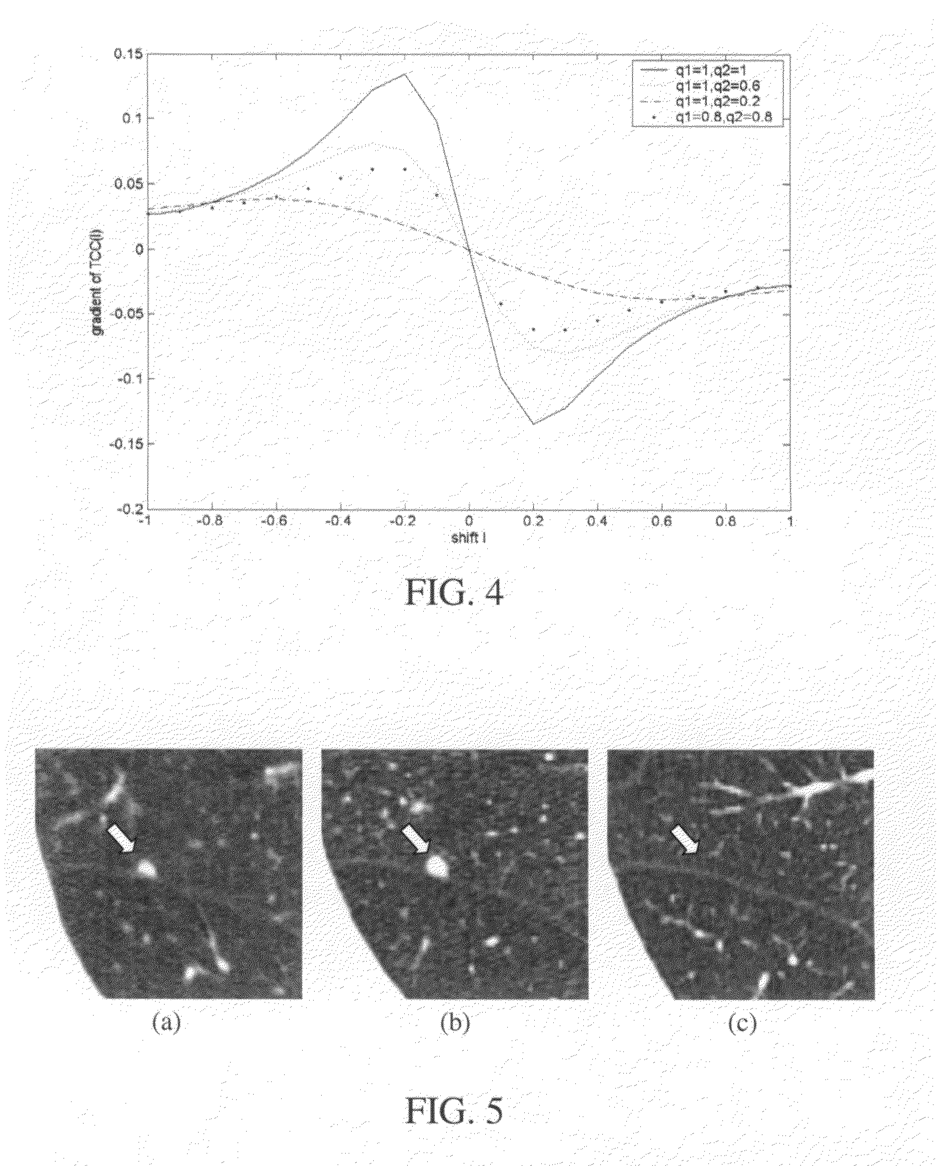
[0008]The Very Fast Simulated Annealing (VFSA) algorithm is a variation of SA. The VFSA uses a new trial generator and cooling schedule to accelerate the convergence. According to the invention, the negative of the Tunable Cross-Correlation (TCC) coefficient is used as the energy function for VFSA. The VFSA algorithm is augmented with a re-annealing schedule and an anti-local-maximum procedure. If the current configuration has remained constant for too long a time, the anti-local maximum procedure replaces the current configuration with any new configuration. This technique advantageously avoids the plateaus in the learning curve.
[0009]The inventive system can register all nodules within their maximum diameter in a fast and accurate manner. In some embodiments, the system achieves, with minimal registration error, an average registration time of 10 seconds or less on a 3.06 GHz computer programmed to implement the present invention.

Problems solved by technology

Unfortunately, CT is not specific for the diagnosis of lung cancer.
Most algorithms suffer from the prohibitive computation and / or errors introduced by unpredictable distortions at differing levels of inspiration.
Although nodule shape and composition may correlate with malignancy / benignity, the correlation is not perfect and, instead, the most reliable means of differentiating benign from malignant nodules is serial CT follow-up and assessment for growth.
Thus, when one or more nodules are found in a CT scan, which, today, may consist of several hundred cross-sectional images, they must be matched with their counterparts in one or more prior scans—a process that is time consuming and error prone.

Method used


Image

  • Registration system and method for tracking lung nodules in medical images
  • Registration system and method for tracking lung nodules in medical images
  • Registration system and method for tracking lung nodules in medical images

Examples


experiment 1

Registration for a Known Non-Affine Transformation

Dataset

[0052]In this experiment, we simulated a second patient scan from a first by transforming the first scan using a non-affine transformation. This scan was acquired using multidetector helical CT and reconstructed with a voxel size of 0.6 mm*0.6 mm*1.25 mm. Because we know the transform beforehand, the true location (ig, jg, kg) in equation (8) for each nodule can be calculated using the known transform.

Simulated Non-Affine Transformation

[0053]In constructing the transformation, we sought to model distortion due to different inspiration points and cardiac pulsation. The non-affine transformation applied consists of a displacement vector field plus a global affine transformation. For an arbitrary point, the transformed coordinate can be calculated through:

(it,jt,kt)=T((i,j,k)+{right arrow over (v)}(i,j,k)),  (10)

where (i, j, k) denotes the original coordinate, (it, jt, kt) denotes the transformed coordinate, {right arrow over (v)...

experiment 2

Registration for Real Patient Data

Dataset

[0061]In this experiment, we used 12 pairs of patient scans, and evaluated registration error with both equations (8) and (9). Two radiologists, each with over 10 years experience with pulmonary CT, determined the true locations of the target nodules and matched them. (One found and matched nodules in 5 pairs of scans and the other found and matched nodules in 7 pairs of scans) Altogether, the radiologists detected 93 nodule pairs in the 12 pairs of scans.

Results

[0062]For all the 93 nodules, the registration error evaluated by equation (8) was 1.2 mm±0.88 mm (s.d.), with a worst case error of 4.3 mm; and the registration error evaluated by equation (9) was 0.27±0.18 (s.d.), with a worst case error of 0.85. The mean absolute error in longitudinal (superior-inferior) coordinate was 0.62 mm±0.72 mm s.d, with a worst case of 3 mm. The average registration time per nodule was 0.16 min±0.17 min (s.d.), with a worst case of 0.87 minutes. FIGS. 9 and...


PUM


Abstract

A nodule registration system useful for tracking lung nodules in computed tomography (CT) scans is presented. The system registers a small sphere centered on a detected nodule in one scan with another scan under a rigid transformation assumption using a fast registration scheme. The registration scheme employs very fast simulated annealing (VFSA) with constraints to maximize a tunable cross-correlation (TCC) coefficient, enabling the system to register, with minimal registration error, all nodules within their maximum diameter. The system achieves an average registration time of 10 seconds or less on a 3.06 GHz computer programmed to implement the present invention.

Description

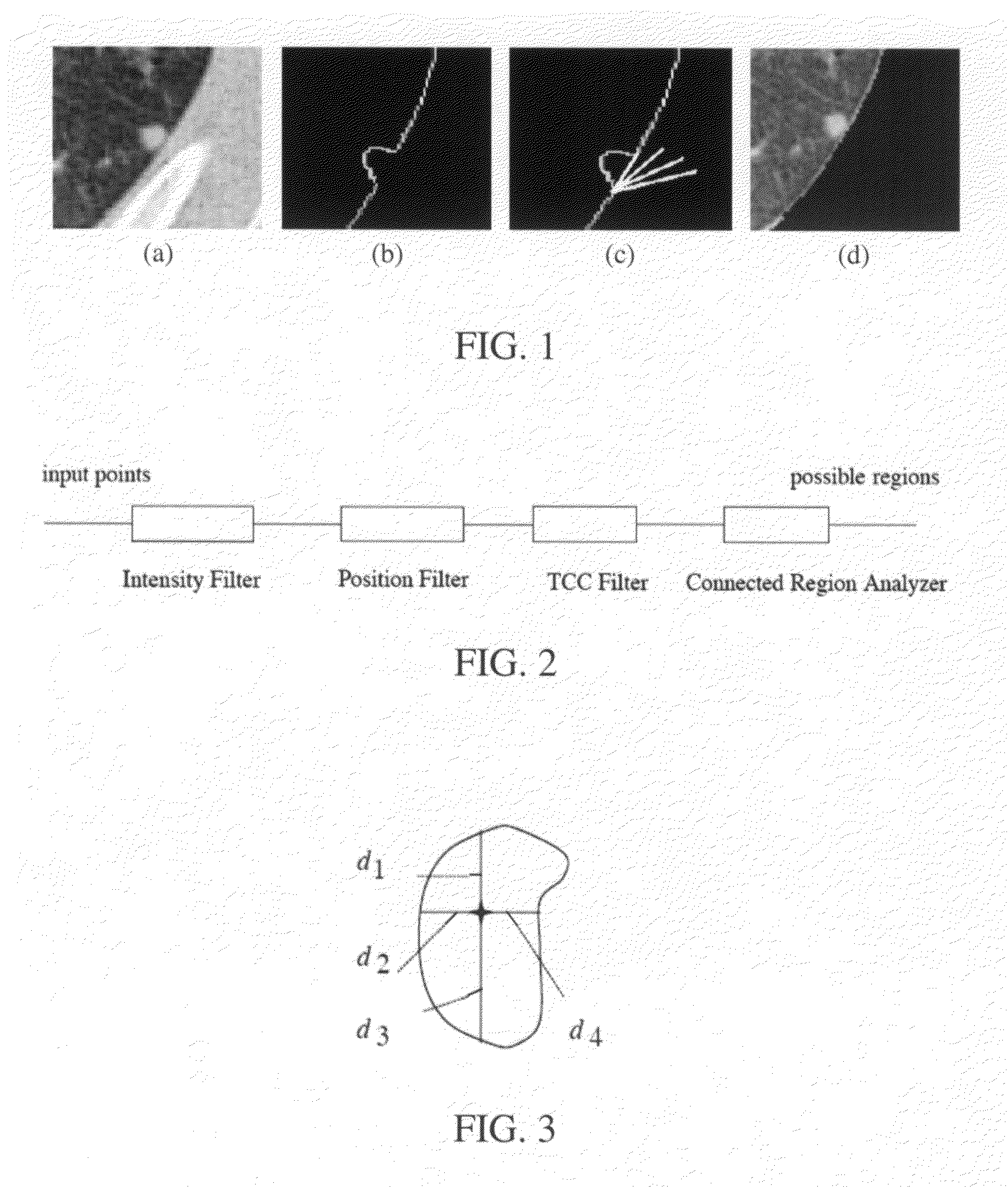
FIELD OF THE INVENTION[0001]The present invention relates generally to the field of medical imaging. More particularly, the present invention relates to lung nodule registration in medical images such as Computed Tomography (CT) scans.BACKGROUND[0002]Lung cancer kills more Americans each year than colorectal, breast, and prostate cancers combined. Over 173,000 new cases of lung cancer will be diagnosed and over 160,000 will die from this disease in 2004. The successful early detection and effective treatment of lung cancer focuses on its identification when it is a small lung nodule. As a result, when lung cancer is suspected, imaging with chest radiography or Computed Tomography (CT) scanning is the first step toward making the diagnosis. The tracking of lung nodules in scans acquired at different times for the same patient is useful in the determination of malignancy.[0003]Because of its superior contrast resolution and cross-sectional acquisition, which effectively eliminates sup...

Claims


Application Information

Patent Timeline
Patent Type & Authority Patents(United States)
IPC IPC(8): G06K9/00
CPCG06T7/0034G06T7/20G06T2207/30061G06T7/35
Inventor SUN, SHAOHUANAPEL, SANDY A.
Owner THE BOARD OF TRUSTEES OF THE LELAND STANFORD JUNIOR UNIV
Learn More