Integrated single photon emission computed tomography (SPECT)/transmission computed tomography (TCT) system for cardiac imaging

a computed tomography and computed tomography technology, applied in tomography, instruments, nuclear engineering, etc., can solve the problems of low spect sensitivity, limited return of heavily attenuated and scattered photons, and less than optimal circular design, etc., to achieve convenient replacement, easy to implement, and easy to handle

Active Publication Date: 2010-03-23
RUSH UNIV MEDICAL CENT
View PDF7 Cites 147 Cited by
  • Summary
  • Abstract
  • Description
  • Claims
  • Application Information

AI Technical Summary

Benefits of technology

[0014]It is a further object of this invention to provide such an integrated SPECT / TCT system for cardiac imaging which provides a plurality of predetermined imaging volumes of various sizes and locations for optimizing acquisition and image quality.

Problems solved by technology

As a result, the long-distance collimation offsets the potential gain in geometric efficiency and renders the circular design less than optimum.
Furthermore, the design devoted considerable collimator and detector area to the patient's right-posterior side, where the heart is too distant from the collimator for effective collimation.
The typical problem of low photon sensitivity in SPECT is further compounded in cardiac imaging where the desirable radiation photons are scarce: only about 2-4% of the injected dose is in the myocardium of the heart.
This previous circular design approach results in a limited return of the heavily attenuated and scattered photons and sub-optimal image quality.
The drawback of this approach is that the photon statistics is quite poor because the finite flux of the relative narrow parallel-beam of radiation has to be spread over a large detector area.
However, the problem with these SPECT / TCT systems is the required special collimation that either cannot be fully utilized in emission imaging of the heart, or the systems require a cumbersome and time-consuming collimator exchange procedure, which disturbs the imaging position of the patient and makes the subsequent matching of SPECT and TCT slices for attenuation correction difficult.
However, such a design compromises either SPECT or TCT imaging.
Changing collimator, not only is awkward, cumbersome, time-consuming, and unrealistic in clinical environment, it is also difficult to reproduce patient imaging position after the collimator has been changed.
The result is conventional SPECT / TCT systems are not flexible in accommodating different SPECT collimation requirements to suit various clinical situations and patients having different sizes and do not provide adequate TCT images needed for attention maps for high quality SPECT imaging.
Such a limitation compromises either SPECT or TCT imaging, which in turn limits the attainable quality of attenuation-corrected SPECT imaging.
The concern of using this approach for cardiac SPECT / TCT is the cost, because an additional dedicated x-ray detector system is required, even for the primitive low-end CTs.
As for the use of an expensive high-end multi-slice Spiral CT for attenuation of cardiac SPECT, it is still controversial and is not cost efficient.
This processing is not trivial and is a potential source of artifacts and sub-optimal correction.
Such a technique is cumbersome, not easily reproducible, prone to both mechanical and electrical errors, slow, inefficient, utilizes expensive hardware to rotate large heavy collimator and detector subsystems, and requires extensive safety measures to protect the patient.
As a result, conventional SPECT images have large variations in image quality and reproducibility, which make comparison of images from different facilities or from different times at the same facility difficult.

Method used


Image

  • Integrated single photon emission computed tomography (SPECT)/transmission computed tomography (TCT) system for cardiac imaging
  • Integrated single photon emission computed tomography (SPECT)/transmission computed tomography (TCT) system for cardiac imaging
  • Integrated single photon emission computed tomography (SPECT)/transmission computed tomography (TCT) system for cardiac imaging

Examples


Embodiment Construction

[0064]Aside from the preferred embodiment or embodiments disclosed below, this invention is capable of other embodiments and of being practiced or being carried out in various ways. Thus, it is to be understood that the invention is not limited in its application to the details of construction and the arrangements of components set forth in the following description or illustrated in the drawings. If only one embodiment is described herein, the claims hereof are not to be limited to that embodiment. Moreover, the claims hereof are not to be read restrictively unless there is clear and convincing evidence manifesting a certain exclusion, restriction, or disclaimer.

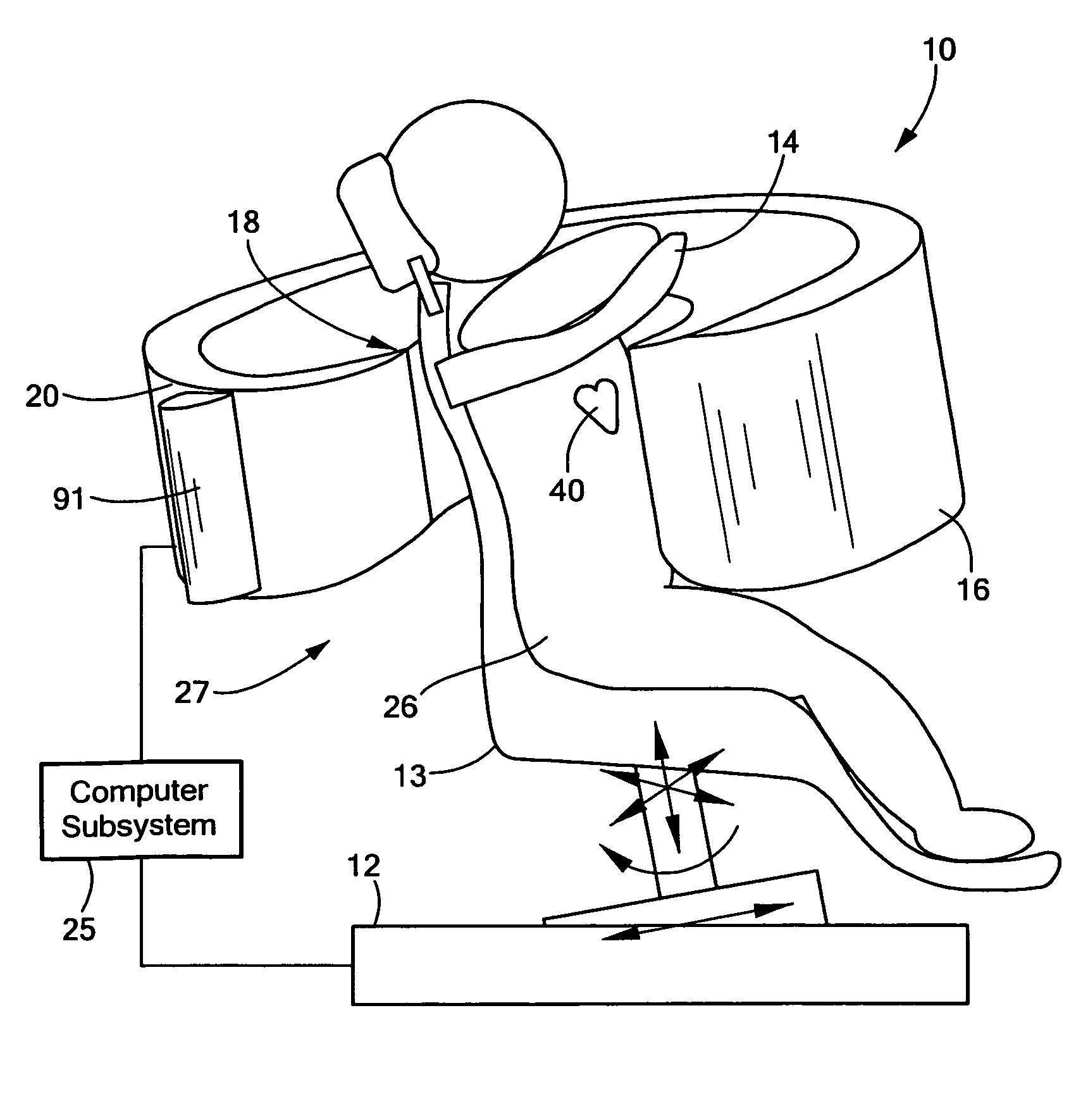
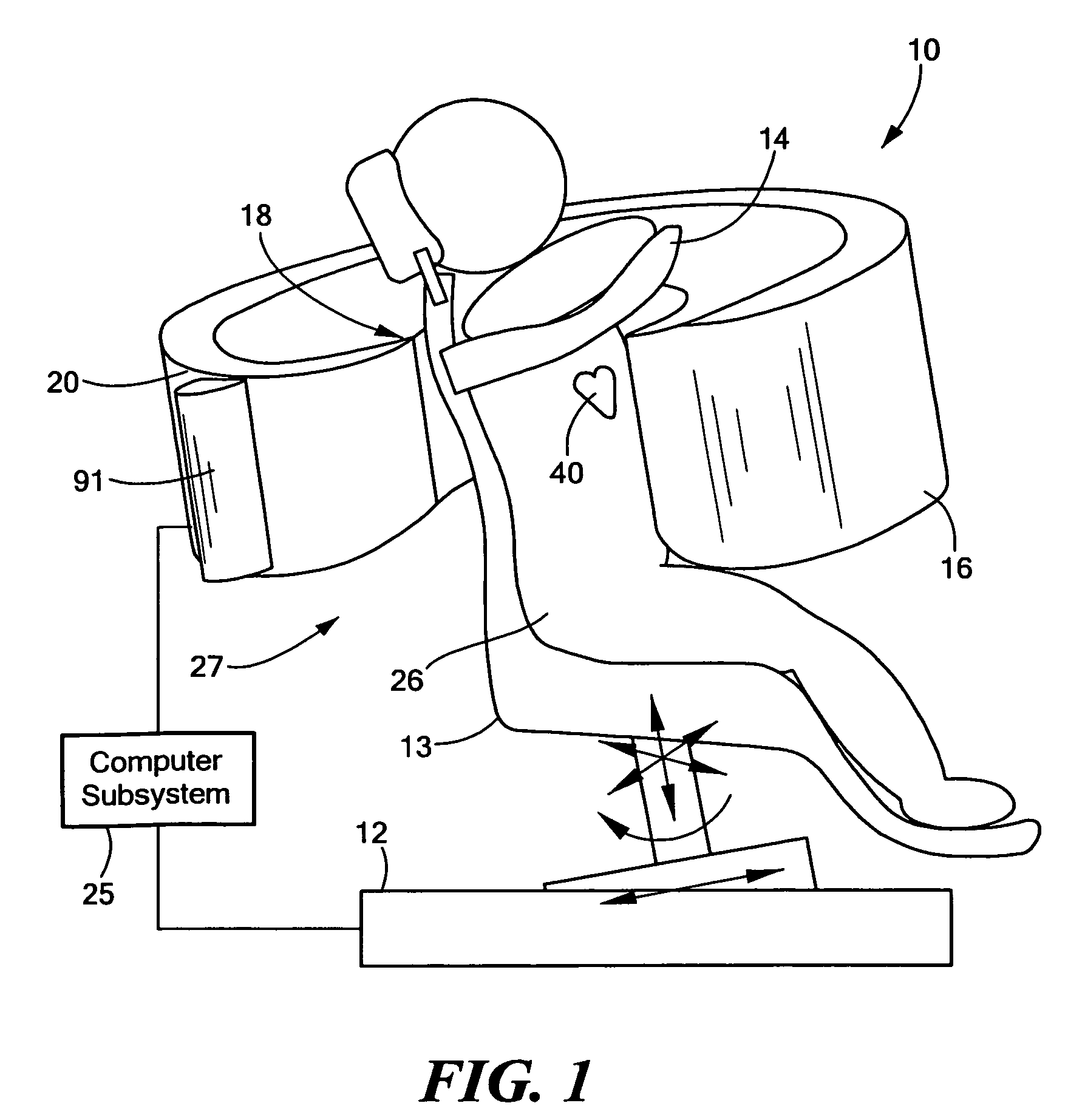
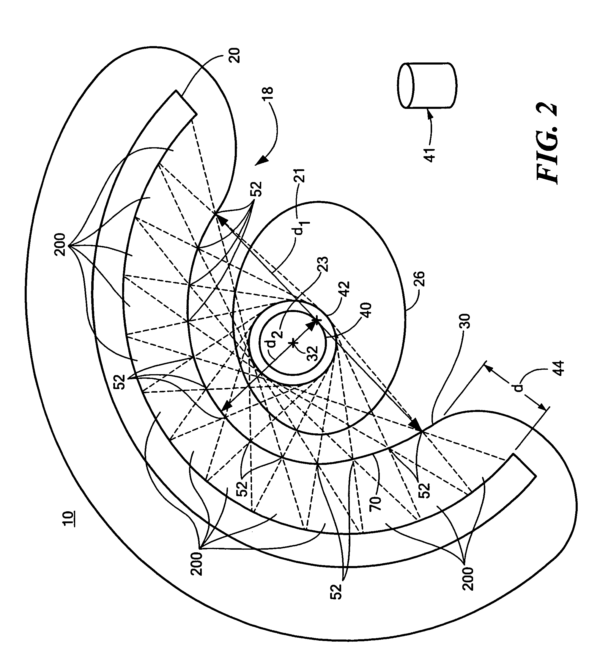
[0065]There is shown in FIG. 1, one embodiment of integrated SPECT / TCT system 10 of this invention. System 10 includes collimator subsystem 18 coupled to frame 16, which is shaped to an open-arc to approximately match the thoracic contour of patient 26, as better shown, e.g., in FIGS. 2 and 3. Collimator subsystem 18, FIGS....


PUM


Abstract

This invention features an integrated single photon emission computed tomography (SPECT) / transmission computed tomography (TCT) system for cardiac imaging including an open arc-shaped frame. A collimator system is shaped to approximately match the thoracic contour of patients having different sizes and weights and shaped to surround and position the collimator closely proximate a heart of a patient of said patients encompassed by at least one predetermined image volume for optimizing collimation of radiation photons emitted from the heart. An arc-shaped detector system is coupled to the collimator subsystem having a shape closely matching the shape of the collimator subsystem for detecting collimated radiation photons from the collimator subsystem and generating output electrical signals. A patient positioning subsystem positions a patient to a predetermined central longitudinal axis of the three-dimensional imaging volume and for intermittently and incrementally rotating the patient about the predetermined central longitudinal axis for generating a plurality of TCT images.

Description

RELATED APPLICATIONS[0001]This application claims the benefit of U.S. Provisional Application No. 60 / 869,274, filed Dec. 8, 2006, which is incorporated by reference herein.FIELD OF THE INVENTION[0002]This subject invention relates to an improved single photon emission computed tomography (SPECT) with an integrated transmission computed tomography (TCT) subsystem for cardiac imaging.BACKGROUND OF THE INVENTION[0003]SPECT systems are often used to show the distribution of a radioactive substance inside a patient's body. A source of penetrating radiation is administered to the patient, which typically consists of a pharmaceutical tagged with a radionuclide which emits radiation photons (radiopharmaceutical). The radiopharmaceutical is designed to be absorbed in a target organ, such as the heart muscle, or other organs or body part of interest. The emitted radiation photons are collimated with a collimator subsystem and detected by a detector subsystem which generates output electrical ...

Claims


Application Information

Patent Timeline
Patent Type & Authority Patents(United States)
IPC IPC(8): G01T1/166
CPCA61B6/037A61B6/032
Inventor CHANG, WEI
Owner RUSH UNIV MEDICAL CENT
Why Eureka
Try Eureka