System and apparatus for rapid stereotactic breast biopsy analysis

a breast biopsy and stereotactic technology, applied in the field of methods, can solve the problems of significant portion of the procedure time consumed by the film development cycle, time-consuming and uncomfortable for the patient, and laborious stereotactic breast biopsy, and achieve the effect of rapid production of specimen radiography

Inactive Publication Date: 2010-05-11
LAFFERTY PETER R
View PDF4 Cites 36 Cited by
  • Summary
  • Abstract
  • Description
  • Claims
  • Application Information

AI Technical Summary

Benefits of technology

[0006]One exemplary embodiment of the present invention is a modification and improvement to the commercially available stereotactic biopsy systems (e.g., LORAD Medical Systems Corp., Danbury, Conn. or Fischer Medical Technologies, Inc., Denver, Colo.). This modification may allow the stereotactic, swing-arm x-ray source (currently used solely to guide the biopsy procedure) to be used in the rapid production of specimen radiography. In one exemplary embodiment, a mechanical track may allow the x-ray source to shift laterally from the working biopsy corridor (occupied by the patient's breast during a procedure) allowing the x-ray source beam to be aligned with an add-on digital image receptor card which may be added to the lateral aspect of the existing image receptor. The harvested biopsy specimen threads may be positioned in, for example, specimen slots on a disposable specimen cassette or holder. The disposable specimen holder may then be attached to the add-on digital image receptor card between the x-ray source and digital image receptor card to allow for instant production of specimen radiographs within the procedure room. Other exemplary embodiments are possible as set forth herein.
[0007]Some examples of the benefits may include, but are not limited to, the following:a) Production of instant digital (rather than analog) specimen radiographs in the procedure room can be achieved. This feature can reduce procedure time up to 30%, thereby improving patient tolerance of the procedure.
[0008]b) Bleeding complications and needle discomfort can be diminished, as the typical number of samples harvested by the operator may decrease with exemplary embodiments of the present invention. There may no longer be a disincentive to “view” the biopsy sample early in the procedure, after a few samples have been retrieved.c) The digital specimen radiograph can be “post-processed” (filtered and windowed) to assure adequate visualization of very small, subtle microcalcifications, (many of which may be less than 0.1 mm in diameter). This feature may improve the accuracy of stereotactic biopsy. With analog specimen radiography, these types of microcalcifications can be very difficult to reliably identify, resulting in the need for additional biopsy retrieval.d) Decreased procedure time may allow for more procedures to be performed within a given time and level of staffing commitment. This may improve the economic viability of this procedure for biopsy centers.

Problems solved by technology

However, though very safe and minimally invasive, stereotactic breast biopsy can be laborious, time-consuming and uncomfortable for the patient.
A significant component of procedure time continues to be consumed by the film development cycles required for specimen radiograph production.

Method used


Image

  • System and apparatus for rapid stereotactic breast biopsy analysis
  • System and apparatus for rapid stereotactic breast biopsy analysis
  • System and apparatus for rapid stereotactic breast biopsy analysis

Examples


Embodiment Construction

)

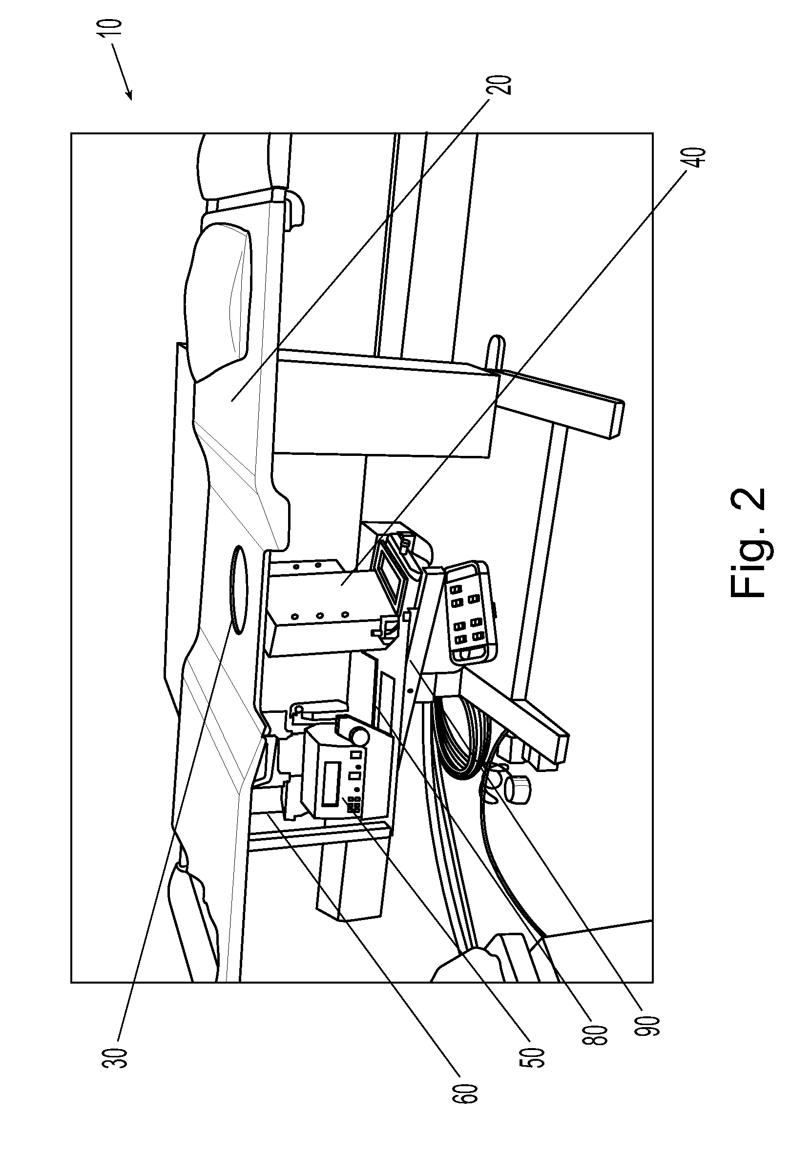
[0030]FIG. 1 illustrates a typical specimen radiograph showing a needle aspirated biopsy specimen 45 and microcalcifications 55. Biopsy specimens similar to 45 may be harvested from a patient's breast 25 typically via a plurality of samples collected from a target area 35. The specimen radiograph is an ex-vivo x-ray picture of the biopsy samples retrieved from the breast, which under conventional circumstances, must be processed outside the procedure room, on a standard mammography x-ray unit or on a separately purchased commercially available unit, such as one produced by Faxitron X-ray Corporation, Wheeling, Ill. In the current state of the art, this picture is required to assure that sufficient quantities of microcalcifications are removed from the groups of calcium targeted within the breast.

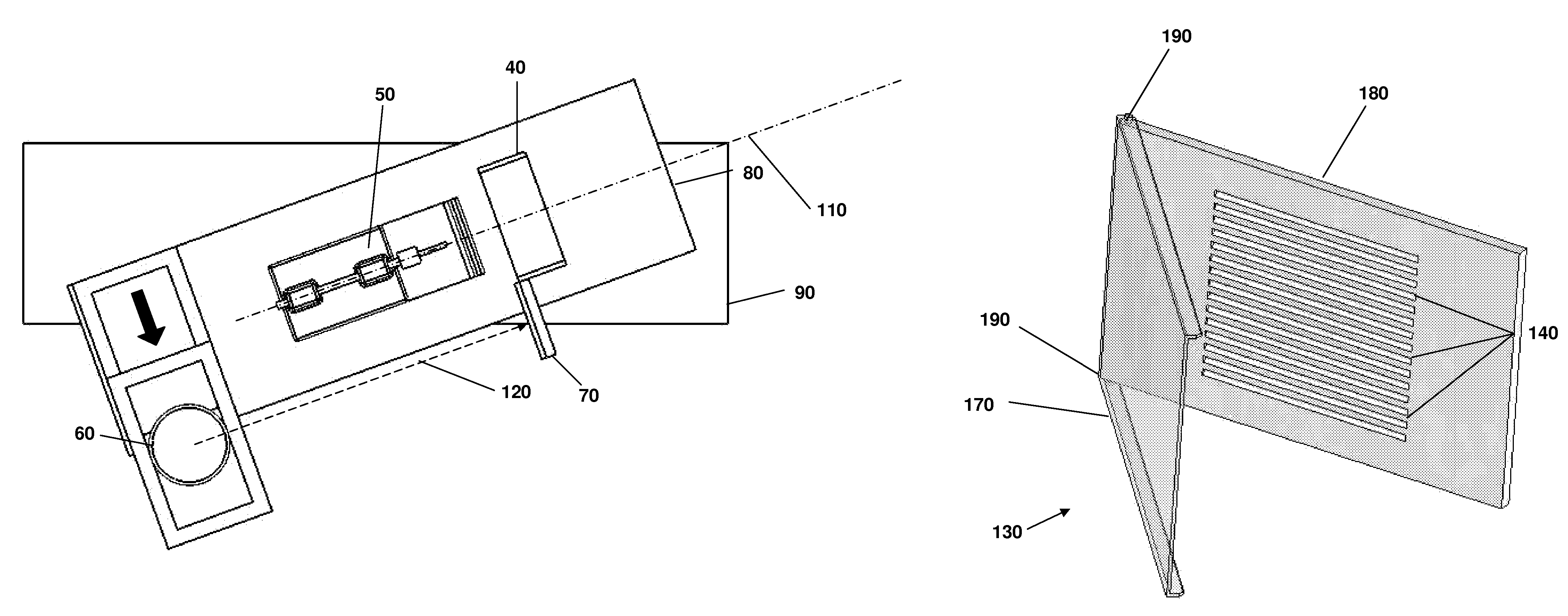
[0031]FIGS. 2, 3, and 4 illustrate a typical example of a commercially available stereotactic biopsy system 10 produced by LORAD Medical Systems Corp., Danbury, Conn. During a typical biops...


PUM


Abstract

A stereotactic breast biopsy apparatus and system that may comprise an x-ray source, a digital imaging receptor, and a biopsy specimen cassette, wherein the x-ray source is provided with a means for displacing the beam axis of the x-ray source from a working biopsy corridor beam axis to permit an unobstructed illumination of the biopsy specimen and thereby produce biopsy x-ray images directly in the procedure room for immediate analysis. Some examples of the benefits may be, but are not limited to, a more rapid analysis of biopsy specimen digital images, post-processing image capability, and decreased procedure time and diminution of patient bleeding complications and needle discomfort.

Description

[0001]This application is a nonprovisional application of, and claims priority to, U.S. Provisional Application No. 60 / 827,327, filed Sep. 28, 2006, which is incorporated by reference as if fully recited herein.TECHNICAL FIELD[0002]Exemplary embodiments of the present invention relate to methods (e.g., medical non-surgical) of diagnosing breast cancer and, more particularly, to a novel apparatus, system, and method which beneficially improves current stereotactic breast biopsy devices and methods.BACKGROUND OF THE ART[0003]Stereotactic breast biopsy has become the method of choice for the non-surgical diagnosis of many forms of breast cancer. Many breast cancers are discovered by the presence of microcalcifications visible on a screening mammogram. Yet, these microcalcifications do not have a corresponding palpable abnormality. Therefore, an image-guided needle biopsy technique must be utilized to determine if early, pre-invasive breast cancer is present. Currently, stereotactically...

Claims


Application Information

Patent Timeline
Patent Type & Authority Patents(United States)
IPC IPC(8): A61B6/00
CPCA61B10/0096A61B19/201A61B10/0266A61B2019/5238A61B2010/0225A61B2019/205A61B90/11A61B90/17A61B2090/376
Inventor LAFFERTY, PETER R.
Owner LAFFERTY PETER R
Why Eureka
Try Eureka