Apparatus and method for three dimensional ultrasound breast imaging

a three-dimensional ultrasound and breast imaging technology, applied in the field of ultrasound medical imaging, can solve the problems of less effective method for finding abnormalities in areas, patient discomfort, and general discomfort of the patient, and achieve the effect of wide variation in patient anatomy, less patient discomfort, and improved capability to image features

Inactive Publication Date: 2010-12-14
SJ STRATEGIC INVESTMENTS LLC
View PDF47 Cites 123 Cited by
  • Summary
  • Abstract
  • Description
  • Claims
  • Application Information

AI Technical Summary

Problems solved by technology

The method is generally uncomfortable to the patient and exposes the patient to X-ray radiation.
Furthermore, it is well known that the method is less effective at finding abnormalities in areas where the breast tissue is particularly dense.
By the nature of their construction and operation, the use of these devices tends to be laborious and generally confined to localized examination of a particular area of interest.

Method used


Image

  • Apparatus and method for three dimensional ultrasound breast imaging
  • Apparatus and method for three dimensional ultrasound breast imaging
  • Apparatus and method for three dimensional ultrasound breast imaging

Examples


Embodiment Construction

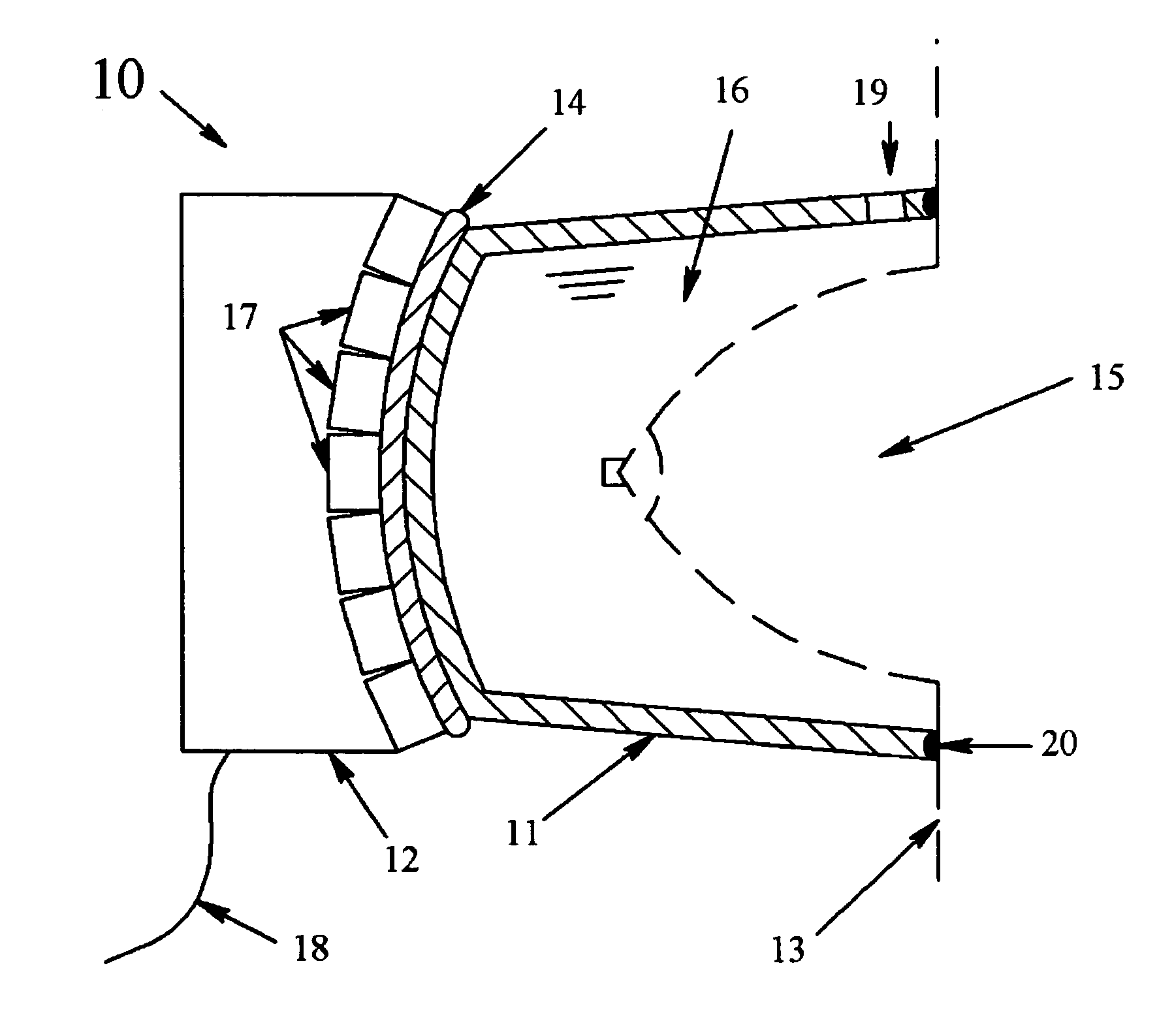
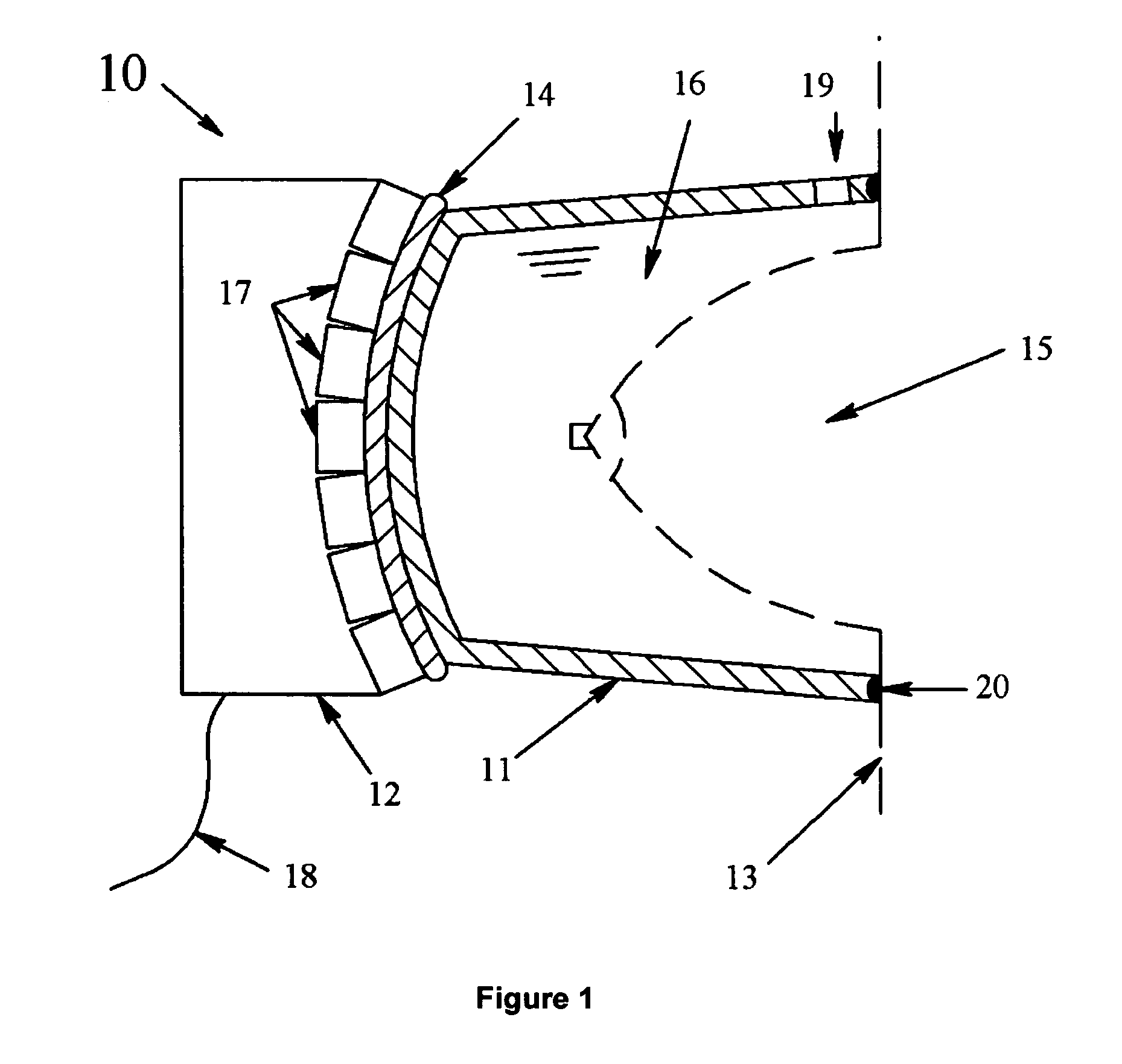
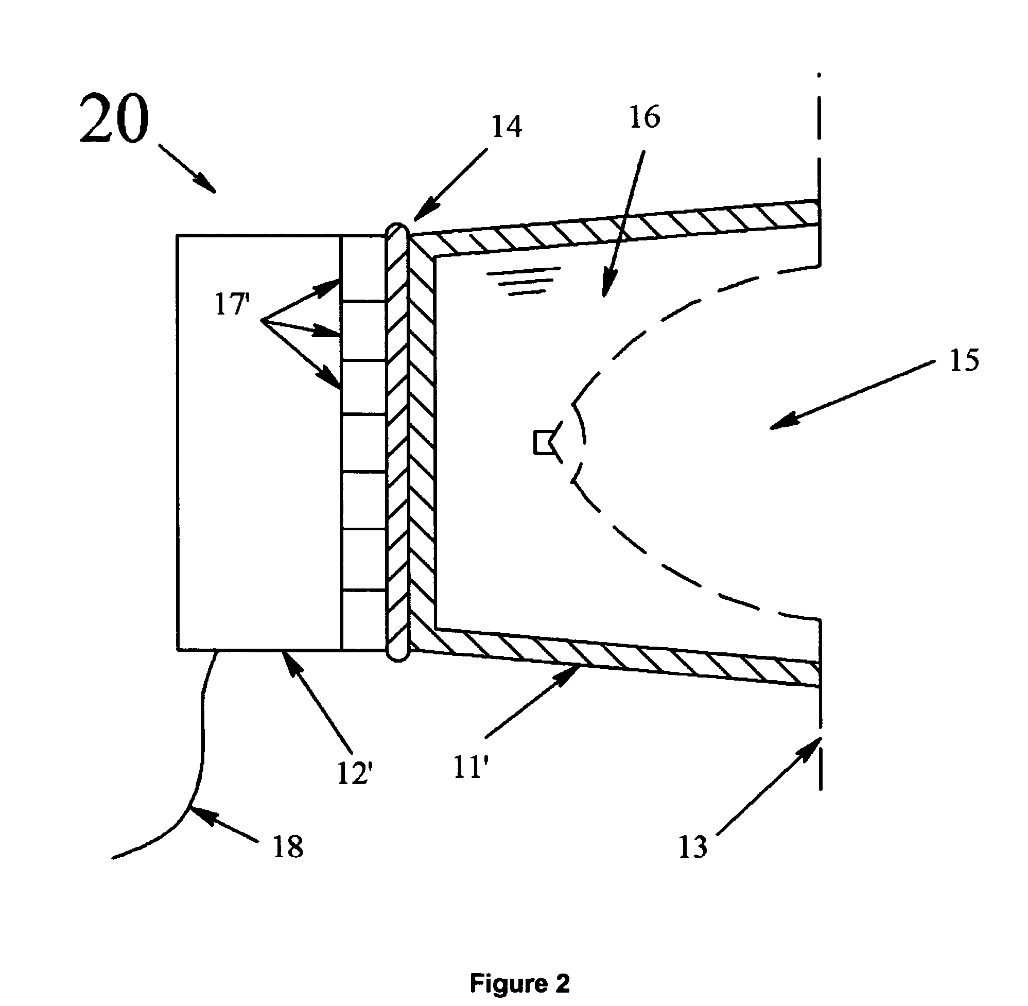
[0020]In its most general embodiment, the invention comprises three basic elements: first, an ultrasonic transducer array capable of generating signals that may be analyzed to produce three-dimensional sonograms; second, an electronic analysis system to convert the ultrasonic signals into various imaging data sets in conjunction with an electronic user interface; and third, an applicator device, preferably disposable, configured to provide good acoustic coupling to breasts of various sizes while eliminating the need to expose the transducer to repeated cleaning.

[0021]Ultrasonic transducers may be designed in various ways, and the present invention is not limited to any particular transducer design but rather may be advantageously applied to adapt many different types of transducers to the problem of breast imaging. Transducer arrays may be curved, with the front surface typically concave, and the individual piezoelectric elements may be substantially square blocks such as described ...


PUM


Abstract

An apparatus for ultrasonic mammography includes: an array of ultrasonic transducers and signal processing means for converting the output of the transducer array into three dimensional renderings of anatomical features; and, an applicator device having a first side conformable to the contour of the transducer array and a second side configured to accept the breast, the applicator device further containing a quantity of fluid sufficient to surround and stabilize the breast during examination without substantially altering the breast from its natural shape.

Description

CROSS REFERENCE TO RELATED APPLICATIONS[0001]This application claims the benefit of Provisional Patent Application Ser. No. 60 / 474,687 filed by the present inventor on May 30, 2003, the entire disclosure of which is incorporated herein by reference.BACKGROUND OF THE INVENTION[0002]1. Field of the Invention[0003]This invention pertains to the field of ultrasonic medical imaging, and more particularly to systems and methods for diagnostic imaging of the breast. More specifically the invention describes a coupling device for an ultrasonic imaging transducer that stabilizes the breast to allow rapid examination of the patient while accommodating anatomical variations, maximizing cleanliness, and minimizing patient discomfort.BACKGROUND[0004]2. Discussion of Prior Art[0005]Mammography is a well-known medical procedure used to diagnose various abnormalities such as cysts, calcifications, and tumors. The standard procedure involves compressing the breast along vertical and horizontal plane...

Claims


Application Information

Patent Timeline
Patent Type & Authority Patents(United States)
IPC IPC(8): A61B8/14A61B8/00A61B8/08
CPCA61B8/0825A61B8/4218A61B8/4281A61B8/483Y10S128/915
Inventor STRIBLING, MARK L.
Owner SJ STRATEGIC INVESTMENTS LLC
Why Eureka
Try Eureka