Displaying breast tomosynthesis computer-aided detection results

a technology of breast tomosynthesis and computer-aided detection, which is applied in the direction of tomography, patient positioning for diagnostics, instruments, etc., can solve the problems of increasing the volume of data requiring radiologist review, and achieve the effect of increasing the likelihood

Inactive Publication Date: 2011-04-19
HOLOGIC INC
View PDF19 Cites 29 Cited by
  • Summary
  • Abstract
  • Description
  • Claims
  • Application Information

AI Technical Summary

Benefits of technology

[0002]Progress toward all-digital medical imaging environments has substantially increased the speed at which large amounts of medical image information can be accessed and displayed to a radiologist. As used herein, radiologist generically refers to a medical professional that analyzes medical images and makes clinical determinations therefrom, it being understood that such person might be titled differently, or might have differing qualifications, depending on the country or locality of their particular medical environment. X-ray based imaging for breast cancer screening / diagnosis is a particularly important field that is experiencing such information-expanding technological progress.
[0009]However, in progressing from conventional x-ray mammography to breast x-ray tomosynthesis imaging, practical issues arise with regard to the rising volume of data requiring review by the radiologist. Whereas there are usually just four conventional x-ray mammogram images per patient, there can be hundreds of tomosynthesis reconstructed image slices (e.g., 40-60 slices for each of the four views). As more visual information becomes available, an important challenge is to present such information to the radiologist effectively and efficiently such that screening for abnormalities can be done thoroughly and effectively, and yet in a reasonable time to be practical, and diagnostic assessment can also be facilitated. Of particular importance is the manner in which an image review workstation displays CAD markers to the radiologist for the large stack of tomosynthesis reconstructed images, it being desirable that the CAD markers not be overly obtrusive on their corresponding image while also not being readily overlooked. The visual solution should simultaneously resolve these goals in conjunction with other beneficial goals, such as limiting the number of attention-sapping off-image gadgets to a workable minimum. Even subtle differences involving a few saved eye movements or a few saved hand strokes, keystrokes, or mouse cursor movements can lead to substantially better efficiency, stamina, and / or accuracy on the part of the radiologist.
[0012]Described in this patent specification are methods, systems, and related computer program products for processing and displaying CAD results to a user (e.g., a radiologist) in conjunction with breast x-ray tomosynthesis data in a manner that resolves several issues facing the user during review of such a large data set, including the need to recognize the existence and locations of CAD markers on a relatively small number of Tr images among a relatively large stack of Tr images, while not having inordinate amounts of attention devoted to searching for those CAD markers, without having an inordinate number of off-image eye movements, and without an inordinate amount of Tr image content obscured by annotation content. For example, in one preferred embodiment, a plurality of Tr images representative of slices of a breast having selected orientations and thicknesses is received, and CAD information is received identifying a detection-containing one of the Tr images and a coordinate location thereon at which a CAD detection marker is to be displayed. An ordered sequence of the Tr images is displayed to the user such that the user is allowed to sequentially page therethrough, the ordered sequence including the detection-containing Tr image. The ordered sequence of Tr images further includes at least one Tr image nearby the detection-containing Tr image with respect to the ordered sequence, the nearby Tr image not being detection-containing with respect to the identified coordinate location. When the detection-containing Tr image is displayed, the CAD detection marker is displayed thereon at the identified coordinate location. When the nearby Tr image is displayed, a CAD proximity marker is displayed thereon at the identified coordinate location, the CAD proximity marker not itself being indicative of a CAD detection on the nearby Tr image, but rather for encouraging user attention toward the identified coordinate location of the detection-containing Tr image, for discouraging user oversight of the CAD detection marker thereon when paging therethrough. Preferably, the CAD proximity has a noticeably different size than the CAD detection marker.
[0014]In other preferred embodiments, further information is conveyed to the user by the CAD proximity markers without additional screen clutter. In one example, a paging behavior of the user, such as the user's paging rate toward or away from the detection-containing Tr image, is detected and instantly used to vary the displayed size of the CAD proximity markers. Thus, for example, if the user is paging very rapidly through the stack of Tr images, thereby increasing the likelihood they might miss the CAD detection marker on the detection-containing Tr image, the CAD proximity markers are made larger and more visible to ensure that the presence of the CAD detection marker is perceived during the fast paging process. However, if the use is paging very slowly, the CAD proximity markers are made smaller and less obtrusive, because it is less likely that the presence of the CAD detection marker will be overlooked. Other useful variations to the size and / or shape of the CAD proximity markers for achieving useful yet non-obtrusive information conveyance, including those described further hereinbelow, are within the scope of the preferred embodiments.

Problems solved by technology

However, in progressing from conventional x-ray mammography to breast x-ray tomosynthesis imaging, practical issues arise with regard to the rising volume of data requiring review by the radiologist.

Method used


Image

  • Displaying breast tomosynthesis computer-aided detection results
  • Displaying breast tomosynthesis computer-aided detection results
  • Displaying breast tomosynthesis computer-aided detection results

Examples


Embodiment Construction

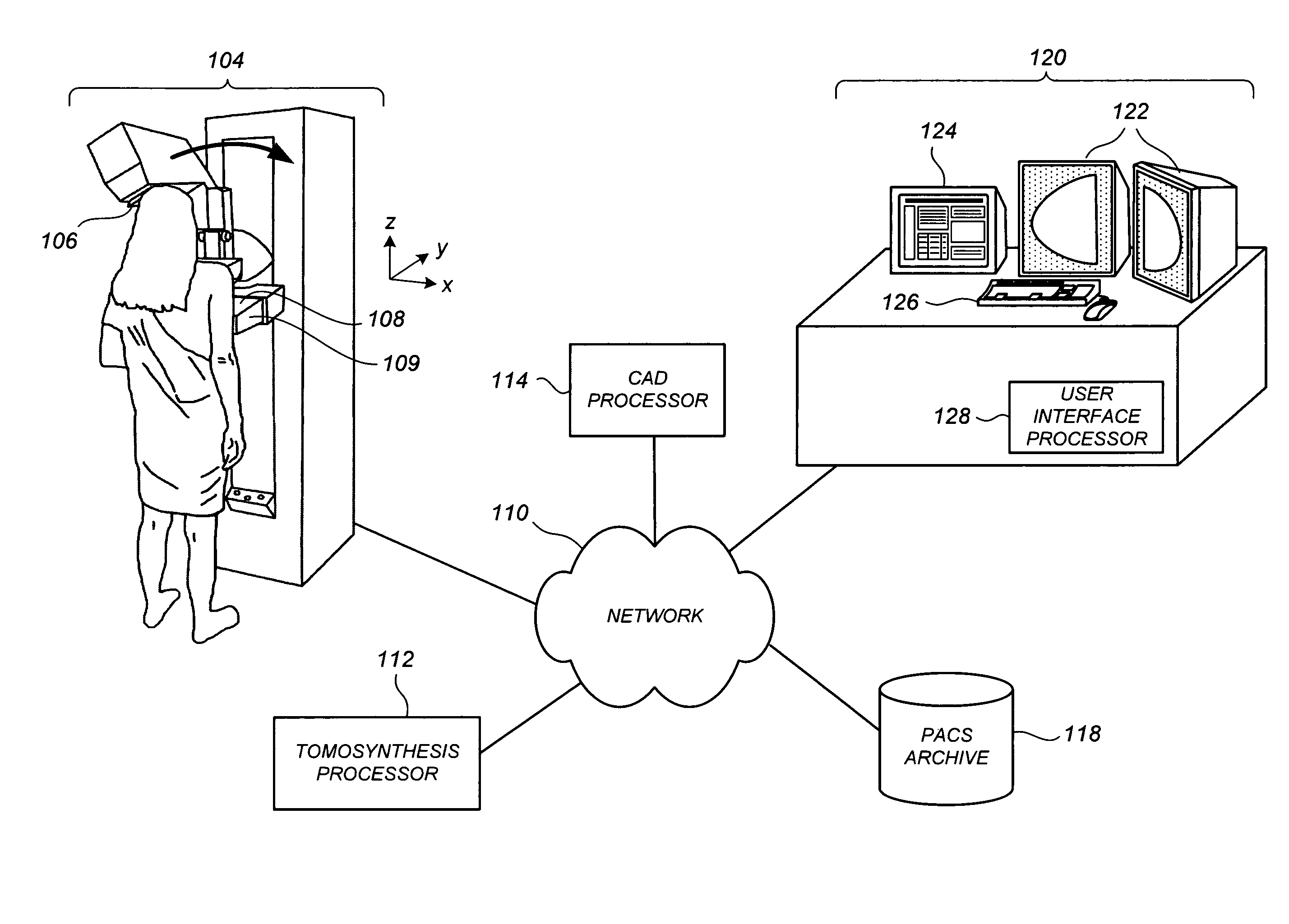
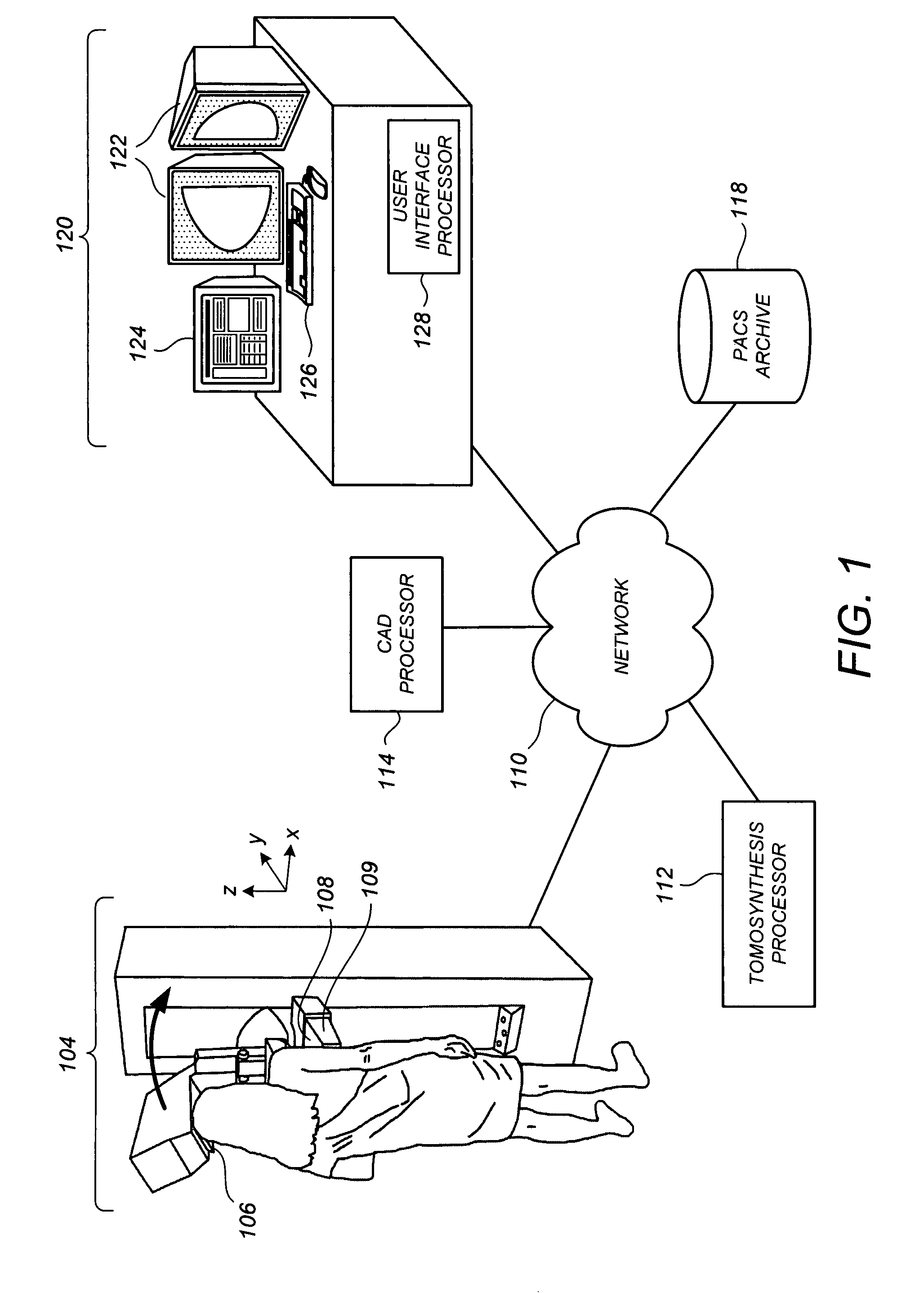
[0022]FIG. 1 illustrates a conceptual diagram of a medical imaging environment for which one or more of the preferred embodiments is particularly suited. Shown in FIG. 1 is a network 110, which may be a HIS / RIS (Hospital Information System / Radiology Information System) network, to which is coupled a breast x-ray tomosynthesis acquisition device 104, which can be dedicated for breast x-ray tomosynthesis or combined with conventional x-ray mammogram functionality. The acquisition device 104 includes an x-ray source 106 projecting x-rays toward a patient's breast that is supported on a breast platform 108. An x-ray imager 109 such as a flat panel, direct conversion imager available from Hologic, Inc. generates projection image data such as data defining images Mp and / or Tp. With respect to a three-dimensional coordinate system as illustrated in FIG. 1, in which an x-z plane corresponds at least roughly to a coronal plane of the patient and the y-direction extends outward from the patie...


PUM


Abstract

Methods, systems, and related computer program products for processing and displaying computer-aided detection (CAD) results in conjunction with breast x-ray tomosynthesis data are described. For one preferred embodiment, as a user pages through a notional stack of tomosynthesis reconstructed slice images (Tr images), including a detection-containing Tr image on which a CAD marker is to be displayed at an identified coordinate location, one or more CAD proximity markers is displayed at that coordinate location on one or more neighboring Tr images. While not themselves indicative of CAD findings on their respective Tr images, the CAD proximity markers encourage user attention toward the coordinate location of the CAD detection marker of the detection-containing Tr image. Preferably, the CAD proximity markers are of noticeably different size from each other and from the CAD detection marker to promote their perception in the peripheral vision of the user during the paging process.

Description

FIELD[0001]This patent specification relates to the computer-aided detection of abnormalities in medical images. More particularly, this patent specification relates to the display of computer-aided detection results for breast tomosynthesis data volumes.BACKGROUND AND SUMMARY OF THE DISCLOSURE[0002]Progress toward all-digital medical imaging environments has substantially increased the speed at which large amounts of medical image information can be accessed and displayed to a radiologist. As used herein, radiologist generically refers to a medical professional that analyzes medical images and makes clinical determinations therefrom, it being understood that such person might be titled differently, or might have differing qualifications, depending on the country or locality of their particular medical environment. X-ray based imaging for breast cancer screening / diagnosis is a particularly important field that is experiencing such information-expanding technological progress.[0003]I...

Claims


Application Information

Patent Timeline
Patent Type & Authority Patents(United States)
IPC IPC(8): G06K9/00A61B6/04G16H30/20G16H30/40
CPCA61B6/025G06F19/321A61B6/502A61B6/03G16H30/40G16H30/20
Inventor KHORASANI, GEORGE ALLEN
Owner HOLOGIC INC
Why Eureka
Try Eureka