Two-piece video laryngoscope

a two-piece, video technology, applied in the field of intubation devices and instruments, can solve the problems of reducing visibility, reducing the possibility of direct visualization of the glottis, and increasing the difficulty of the procedure, so as to reduce the potential of cross contamination, reduce the risk, and reduce the effect of invasiveness

Inactive Publication Date: 2011-05-24
CUBB ANTHONY
View PDF27 Cites 179 Cited by
  • Summary
  • Abstract
  • Description
  • Claims
  • Application Information

AI Technical Summary

Benefits of technology

[0028]The endotracheal tube receptacle channel has a transversely generally arcuate or half-ovoid bottom segment that extends along the dorsal surface of the handle / blade unit starting a short distance from the handgrip portion and along the curved distal end portion to the terminal end, and at least one transversely curved generally arcuate top segment that extends partially over, and a distance along, the bottom segment of the channel. A plastic endotracheal tube is slidably received and releasably retained in the channel in a preloaded condition such that the exit direction of the tube is predictable during insertion of the handle / blade unit into the patient's mouth and throat. An air passageway in fluid communication with a vacuum / oxygen connector and control orifice beneath the cavity extends through the interior of the handle / blade unit to the terminal end for providing suction or oxygen.
[0044]A still further feature and advantage is that the video laryngoscope is user friendly and can be easily and efficiently used by a practitioner with basic training.

Problems solved by technology

Additionally, if there is concern that the patient may have suffered spinal injuries, the technique for possible direct visualization of the glottis involves risk due to the potential for increased spinal injury from this procedure.
This procedure is often made more difficult by the presence of bodily fluids on or in the larynx and trachea, which significantly reduces visibility.
Without visualization of anatomical structure, intubation of a patient during an emergency situation may require blind placement of an endotracheal tube based on freehand trial and error.
Without proper positioning and guidance, the tubular members often cause trauma or injury to anatomical tissue, or missed intubation into the esophagus with potential fatal consequences.
Such devices obviously require the use of relatively large diameter endotracheal tubes in order to be carried on the tubular probe, and their use necessarily is limited to patients with sufficiently large airway passages to accommodate the combined size of the probe and endotracheal tube.
Additionally, due to the flexible nature of the probes, it is difficult to manipulate the probe to displace the tongue and epiglottis to permit guidance during insertion of the tube into the trachea.
These instruments require a high degree of skill and a concomitant degree of training to perform the procedure quickly, without injuring the patient.
Additionally, because of expense, lack of portability, and sterilization requirements to prevent cross contamination among patients, these instruments are generally not available in a non-hospital setting.

Method used


Image

  • Two-piece video laryngoscope
  • Two-piece video laryngoscope
  • Two-piece video laryngoscope

Examples


Embodiment Construction

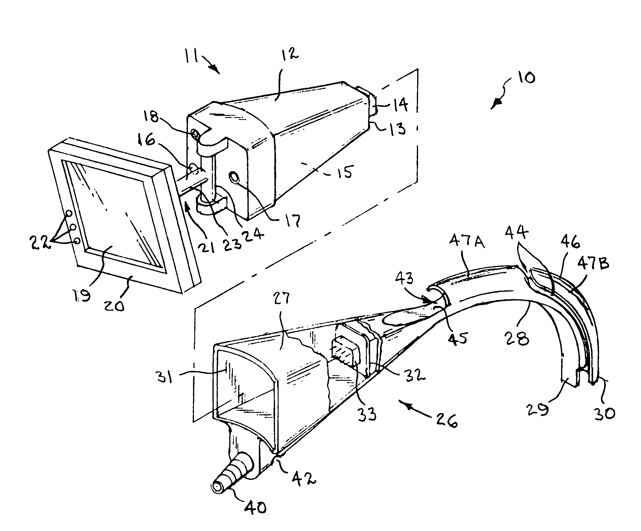
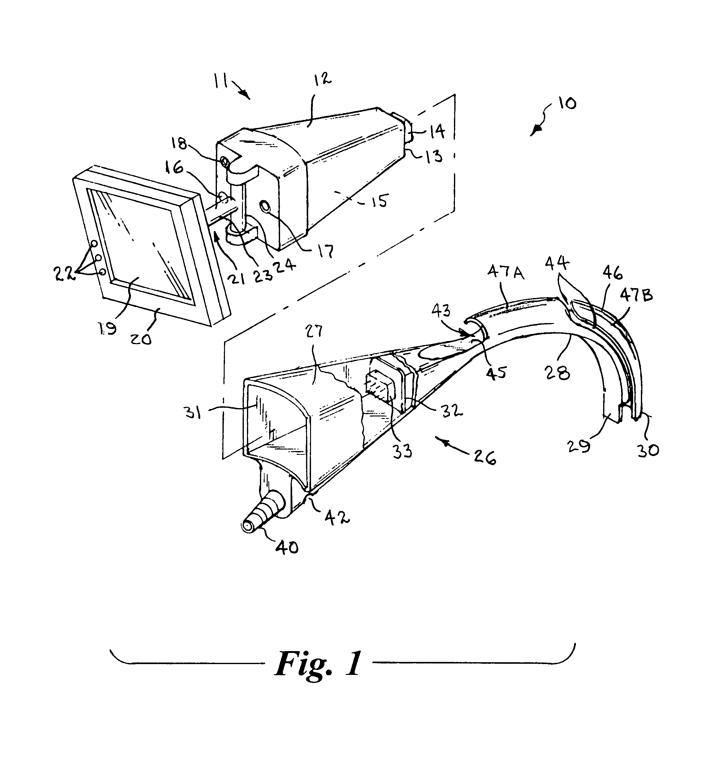
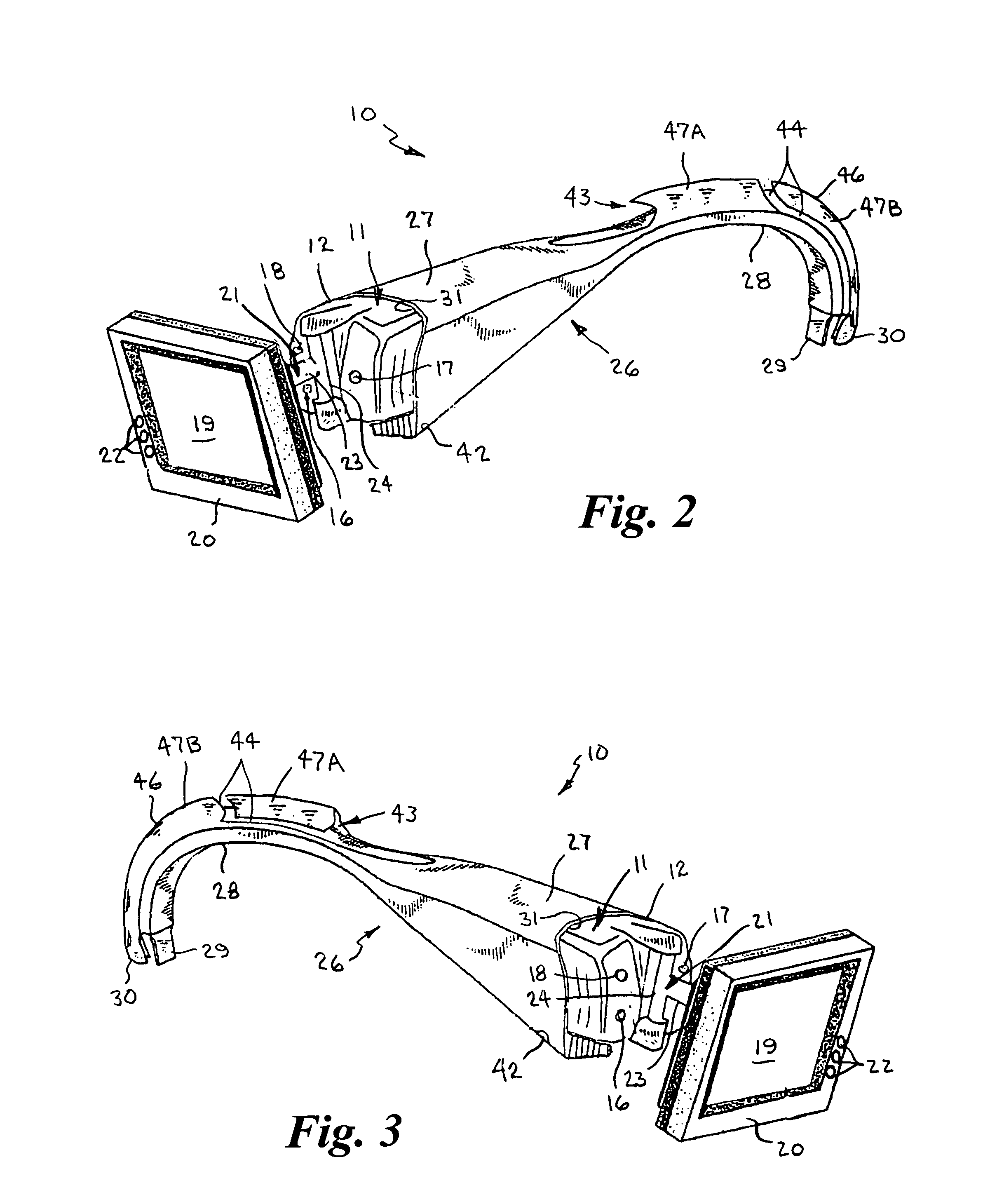
[0052]Referring now to FIGS. 1-6, the present invention is a two-piece video laryngoscope 10 for use during endotracheal intubation that enables indirect external visualization of a patient's upper airway from various operator positions around the patient. The two-piece video laryngoscope 10 includes a lightweight disposable handle / blade unit 26 and a power / video module 11 with a pivotally mounted flat panel display viewing screen 19 at the proximal end thereof, which is releasably engaged in the proximal end of the handle / blade unit. FIG. 1 shows the components in a disconnected condition, and FIGS. 2-5 show the components in the connected condition.

[0053]The power / video module 11 has generally rectangular outer housing 12 that slightly tapers rearwardly and terminates in a rear wall 13 having a first female or male pin connector 14 mounted thereon. As represented by the reference numeral 15, the housing 12 of the power / video module 11 contains a rechargeable battery power supply a...


PUM


Abstract

A two-piece video laryngoscope includes a disposable handle / blade unit having a handgrip portion with a cavity at the proximal end, a curved distal end portion extending from the handgrip portion terminating in a terminal face containing a LED and a lens and digital image sensor connected with a first connector in the cavity, and a tube receptacle channel extending distally along the dorsal surface of and a vacuum / oxygen passageway extending through the curved distal end portion; and a power / video module releasably engaged in the cavity having a flat panel display pivotally mounted at the proximal end thereof and containing a rechargeable battery and electrical and video circuitry connected with a second connector. An endotracheal tube is received and releasably retained in the tube receptacle channel in a preloaded condition. When assembled, the connectors are engaged to complete the electrical and video circuits and allow viewing of insertion and intubation.

Description

CROSS REFERENCE TO RELATED APPLICATION[0001]This application is a Continuation-In-Part and claims the benefit of priority under 35 U.S.C. 120 of U.S. patent application Ser. No. 10 / 693,206 filed Oct. 23, 2003 now abandoned, and which is expressly incorporated herein by reference in its entirety.BACKGROUND OF THE INVENTION[0002]1. Field of the Invention[0003]The present invention generally relates to intubation devices and instruments for accessing the laryngeal area of the human body and, more particularly, to a two-piece video laryngoscope having a power / video module with a small color flat panel display pivotally mounted at the proximal end thereof and a replaceable handle / blade unit connected thereto, that allows indirect external visualization of a patient's upper airway, during endotracheal intubation.[0004]2. Background Art[0005]Laryngoscopes are widely known and used in the medical field to facilitate endotracheal intubation of a patient during a respiratory emergency situati...

Claims


Application Information

Patent Timeline
Patent Type & Authority Patents(United States)
IPC IPC(8): A61B1/267
CPCA61B1/00052A61B1/00094A61B1/00103A61B1/00105A61M16/0488A61B1/0676A61B1/0684A61B1/267A61B1/05A61B1/015A61M16/0495
Inventor CUBB, ANTHONY
Owner CUBB ANTHONY
Why Eureka
Try Eureka