
Systems for and methods of repair of atrioventricular valve regurgitation and reversing ventricular remodeling

a technology of atrioventricular valve and ventricular remodeling, which is applied in the direction of heart stimulators, prostheses, therapy, etc., can solve the problems of not necessarily complete restoration of normal cardiac geometry, and move towards normal, and achieve the effect of improving leaflet coaptation

Inactive Publication Date: 2011-06-07
THE GENERAL HOSPITAL CORP
View PDF32 Cites 84 Cited by
  • Summary
  • Abstract
  • Description
  • Claims
  • Application Information

AI Technical Summary

Benefits of technology

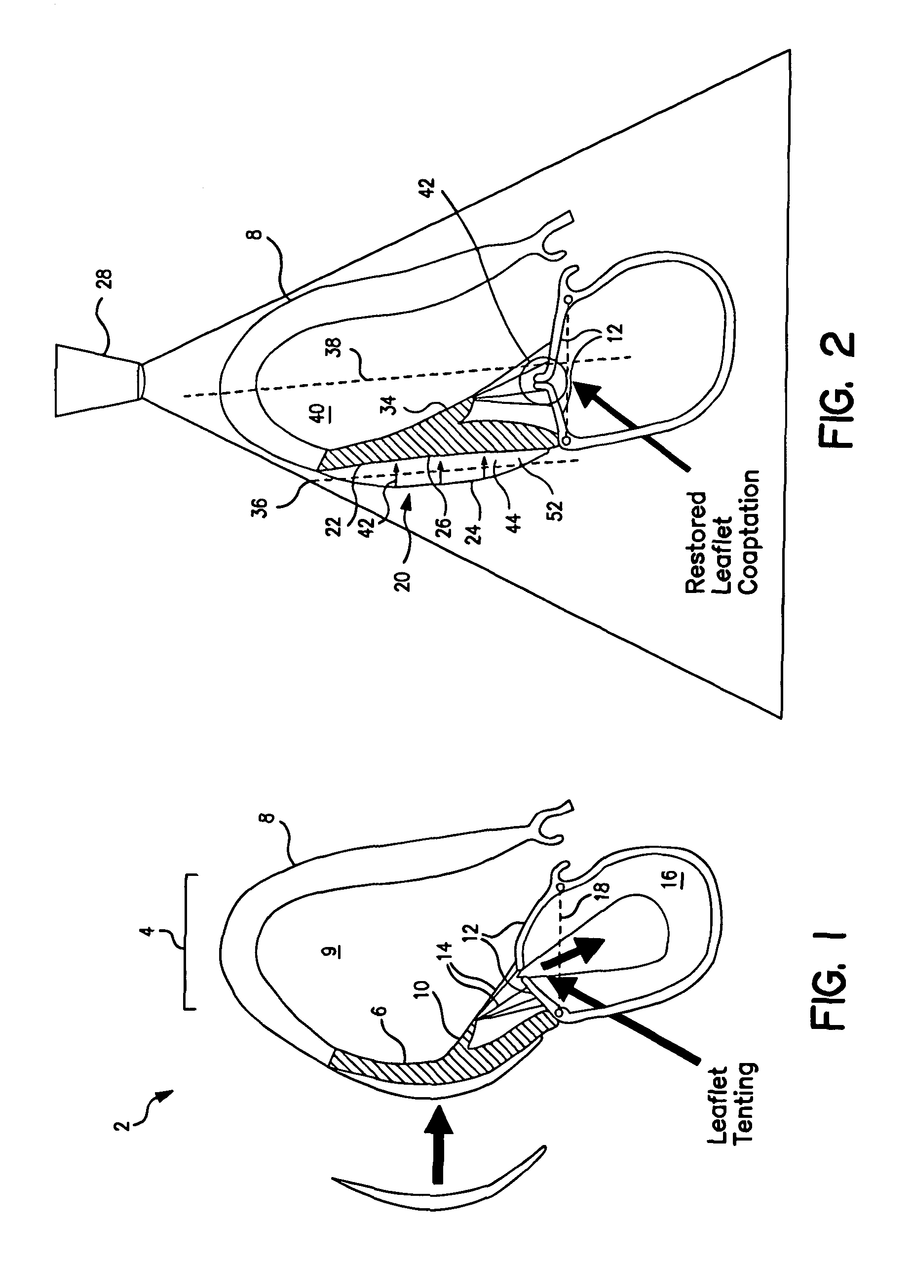
[0017]Intraoperative observations suggest that force or pressure applied outside papillary muscle can eliminate atrioventricular valve regurgitation (AVR). The present invention provides devices for placement in a longitudinal plane along a portion of the papillary muscle that exert such forces in three-dimensional space (i.e., causing displacement both normal to and radially from said longitudinal plane.)
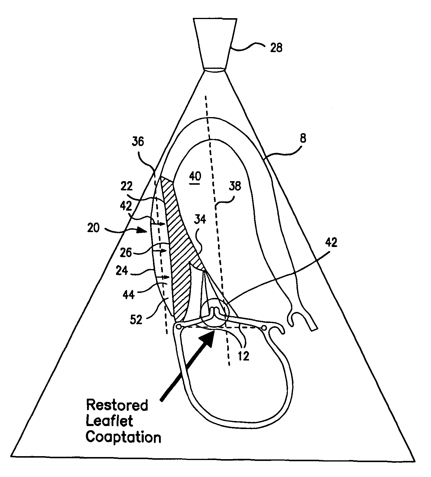
[0018]The devices for treating AVR generally include a compression member acting on an exterior wall segment of the heart to achieve the displacement of the exterior wall segment inward and toward the center line of the ventricular cavity of the heart so as to normalize papillary muscle geometry and improve leaflet coaptation. In preferred embodiments, the devices are reversibly collapsible or furlable for easier delivery to the heart wall attachment site.
[0020]Echocardiographic imaging may aid in performance of any of the procedural steps described herein, such as guiding the placement of the devices, or monitoring and / or adjusting the displacement of the wall segment the devices effect. Such imaging is also useful in designing the device to have a compressive member contoured to achieve a displacement unique to the portion of the exterior wall segment intended to be displaced.
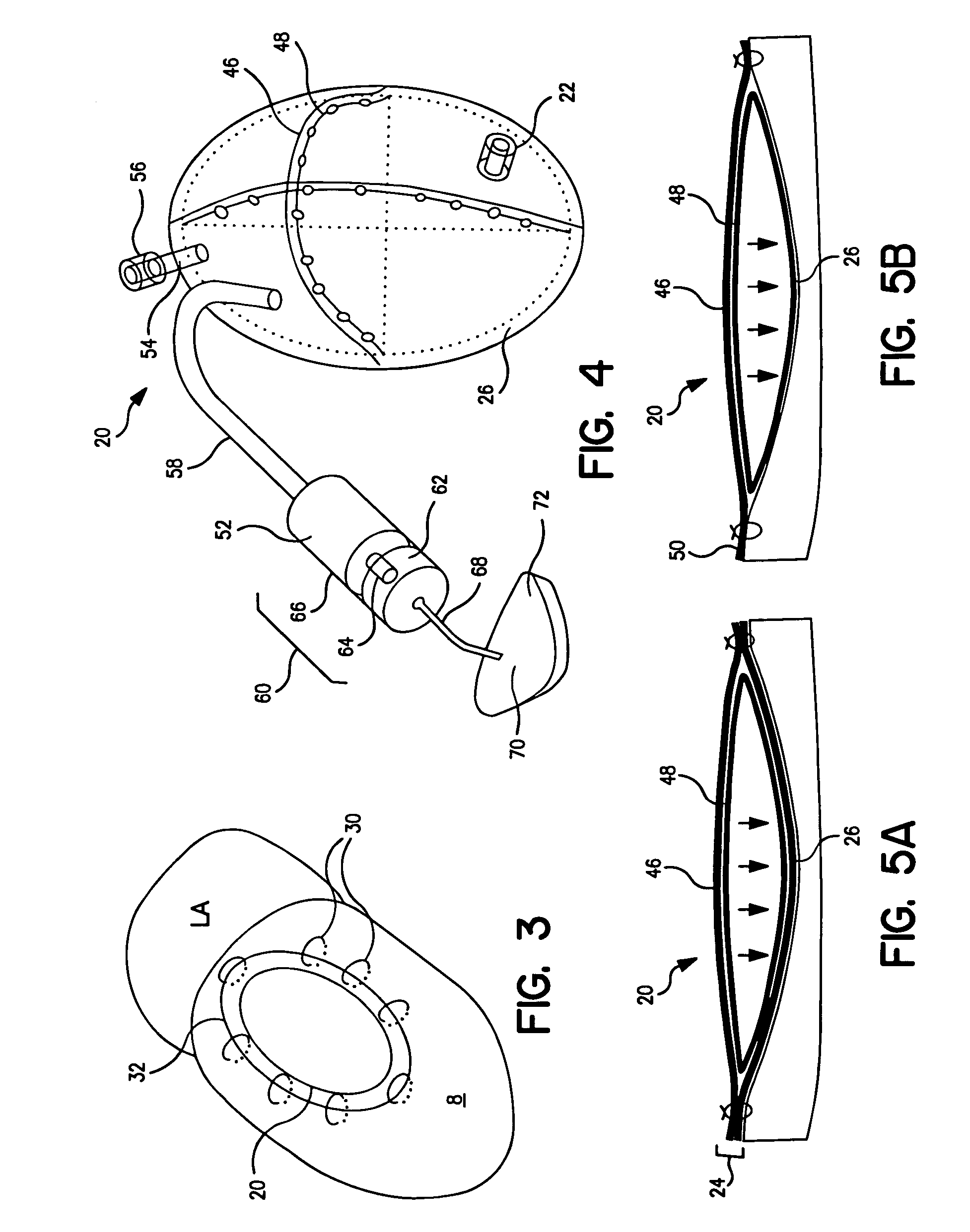
[0021]In another embodiment, the displacement is achieved by filling a fillable region between the compressive member and the buttressing portion with a stuffing. The stuffing may comprise engineered tissue that stiffens in the fillable region (e.g, bone or cartilage forming tissues), thereby maintaining the compressive force on the exterior wall segment. A scaffold structure may be employed to initially receive the engineered tissue. A portion of the apparatus may biodegrade over a time period comparable to that required for the engineered tissue to stiffen to the point of independently providing the displacement force required to move the heart wall segment so as to normalize the papillary muscle geometry and improve leaflet coaptation.
[0024]The present invention further provides an apparatus and method for reducing regurgitation of an atrioventricular valve of a heart by delivering a material into a muscle wall region of a heart proximate the papillary muscle. The material displaces of a portion of said muscle wall region inward and toward the center line of the ventricular cavity and thus normalizes papillary muscle geometry and improves leaflet coaptation. Suitable materials include hydrogels or nickel titanium alloys (e.g., Nitinol™) that substantially retain their shape after delivery. The material may be encapsulated in a structure implanted within the muscle wall, such as, for example, a balloon or a cellular matrix comprised of fibroblasts.

Problems solved by technology

Also, use of the ‘normalize’ with respect to cardiac geometry means moving towards normal and not necessarily complete restoration of normal cardiac geometry.

Method used


Image

  • Systems for and methods of repair of atrioventricular valve regurgitation and reversing ventricular remodeling
  • Systems for and methods of repair of atrioventricular valve regurgitation and reversing ventricular remodeling
  • Systems for and methods of repair of atrioventricular valve regurgitation and reversing ventricular remodeling

Examples


Embodiment Construction

[0043]Preferred embodiments of the present invention will now be described with reference to the several figures of the drawing.

[0044]In one embodiment, illustrated in FIG. 2, the present invention provides a device 20 that is secured to an exterior wall segment 22 of the epicardium of a left ventricle 8. Device 20 includes a pouch 24 or a patch (not shown) composed of expanded PTFE or a polyester such as Dacron™ or another known biocompatible material, provided it displays sufficient buttressing properties as described below. Device 20 includes a compression member 26 (e.g., a surface) in contact with exterior wall segment 22. The compression member may comprise a surface of the pouch 24. Alternatively, in patch configurations, the compression member may be an outer surface of an inflatable balloon reservoir. In various embodiments of the present invention described below, the compression member 26 comprises any surface, including non-contiguous surfaces and surfaces defined by eng...


PUM

PropertyMeasurementUnit
volumeaaaaaaaaaa
body temperatureaaaaaaaaaa
structureaaaaaaaaaa

Abstract

Methods of and devices for restoring the normal geometry of a heart, including but not limited to the structures supporting the atrioventricular valves. The techniques and devices described herein operate on the principle of displacement, both active and passive, to reverse cardiac remodeling and limit ischemic atrioventricular valve regurgitation.

Description

CROSS-REFERENCE TO RELATED APPLICATIONS[0001]This application claims priority of International Application No. PCT / US2003 / 020450 filed on Jun. 27, 2003, and U.S. Provisional Application No. 60 / 392,332 filed on Jun. 27, 2002, the entirety of which are incorporated herein by reference.FIELD OF THE INVENTION[0002]The present invention relates generally to methods and devices for reversing ventricular remodeling and correcting valvular dysfunction and, more specifically, for locally addressing and reorienting ventricular tissues. Several minimally invasive techniques and adjustable devices are described.BACKGROUND OF THE INVENTION[0003]Patients with ischemic heart disease experience a wide range of pathological changes. Some of these include ventricular dilatation, dilatation of the mitral annulus, papillary muscle displacement and tethering, papillary infarcts, segmental and global ventricular dysfunction. The mitral valve function and competency rests on the fine geometric and functio...

Claims


Application Information

Patent Timeline
Patent Type & Authority Patents(United States)
IPC IPC(8): A61N1/362A61L27/00A61B17/00A61FA61F2/00A61F2/24
CPCA61B17/00234A61M1/1068A61F2/2478A61F2/2481A61B2017/00243A61B2017/00495A61F2002/249A61F2/2442B33Y80/00A61M60/161A61M60/497
Inventor LEVINE, ROBERT A.HUNG, JUDY W.GUERRERO, J. LUISVLAHAKES, GUS J.
Owner THE GENERAL HOSPITAL CORP
Learn More