Method and device for registering an anatomical structure using markers

a technology of anatomical structure and registration method, which is applied in the field of medical registration of anatomical structure using markers, can solve the problems of difficult or impossible to identify a characteristic or unique contour of the pelvis, difficult to detect a contour or characteristic point, etc., and achieve the effect of easy registration of a structur

Inactive Publication Date: 2011-10-18
BRAINLAB
View PDF13 Cites 28 Cited by
  • Summary
  • Abstract
  • Description
  • Claims
  • Application Information

AI Technical Summary

Benefits of technology

[0009]In particular, when, prior to performing the method in accordance with the invention, the structure to be registered has been recorded with attached or inserted markers, then, in order to generate a three-dimensional computer model of the structure with the inserted markers, a known matching method can quickly and easily ascertain how the markers, which are visible on the at least two two-dimensional recordings of the structure, are assigned to the three-dimensional computer model. From this, it is then possible to ascertain the position of one or more characteristic points or landmarks that have a locational relationship with respect to the markers that are known from the three-dimensional recording of the structure. It is thus possible to determine known geometries, lines or points of a structure by means of two fluoroscopic images, for example, without using a pointer or the like to identify the characteristic points or landmarks on the structure.
[0011]If a calibrated recording method or a calibrated recording device (e.g., a calibrated C-arm to which a number of markers are attached that can be identified by a navigation system) is used to generate the at least two two-dimensional recordings of the structure, then it is possible to determine or calculate the three-dimensional positions of the markers from the two-dimensional marker positions in the recordings or images, for example by rear-projection. This can be accomplished, for example, based on the registered recordings or fluoroscopic images taken from at least two different viewing directions, and in which markers connected to the structure are visible. The three-dimensional positions of the markers can thus be spatially ascertained. Further, and after a matching method using a previously obtained three-dimensional recording of the structure, the position and orientation of the three-dimensional structure can be spatially determined from the marker positions ascertained in this way, thus enabling the structure to be automatically registered. Using the method described herein, it is thus possible to easily register a structure, since only the position of one or more markers are ascertained. It is not necessary to detect a contour, which for example changes in accordance with the direction of the respective two-dimensional recording, and match it to a three-dimensional computer model.

Problems solved by technology

If, in a hip operation, recordings are taken of the pelvis using a C-arm, then it is difficult or impossible to identify a characteristic or unique contour of the pelvis due to the different types of tissues and operating instruments that may be present.
It also can be difficult to detect a contour or characteristic point since the anatomical structures are different for different patients, such that when registering the anatomical structure inaccuracies may be introduced.
This can cause problems during or after surgery.

Method used


Image

  • Method and device for registering an anatomical structure using markers
  • Method and device for registering an anatomical structure using markers
  • Method and device for registering an anatomical structure using markers

Examples


Embodiment Construction

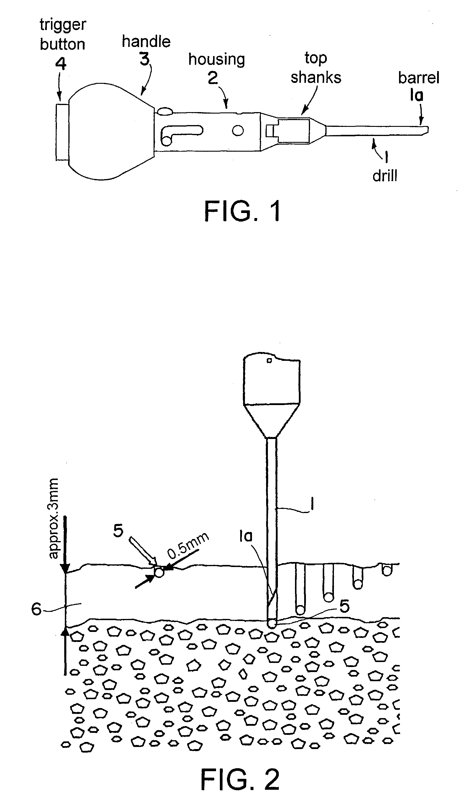
[0024]FIG. 1 illustrates an exemplary injection device for inserting spherical tantalum markers into a bone. The injection device includes a hollow drill 1 having an opening 1a at its tip. One or more tantalum spherule 5 (FIG. 2) can be dispensed through the opening 1a once the drill 1 has drilled a recess or hole into a bone 6 (e.g., to a depth of about 3 mm). The drill 1 can be detachably fixed on a housing 2, which includes a handle 3 and a trigger button 4. The drill 1 can be rotated either by manually operating the handle 3 or by a motor provided in the housing 2 of the instrument. At the desired depth of penetration, the button 4 can be operated, which inserts the tantalum sphere 5 into the bone 6.

[0025]FIG. 2 illustrates a cross-sectional view of the drill 1 penetrating an approximately 3 mm thick compact bone layer in order to insert the spherical tantalum marker 5 (which has a diameter of about 0.5 mm) into the bone 6. A marker 5 inserted in this way sits securely in the bo...


PUM


Abstract

A method for registering an anatomical structure using at least one marker attached to the structure includes: obtaining a three-dimensional model of the structure via an imaging method; obtaining at least two two-dimensional recordings of the structure from different angles; and ascertaining a spatial position and location of the three-dimensional model or a position and location of the three-dimensional model in a patient coordinate system based on a matching method that uses the position of the at least one marker in the at least two two-dimensional mappings such that the three-dimensional model of the structure matches the structure.

Description

RELATED APPLICATION DATA[0001]This application claims priority of U.S. Provisional Application No. 60 / 867,613 filed on Nov. 29, 2006, which is incorporated herein by reference in its entirety.FIELD OF THE INVENTION[0002]The present invention relates to medical registration of anatomical structures using markers.BACKGROUND OF THE INVENTION[0003]Prior to surgery, it is often necessary to register an anatomical structure within a medical workspace, i.e., to determine a spatial location of the structure or of a partial region of the structure, such as a three-dimensional position of characteristic points or landmarks on the structure. These characteristic points or landmarks can be used in a hip operation, for example, to determine the spatial location of characteristic planes. From the location of the characteristic planes, the inserted position of a hip joint cavity can be determined.[0004]If, in a hip operation, recordings are taken of the pelvis using a C-arm, then it is difficult o...

Claims


Application Information

Patent Timeline
Patent Type & Authority Patents(United States)
IPC IPC(8): G06K9/00
CPCA61B6/022A61B6/032A61B6/5235A61B6/5247A61B19/5244A61B19/54G06K9/3216A61B6/12A61B2019/505A61B2019/5238A61B2019/524A61B2019/5289A61B2019/5416A61B2019/5454A61B2019/566G06K2209/051A61B2090/3916A61B2034/256A61B34/20A61B2090/364A61B2090/376A61B2034/105A61B2090/3762A61B90/39A61B2090/3954G06V10/245G06V2201/031
Inventor DRUMM, PETERHEPKE, MARKUS
Owner BRAINLAB
Why Eureka
Try Eureka