Single molecule-format real-time bioluminescence imaging probe

a single-molecule, luminescence imaging technology, applied in the direction of animal/human proteins, peptides, enzyme stabilisation, etc., can solve the problems of lack of selectivity to target ligands, cytotoxicity, and difficulty in living subjects to perform bioanalysis, etc., to achieve convenient bioluminescence analysis, wide range of bioanalysis, and efficient

Inactive Publication Date: 2011-10-25
NAT INST OF ADVANCED IND SCI & TECH
View PDF3 Cites 13 Cited by
  • Summary
  • Abstract
  • Description
  • Claims
  • Application Information

AI Technical Summary

Benefits of technology

[0061]The fusion protein of the present invention enables luminescence imaging on a quasi-real-time basis in both living cell systems and cell-free systems, and provides a real-time bioluminescence imaging probe having the following advantages.
[0062](1) The probe overcomes the limits of fluorescence, and provides a very convenient bioluminescence analysis method.
[0063](2) The probe makes possible of bioimaging of observing, on a real-time basis, biological phenomena which occur on a time scale of seconds.
[0064](3) In contrast to the conventional probes sensing only membrane-permeable ligands, the probe in the present invention can sensitively measure even an exogenous ligand which is incapable of permeating the cell membrane (insulin, growth hormones and the like). Namely, the measurement of membrane-permeable ligands and native, endogenous ligands inside cells can be achieved with present probes. Therefore, practically all ligands can be taken as the subject of analysis, and it is now possible to achieve a wide spectrum of bioanalysis.
[0065](4) Since it is possible to design both luminescence reduction-type and luminescence enhancement-type probes for the same ligand stimulation, the users can choose one according to their needs.
[0066](5) The probe was devised to more efficient, simpler and more convenient by integrating ion-sensing and luminescence emission components in a single molecule. Particularly, for a cell-free system, there can be provided a luminescent probe test strip (luminescence-type ligand sensing means), which is produced by immobilizing and drying the present luminescent probe at an edge of a paper strip.

Problems solved by technology

Also, since excitation by short wavelength light from an external source is required, it has been very difficult to perform bioanalysis in living subjects where the absorption of short wavelength light is significant.
1)), but these synthetic substances have problems in that they cause cytotoxicity, accumulation in some parts of cells, and lack selectivity to target ligands.
It was also strongly feared that such stoichiometric variance causes inefficiency of the probe in sensing a ligand.
However, the probe takes approximately 10 minutes to 20 minutes from the recognition of a ligand to the light emission.
The large molecular weight (92 kD) of the conventional probes imposes a burden on the cells, and thus is not adequate to trace the molecular phenomena which take short time, in the living cells.
Furthermore, the target ligand must permeate the cell membrane and reach the probe in the cytoplasm, but in the case of insulin, various growth hormones, cytokines and the like, since these molecules originally cannot permeate the cell membrane, they cannot be detected with conventional probes expressed within the cells.
Even in the case of ligands which are capable of permeating the cell membrane, since the step of membrane permeation is the rate-limiting step, thus requiring a measurement time of about several ten minutes, the real time observation of various life phenomena in a cell cannot be made by any conventional means.
However, fluorescent proteins also have problems that the proteins are generally sensitively to pH, show high background fluorescence due to autofluorescence, and have larger molecular weights because two fluorescent proteins are linked to both termini of the target protein.
Moreover, there is also a problem in terms of use, such as that a large instrumentation is required to detect fluorescence (JP-A No. 2007-49943).
However, the probe has the same intrinsic limitations of general fluorescent probes such as that due to the poor reversibility, when bisected GFP fragments are re-bound, the probe becomes inadequate for repeated use, and that GFP requires a long time (about 6 hours) for protein folding, and is prone to undergo misfolding.

Method used


Image

  • Single molecule-format real-time bioluminescence imaging probe
  • Single molecule-format real-time bioluminescence imaging probe
  • Single molecule-format real-time bioluminescence imaging probe

Examples


example 1

Construction of Plasmid Carrying Nucleic Acid Molecules which Encode Various Fusion Proteins for Single-molecule-format Luminescent Probe Containing Calmodulin (CaM) for Ca2+ Recognition

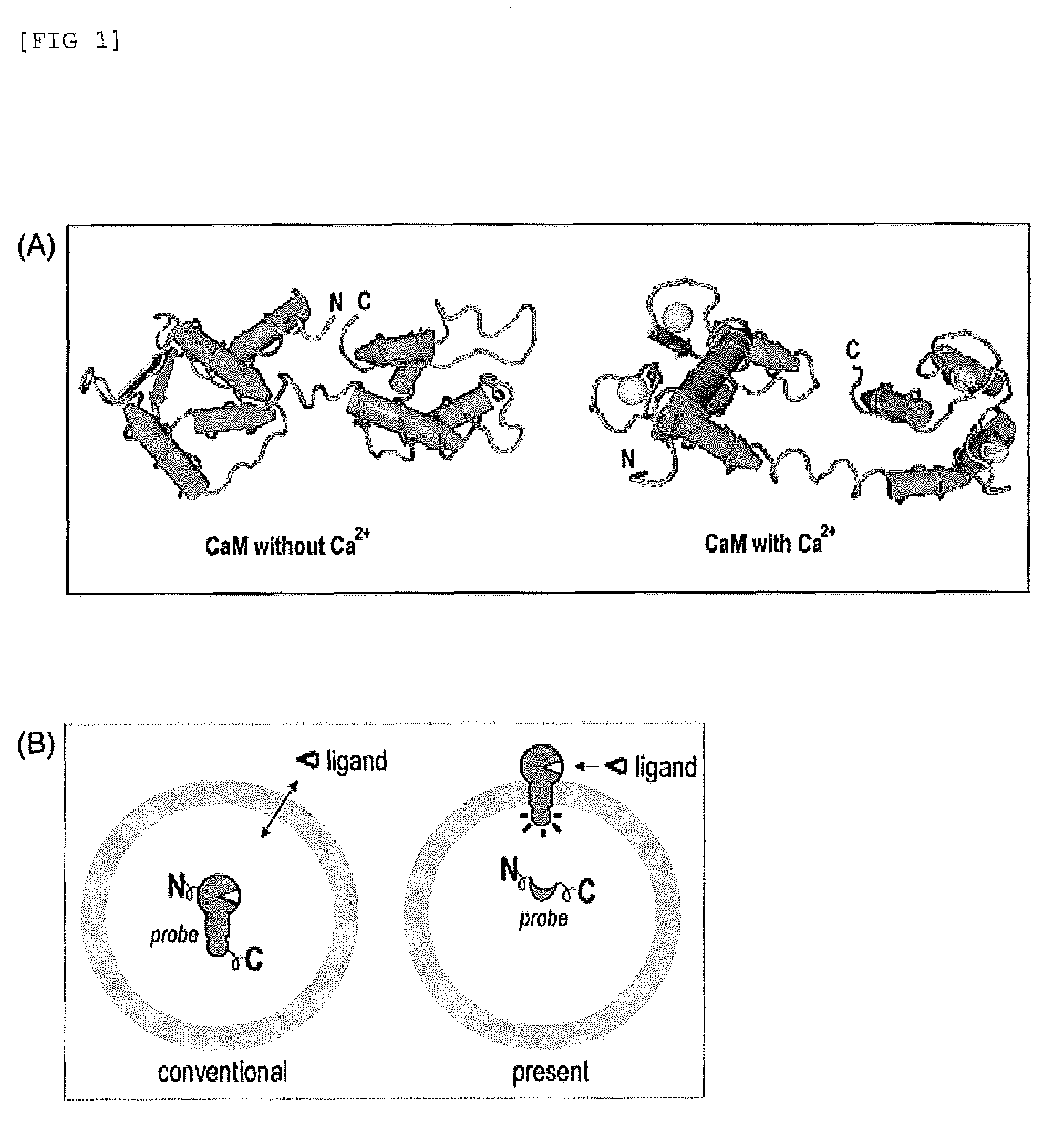
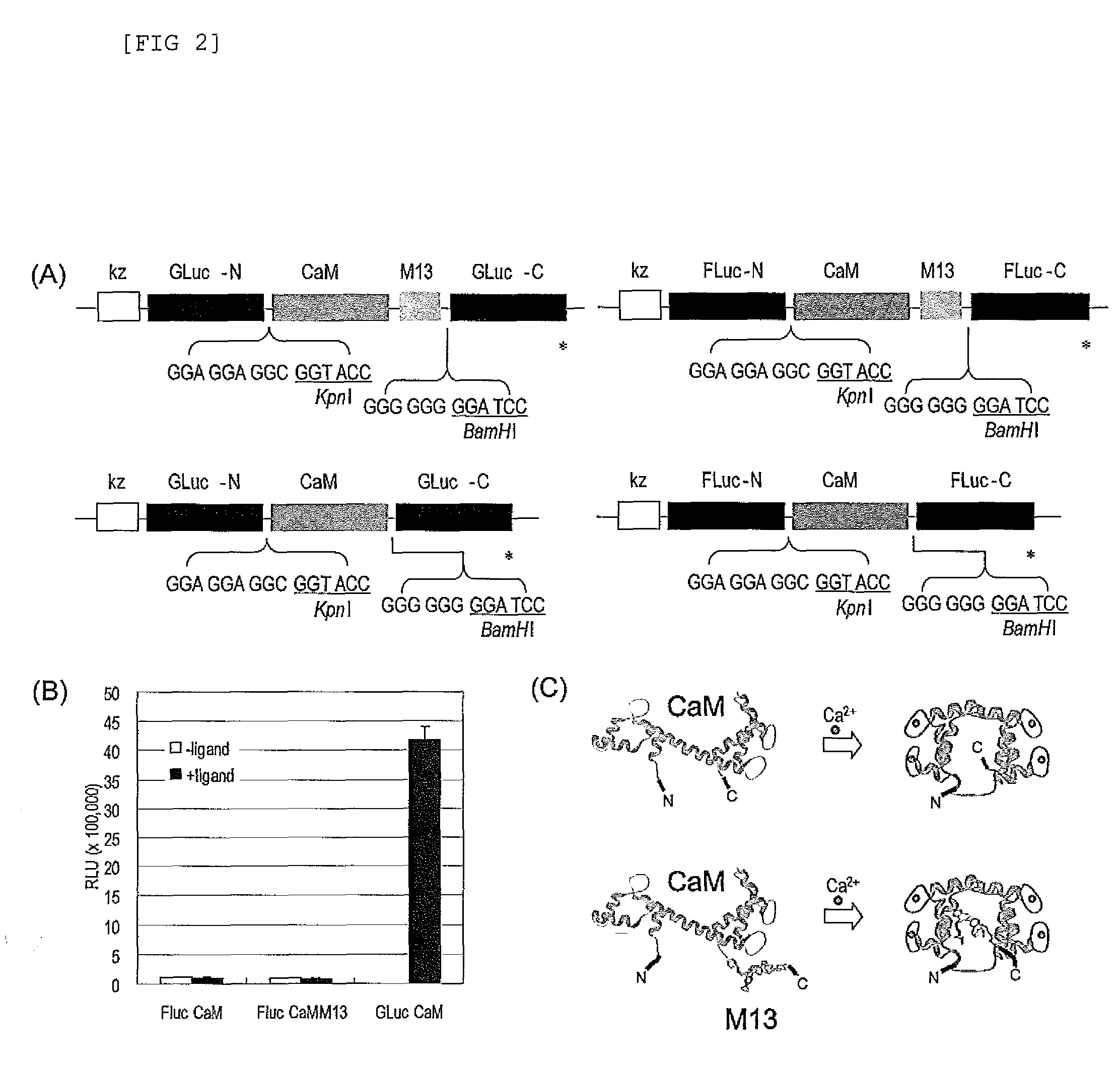
[0126]The subject probe which is capable of imaging, on a real-time basis, the response phenomena induced by external stimuli in living cells, will be designated as Simor probe, based on its name in English (a SIngle-MOlecule-format bioluminescent probe for Realtime imaging). A probe comprising calmodulin (CaM) will be designated as Sica. On the other hand, a probe comprising calmodulin as well as calmodulin recognition peptide sequence (M13), will be designated as Sicam. The eukaryotic expression vectors incorporated with the respective probes will be respectively designated as pSica and pSicam.

[0127]PCR amplification was carried out to introduce KpnI and BamHI, which are restriction enzyme sites, into the two termini of a cDNA encoding calmodulin, which is a Xenopus laevis (African clawed frog)-der...

example 2

Examination of Partition Site of GLuc

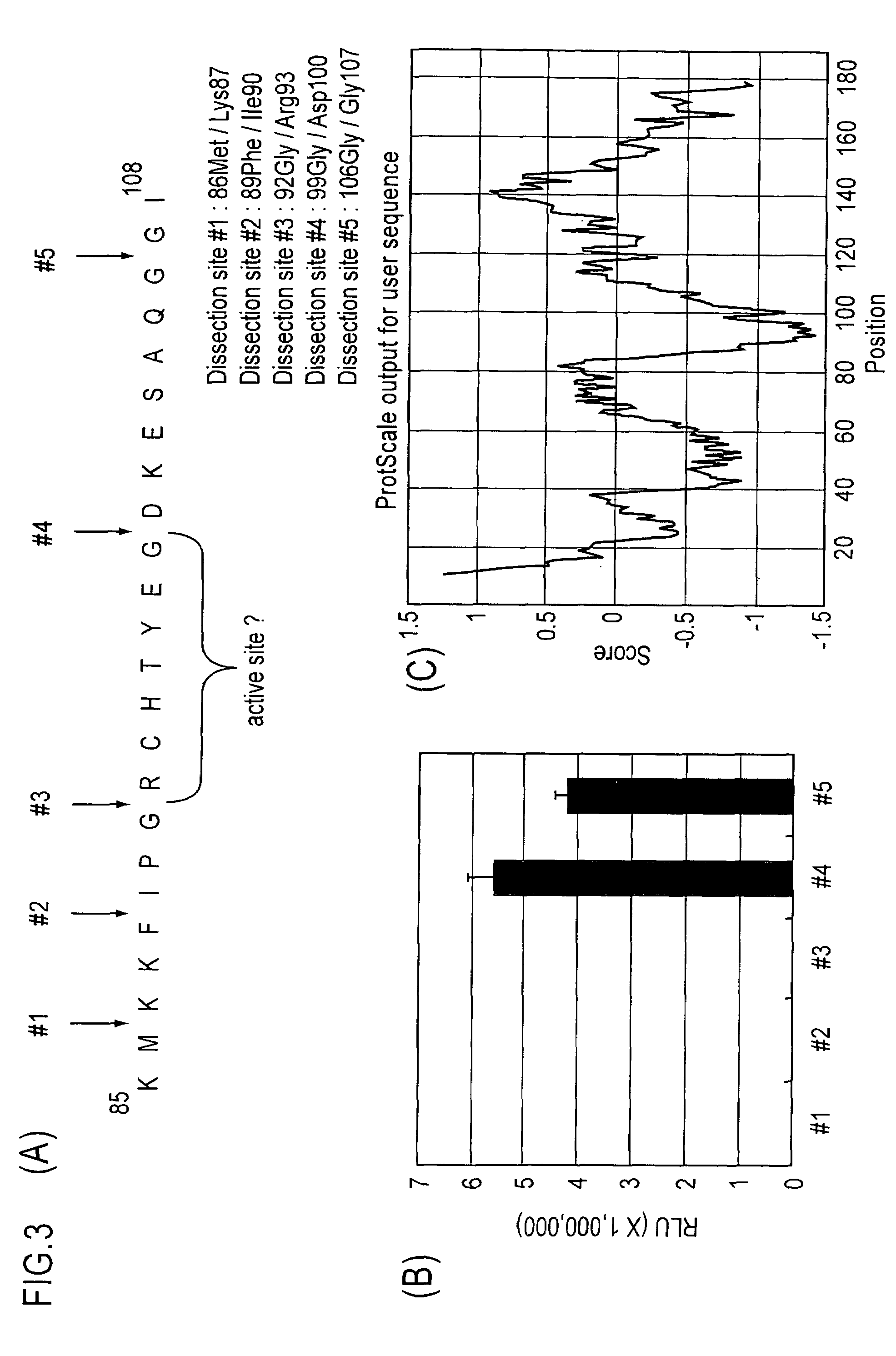
[0132]The inventors of the present invention presupposed, on the basis of the analysis of hydrophilicity distribution search in the total amino acid sequence of GLuc, that a hydrophilic region of amino acid residues at positions 90 to 108 in the amino acid sequence of GLuc was the region between the two protein domains constituting GLuc. Thus, the inventors determined five partition sites inside the above-mentioned region, and produced five fragment pairs by fragmentizing five sites of the cDNA of GLuc by PCR reaction, as previously described (FIG. 3).

[0133]The plasmids carrying only CaM between these five pairs of N-terminal fragment and C-terminal fragment of GLuc, were referred to as pSica series, and the plasmids were respectively designated as pSica-1 to pSica-5 in accordance with the different partition sites.

[0134]The luminescence values of the respective probes were compared, and the luminescence values of the probes having partition site...

example 3

Production of Point-mutated Type Probes and Determination of Luminescence Spectrum Thereof

[0136]There exists a sequence of seven amino acid residues (93RCHTYEG99) between the dissection sites #3 and #4 of GLuc. In this experiment, N-terminal fragments each having a single point-mutated to glycine (G), were produced in order to search, among those seven amino acid residues, for the amino acid which is responsible for the luminescence activity of GLuc. Thus, six kinds of plasmids were produced using the nucleic acids which respectively encode the fusion proteins comprising a single point-mutated amino acid in the GLuc-N-terminal fragments.

[0137]To produce these point mutants, the following procedure was conducted. PCR-based synthesis of a cDNA encoding RCHTYEG, which is a portion of the amino acid sequence of the N-terminal fragment of GLuc, was carried out using pSica-4 as a template, and primer systems which generate the respective point mutations. The mutated N-terminal fragments t...


PUM

PropertyMeasurementUnit
timeaaaaaaaaaa
diameteraaaaaaaaaa
conformationalaaaaaaaaaa

Abstract

The present invention provides an “in vivo and in vitro real-time bioluminescence imaging means,” which can transmit a detection signal promptly in response to an external signal, while taking advantage of a single-molecule-format luminescent probe as a bioluminescent means.The present invention is characterized by using, as a single-molecule-format luminescent probe utilizing the increase and decrease of a second messenger level as an index, a fusion protein including a single-chain protein containing a second messenger recognition protein and optionally a peptide which is capable of binding with the protein, and linked respectively to the N-terminus and the C-terminus thereto, an N-terminal fragment (N-LE) and a C-terminal fragment (C-LE) generated by dissecting a luminescent enzyme (LE). A single-molecule-format luminescent probe could be provided, which makes it possible to observe light emission (extinction) in vivo as well as in vitro, as a result of a conformational change of the second messenger recognition protein induced by the binding (unbinding) with a second messenger, and the subsequent association or dissociation between the N-LE and the C-LE flanked at both termini of the recognition protein.

Description

TECHNICAL FIELD[0001]The present invention relates to a fusion protein useful as a real-time luminescence imaging probe, which utilizes a conformational change of a second messenger recognition protein induced by its association or dissociation with a second messenger. The present invention also relates to a fusion protein useful as a versatile single-chain luminescent probe utilizing a Gaussia luciferase bisected at an optimal position.BACKGROUND ART[0002]The realm of nature in which we are dwelling is inhabited by innumerable higher and lower organisms, and these organisms are maintaining their life phenomena on the basis of common molecular mechanisms such as expression of proteins by means of DNAs in response to external stimuli.[0003]In both eukaryotic cells and prokaryotic cells, rapidly responding to the temperature, signal transducers, the presence or absence of nutrients, and the like in the breeding environment is directly related to the matter of life and death of the org...

Claims


Application Information

Patent Timeline
Patent Type & Authority Patents(United States)
IPC IPC(8): C12Q1/66C12P21/06C12N15/00C12N9/02C12Q1/68C07H21/04
CPCC07K14/4728C07K14/721C12N9/0069C12N9/1205C12N15/62
Inventor KIM, SUNG-BAETAO, HIROAKISATO, MORITOSHI
Owner NAT INST OF ADVANCED IND SCI & TECH
Why Eureka
Try Eureka