Method and apparatus for real time imaging and monitoring of radiotherapy beams

a radiation beam and real-time monitoring technology, applied in the field of methods and apparatus for imaging and monitoring the delivery of therapeutic radiation beams, can solve the problems of no standard technique available and time for real-time monitoring

Inactive Publication Date: 2011-11-01
JEFFERSON SCI ASSOCS LLC
View PDF5 Cites 18 Cited by
  • Summary
  • Abstract
  • Description
  • Claims
  • Application Information

AI Technical Summary

Problems solved by technology

Verification of the location and magnitude of the radiation dose delivered to target volume in the body in such treatments is an important requirement and in-situ / in-vivo tracking of these beams is an important challenge for radiation therapy today.
While radiation therapy procedures are very carefully planned, specifically addressing a selected target volume in each patient, and each radiation session planned using available simulation tools and prior experimental clinical data, there is no standard technique available at this time to provide real time monitoring and assurance that the procedure is indeed delivering the desired / lethal dose to the cancer tissue, while sparing the neighboring organs and tissues that might be at risk.

Method used


Image

  • Method and apparatus for real time imaging and monitoring of radiotherapy beams
  • Method and apparatus for real time imaging and monitoring of radiotherapy beams
  • Method and apparatus for real time imaging and monitoring of radiotherapy beams

Examples


Embodiment Construction

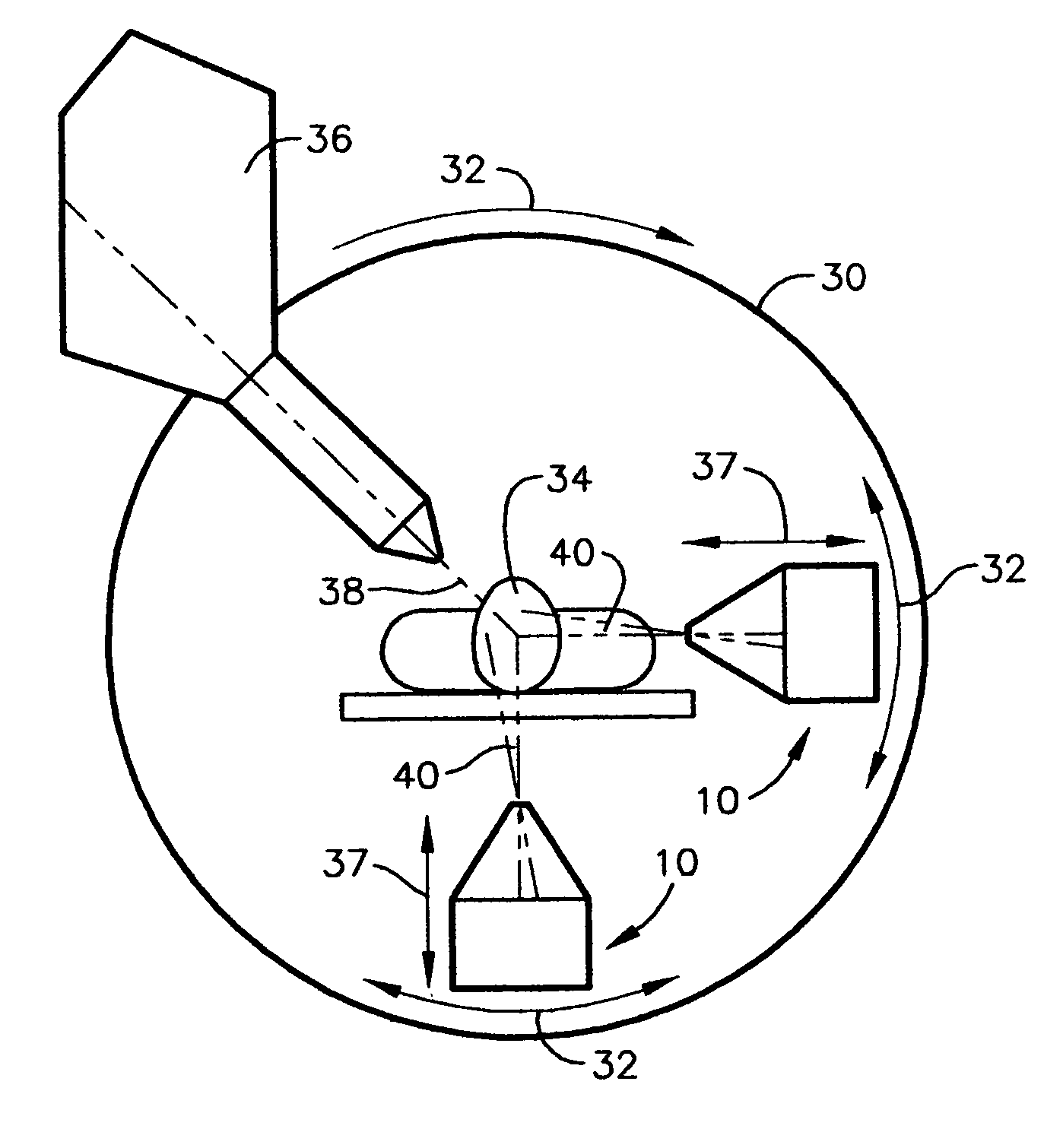
[0012]As a result of energetic multi-MeV radiation beams interacting with the tissue / bone target, as occurs during brachytherapy treatment, different excited nuclear states are produced with the highest representation from species produced from naturally occurring nuclei in the organic target such as Oxygen, Carbon, etc. Some of these excited states de-excite with prompt emission of characteristic high energy gamma rays. These signature gamma lines can be used to identify the specific excited species and to study and establish the correlation between the emission of these gamma lines and therapeutically relevant energy deposit in the target.

[0013]However, imaging these multi-MeV gamma lines is very difficult because collimators must be used to produce meaningful images of the beam in the target area.

[0014]In the recent study (Chul-Hee Min et al., “Prompt Gamma Measurements for Locating the dose Falloff Region in ProtonTherapy”, Applied Physics Letter 89, 183517, 2006) it was shown t...


PUM


Abstract

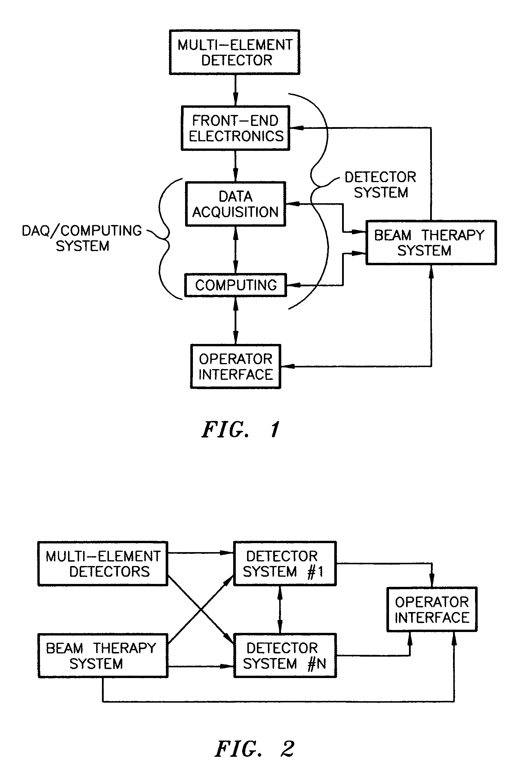
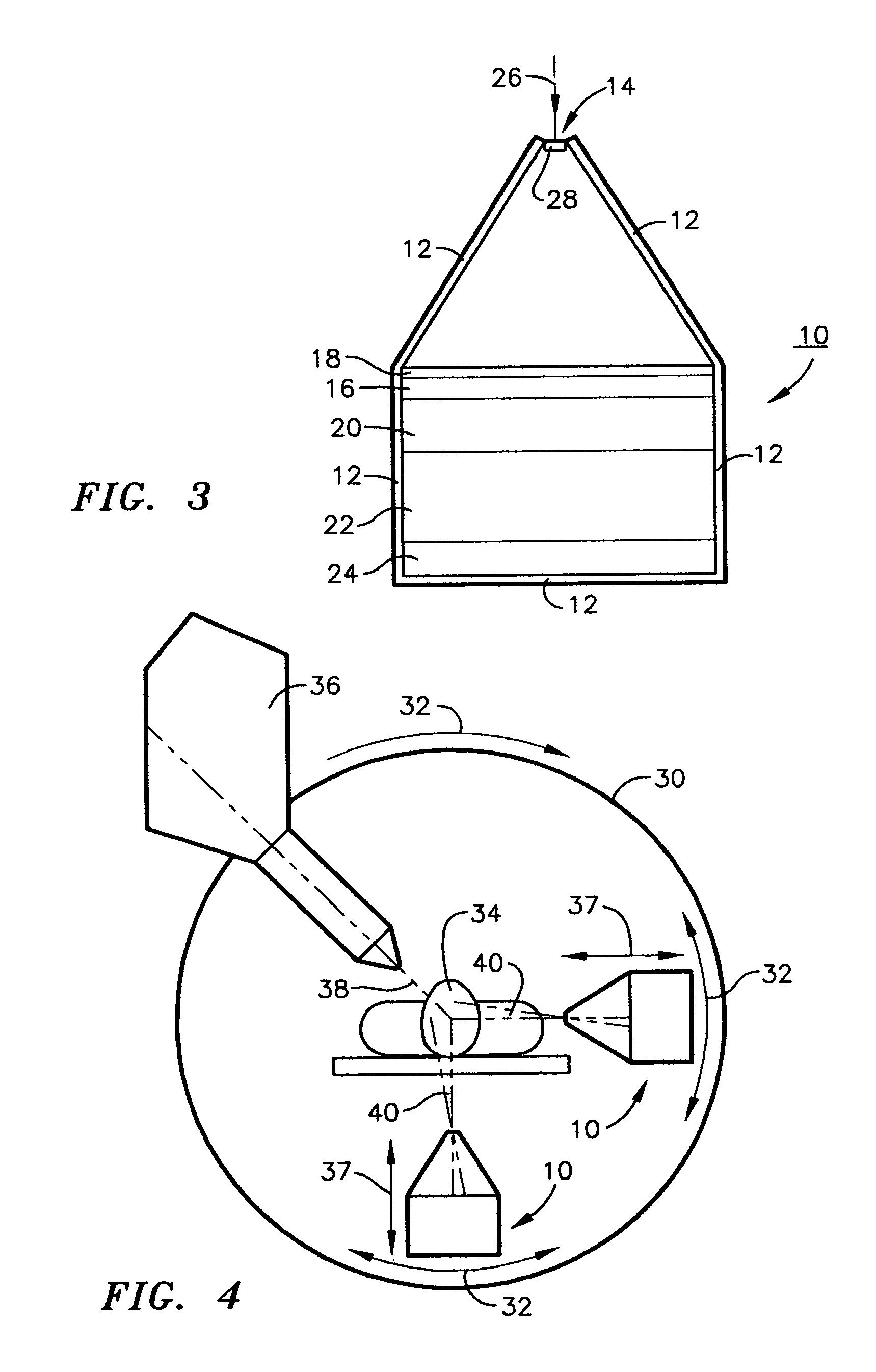
A method and apparatus for real time imaging and monitoring of radiation therapy beams is designed to preferentially distinguish and image low energy radiation from high energy secondary radiation emitted from a target as the result of therapeutic beam deposition. A detector having low sensitivity to high energy photons combined with a collimator designed to dynamically image in the region of the therapeutic beam target is used.

Description

[0001]The United States of America may have certain rights to this invention under Management and Operating Contract DE-AC05-060R23177 from the United States Department of Energy.FIELD OF THE INVENTION[0002]The present invention relates to methods and apparatus for imaging and monitoring the delivery of therapeutic radiation beams and more particularly to in-situ / in-vivo tracking of these beams.BACKGROUND OF THE INVENTION[0003]Different types of radiation therapy beams, including electron beams, gamma rays, high energy X-rays and protons are used in the treatment of various cancer states. Verification of the location and magnitude of the radiation dose delivered to target volume in the body in such treatments is an important requirement and in-situ / in-vivo tracking of these beams is an important challenge for radiation therapy today.[0004]While radiation therapy procedures are very carefully planned, specifically addressing a selected target volume in each patient, and each radiatio...

Claims


Application Information

Patent Timeline
Patent Type & Authority Patents(United States)
IPC IPC(8): G01T1/16
CPCA61N5/1049A61N5/1071A61N2005/1052A61N2005/1054
Inventor MAJEWSKI, STANISLAWPROFFITT, JAMESMACEY, DANIEL J.WEISENBERGER, ANDREW G.
Owner JEFFERSON SCI ASSOCS LLC
Why Eureka
Try Eureka