Orthopedic fixation mechanism

a fixation mechanism and orthopedic technology, applied in the field of prostheses for treating spinal pathologies, can solve the problems of severe limitations on a person's functional ability and quality of life, damage to the articular surfaces of one or more vertebral bodies, back pain,

Active Publication Date: 2012-02-07
LANTERNA MEDICAL TECH
View PDF12 Cites 48 Cited by
  • Summary
  • Abstract
  • Description
  • Claims
  • Application Information

AI Technical Summary

Problems solved by technology

In many cases, the pain severely limits a person's functional ability and quality of life.
A variety of spinal pathologies can lead to back pain.
Through disease or injury, the laminae, spinous process, articular processes, or facets of one or more vertebral bodies can become damaged, such that the vertebrae no longer articulate or properly align with each other.
This can result in an undesired anatomy, loss of mobility, and pain or discomfort.
These conditions induce lower back and nerve roots pain which affect a large part of the population.
The vertebral facet joints, for example, can be damaged by either traumatic injury or by various disease processes, such as osteoarthritis, ankylosing spondylolysis, and degenerative spondylolisthesis.
The damage to the facet joints often results in pressure on nerves, also called a “pinched” nerve, or nerve impingement.
The result is pain, malaligned anatomy, and a corresponding loss of mobility.
The locking of vertebral motions by spinal arthrodesis or ligamentoplasty induces, beyond a spinal stiffness, an increased force on the joint facets of the adjacent vertebrae above and below the fusion, usually sustained by the considered intervertebral space resulting in an increase of degeneration of these joint facets.
This solves the aging intervertebral disc problem but does not function to reduce the force on posterior joint facets.
These traditional treatments are subject to a variety of limitations and varying success rates.
For example, traditional treatments may result in limiting the patient's mobility or spontaneous fusion.

Method used


Image

  • Orthopedic fixation mechanism
  • Orthopedic fixation mechanism
  • Orthopedic fixation mechanism

Examples


Embodiment Construction

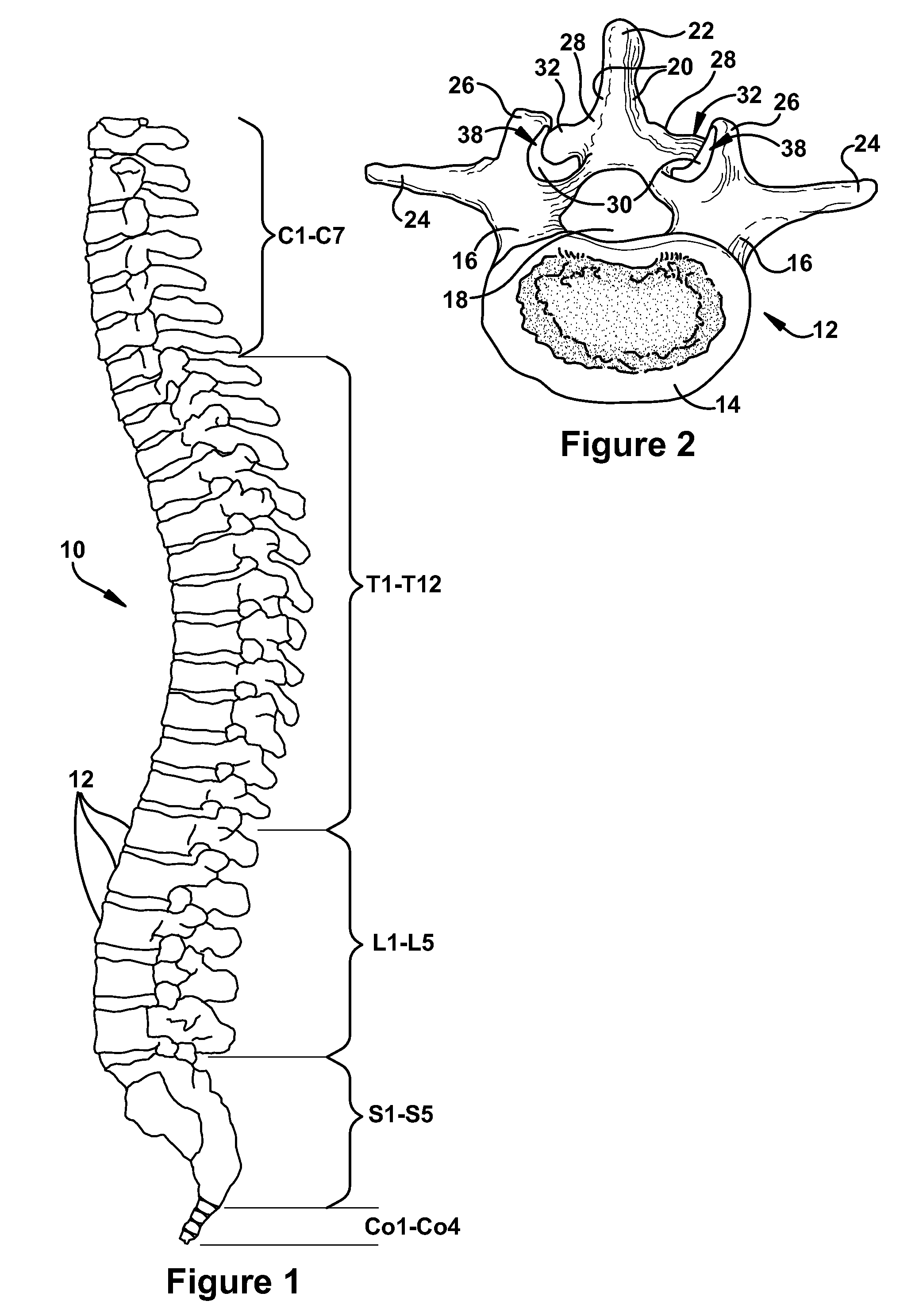
[0035]Referring initially to FIG. 1, the human spinal column 10 is illustrated. The spinal column 10 is comprised of a series of thirty-three stacked vertebrae divided into five regions. The cervical region includes seven vertebrae, known as C1-C7. The thoracic region includes twelve vertebrae, known as T1-T12. The lumbar region contains five vertebrae, known as L1-L5. The sacral region is comprised of five vertebrae, known as S1-S5. The coccygeal region contains four vertebrae 12, known as Co1-Co4.

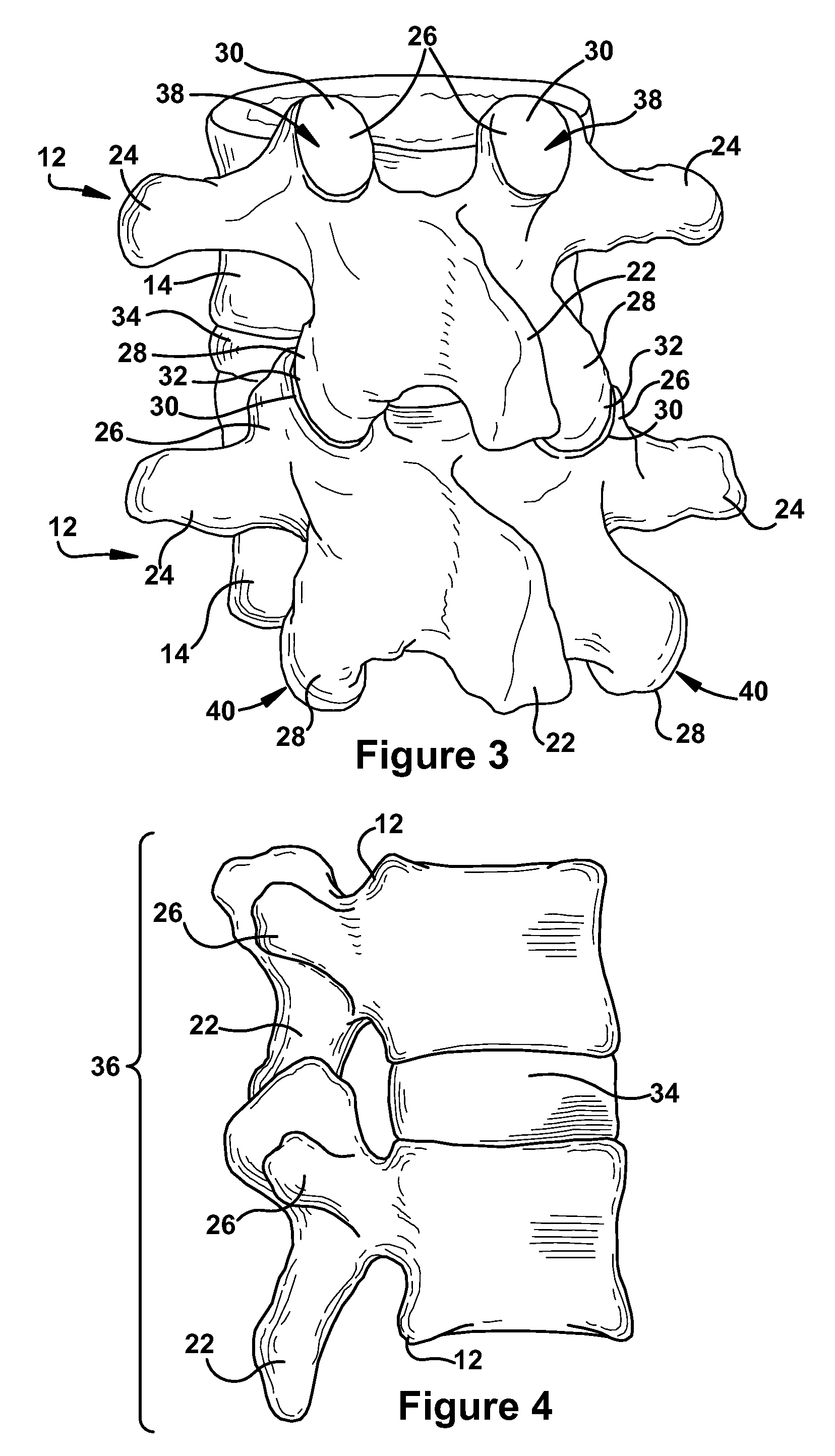
[0036]Turning now to FIGS. 2 and 3, normal human lumbar vertebrae 12 are illustrated. It will be understood by those skilled in the art that while the lumbar vertebrae 12 vary somewhat according to location, they share many features common to most vertebrae 12. Each vertebra 12 includes a vertebral body 14. Two short bones, the pedicles 16, extend backward from each side of the vertebral body 14 to form a vertebral arch 18. At the posterior end of each pedicle 16, the vertebral arch 18 fl...


PUM


Abstract

A fixation mechanism and method of fixation, such as the fixation of a facet resurfacing implant to an articular facet of a spinal vertebra. The fixation mechanism includes a securing mechanism having a socket configured to receive a knob of a biologic implant. The fixation mechanism further includes a locking mechanism for limiting expansion of the socket following engagement with the knob of the implant.

Description

FIELD OF THE INVENTION[0001]The present invention relates generally to prostheses for treating spinal pathologies, and more specifically to a system and method for treating articulating surfaces of facet joints.BACKGROUND OF THE INVENTION[0002]Back pain, such as in the “small of the back”, or lumbosacral (L1-S1) region, is a common ailment. In many cases, the pain severely limits a person's functional ability and quality of life. A variety of spinal pathologies can lead to back pain.[0003]Through disease or injury, the laminae, spinous process, articular processes, or facets of one or more vertebral bodies can become damaged, such that the vertebrae no longer articulate or properly align with each other. This can result in an undesired anatomy, loss of mobility, and pain or discomfort. With respect to vertebral articular surface degeneration, facet joints may show a reduced thickness of cartilage and may advance to entire disappearance thereof. Furthermore, surrounding the degenerat...

Claims


Application Information

Patent Timeline
Patent Type & Authority Patents(United States)
IPC IPC(8): A61B17/70
CPCA61B17/1659A61B17/1671A61F2/4405A61B17/1757A61F2002/30378A61F2002/30405A61F2002/305A61F2002/30507A61F2002/30538A61F2002/30604A61F2002/30841A61F2002/30879A61F2002/4631A61F2220/0025A61F2220/0033A61F2250/0006A61F2310/00011A61F2310/00023A61F2310/00179
Inventor HALE, HORACE WINSTON
Owner LANTERNA MEDICAL TECH
Why Eureka
Try Eureka