

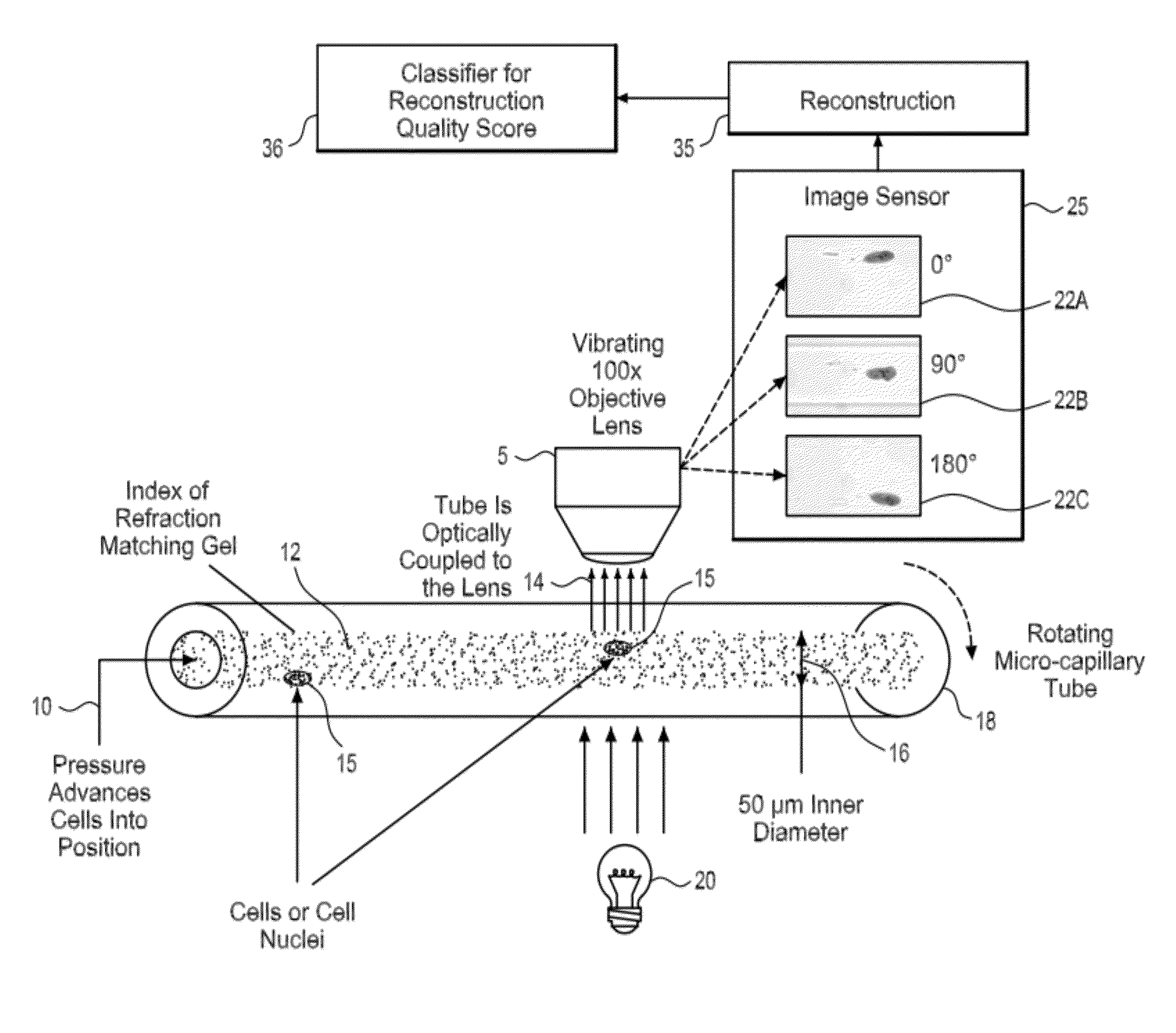
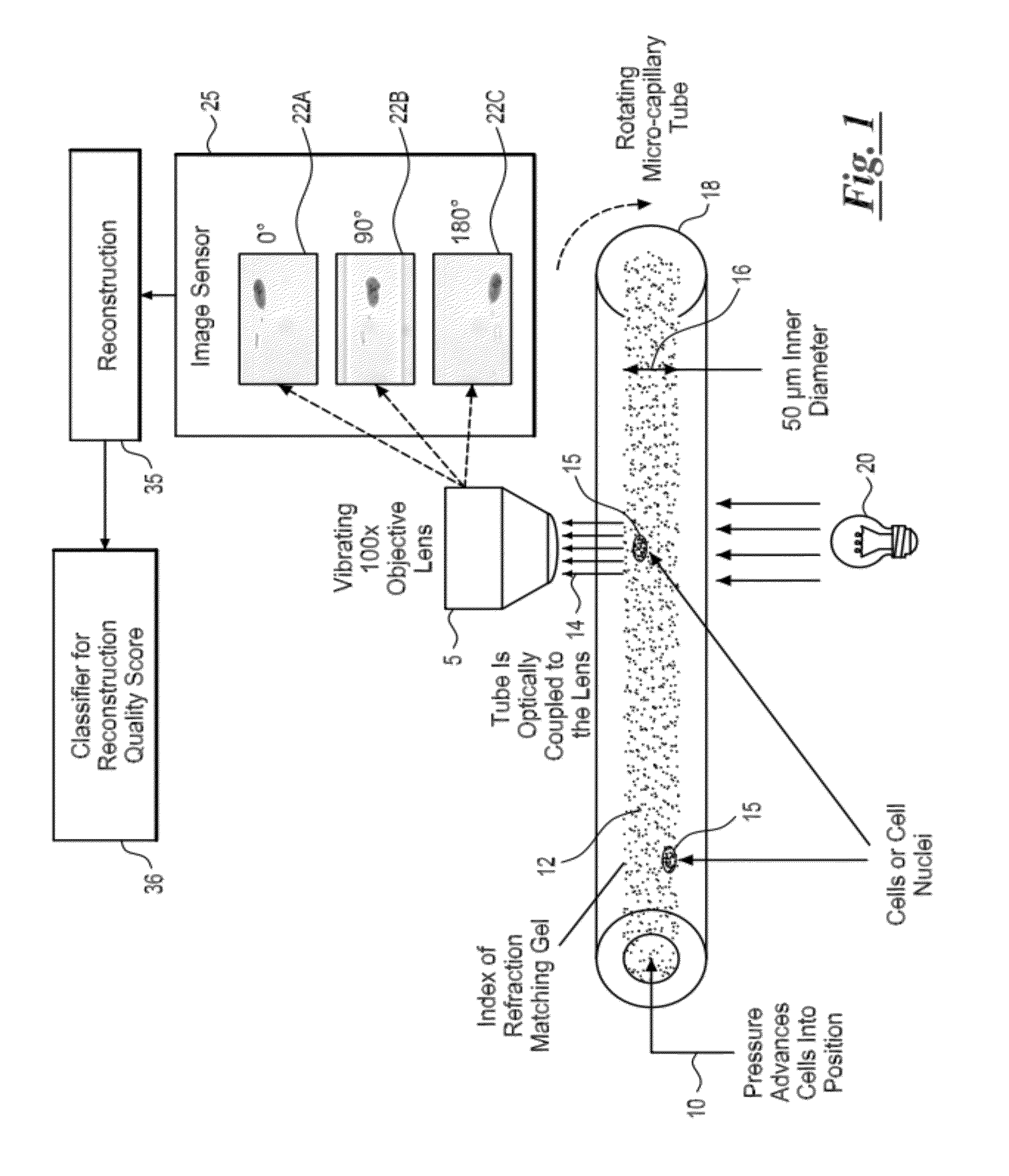
System and method for detecting poor quality in 3D reconstructions
a three-dimensional reconstruction and poor quality technology, applied in the field of medical imaging data analysis, can solve the problems of unfavorable refraction effect, inability to form standard projection images, and lack of an automated system for detecting such poor quality 3d reconstructions
- Summary
- Abstract
- Description
- Claims
- Application Information
AI Technical Summary
Benefits of technology
Problems solved by technology
Method used
Image

Examples
Embodiment Construction
[0019]The following disclosure describes several embodiments and systems for imaging an object of interest. Several features of methods and systems in accordance with example embodiments of the invention are set forth and described in the figures. It will be appreciated that methods and systems in accordance with other example embodiments of the invention can include additional procedures or features different than those shown in figures.
[0020]Example embodiments are described herein with respect to biological cells. However, it will be understood that these examples are for the purpose of illustrating the principles of the invention, and that the invention is not so limited. Additionally, methods and systems in accordance with several example embodiments of the invention may not include all of the features shown in these figures. Throughout the figures, like reference numbers refer to similar or identical components or procedures.
[0021]Unless the context requires otherwise, through...
PUM
 Abstract
Description
Claims
Application Information