Endoscope having detachable imaging device and method of using

an endoscope and imaging device technology, applied in the field of detachable imaging device and detachable imaging device, can solve the problems of endoscope camera, potential malignant (cancerous) polyps to be missed, added discomfort, complications, etc., and achieve the effect of avoiding the cost of modifying the endoscop

Inactive Publication Date: 2012-05-22
PSIP LLC
View PDF342 Cites 887 Cited by
  • Summary
  • Abstract
  • Description
  • Claims
  • Application Information

AI Technical Summary

Benefits of technology

[0013]According to some aspects of the present invention, a retrofit auxiliary camera is provided that does not require significant modification of an existing endoscope or use of a channel of the endoscope, thereby avoiding the costs of modifying the endoscope and preserving all of the endoscope's designed functions.
[0021]In accordance with still another embodiment of the invention, the detachable imaging device includes a support mechanism that increases the rigidity of the detachable imaging device and reduces the bending of the link.
[0022]In accordance with yet still another embodiment of the invention, the wireless imaging element includes an imaging unit and / or a light source. The imaging unit may be mounted on the proximal end of the wireless imaging element and faces towards a main imaging device mounted on the distal end of the insertion tube. Preferably, the imaging unit and the main imaging device provide different views of the same area. To reduce light interference, the imaging element and the main imaging device and their light sources may be turned on and off alternately. Preferably the imaging element and the main imaging device and their light sources are turned on and off at a sufficiently high frequency that the eyes do not sense that the imaging element and the main imaging device and their light sources are intermittently turned on and off.

Problems solved by technology

Repositioning and movement of the endoscope prolongs the procedure and causes added discomfort, complications, and risks to the patient.
Additionally, in an environment similar to the lower gastro-intestinal tract, flexures, tissue folds and unusual geometries of the organ may prevent the endoscope's camera from viewing all areas of the organ.
The unseen area may cause a potentially malignant (cancerous) polyp to be missed.
Unfortunately, most of the endoscopes currently in use do not have such an auxiliary camera.
To replace these existing endoscopes with new endoscopes with auxiliary cameras is expensive.
Although a channel of an endoscope can be used to accommodate an auxiliary camera that does not require modification of the endoscope, the loss of a channel may impair the endoscope's ability to perform all of its designed functions.

Method used


Image

  • Endoscope having detachable imaging device and method of using
  • Endoscope having detachable imaging device and method of using
  • Endoscope having detachable imaging device and method of using

Examples


embodiment 80

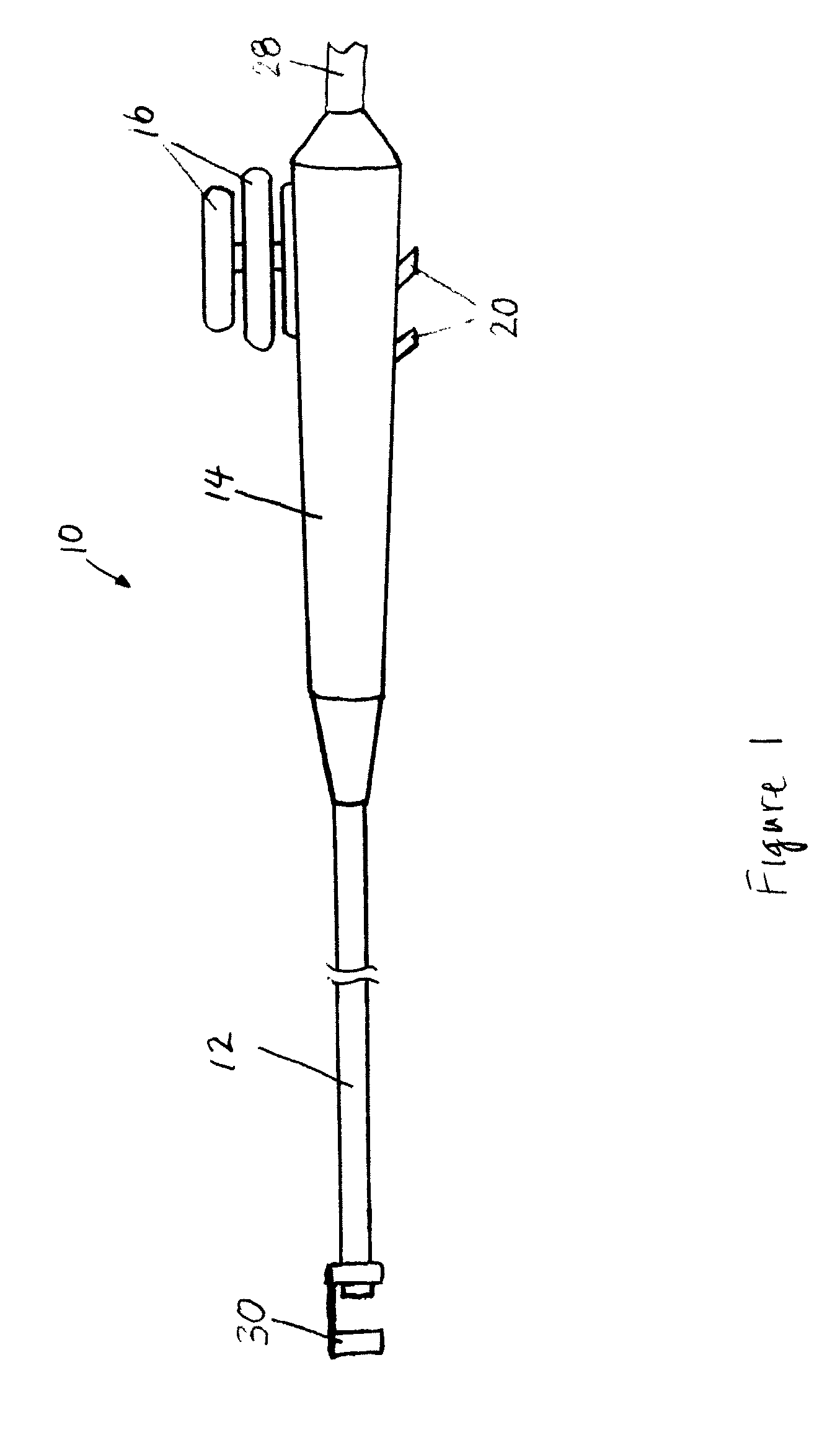
[0073]The endoscope 10 may further include a support mechanism, which increases the rigidity of the detachable imaging device 30 during insertion of the endoscope 10 into the body. This support mechanism preferably reduces or eliminates the bending of the link 36 of the detachable imaging device 30 during insertion. An embodiment 80 of the support mechanism is shown in FIGS. 11a, 11b, 12a, and 12b. The exemplary support mechanism 80 includes a rod 82 that is rigid at its distal end region 84 but is otherwise flexible. The exemplary support mechanism 80 may further include a locking mechanism 86 that locks the distal end of the rod 82 to the wireless imaging element 34. As shown in FIGS. 11b and 12b, the lock mechanism 86 includes mating grooves 88, 90 that are disposed on the distal end of the rod 82 and the wireless imaging element 34, respectively. The grooves 88, 90 can be interlocked by applying a torque to turn the rod 82 at the proximal end of the insertion tube 12, and can be...

embodiment 200

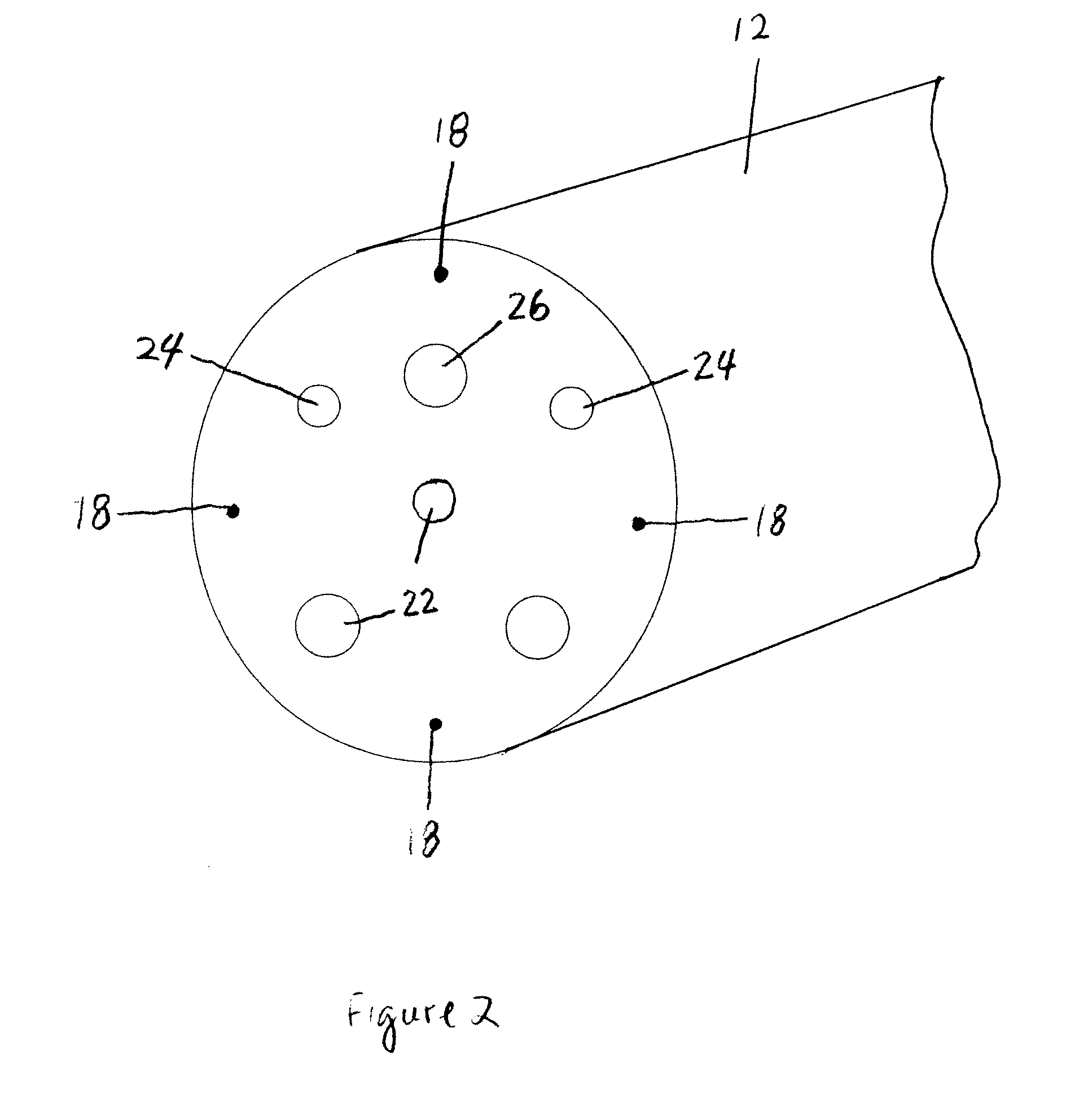
[0076]FIG. 14 illustrates additional embodiment 200 of the present invention that includes an insertion tube 212 and an attachment 232 mounted on the distal end region of the insertion tube 212. This attachment 232 may have some or all of features of the attachment 32 shown in FIGS. 3 and 5. Additionally, the attachment 232 is configured to accommodate one or more imaging units 242 and light sources 244 of the endoscope 200. In other words, the entire detachable imaging device 230 is mounted on the distal end region of the insertion tube 212 and does not extend beyond the distal end of the insertion tube 212. The imaging units 242 and light sources 244 may be mounted at any suitable locations on the attachment 232 and may be oriented in any directions. In this embodiment, the imaging units 242 and light sources 244 are placed on a proximal end of the attachment 232 and face backwards, although they may be alternatively or additionally placed on a distal end and / or side of the attach...


PUM


Abstract

An endoscope assembly with a main imaging device and a first light source is configured to provide a forward view of a body cavity, and further includes a detachable imaging device with an attachment member engageable with the distal end region of the endoscope, a linking member connected to the attachment member, and an imaging element with a second light source, wherein the detachable imaging device provides a retrograde view of the body cavity and the main imaging device. Light interference is reduced by using polarizing filters or by alternating the on / off state of the main imaging device, the first light source, the imaging element and the second light source so that the main imaging device and first light source are on when the imaging element and second light source are off and the main imaging device and first light source are off when the imaging element and second light source are on.

Description

[0001]This application claims the benefit of U.S. Provisional Patent Application No. 60 / 750,325, filed Dec. 13, 2005, the entire disclosure of which is incorporated herein by reference.[0002]This application also claims the benefit of U.S. Provisional Patent Application No. 60 / 761,475, filed Jan. 23, 2006, the entire disclosure of which is incorporated herein by reference.[0003]This application also claims the benefit of U.S. Provisional Patent Application No. 60 / 772,442, filed Feb. 9, 2006, the entire disclosure of which is incorporated herein by reference.[0004]This application further claims the benefit of U.S. Provisional Patent Application No. 60 / 802,056, filed May 19, 2006, the entire disclosure of which is incorporated herein by reference.[0005]The entire disclosure of U.S. patent application Ser. No. 11 / 215,660, filed Aug. 29, 2005, is incorporated herein by reference.TECHNICAL FIELD OF THE INVENTION[0006]The present invention relates to a detachable imaging device, an endos...

Claims


Application Information

Patent Timeline
Patent Type & Authority Patents(United States)
IPC IPC(8): A61B1/05A61B1/06
CPCA61B1/00016A61B1/00101A61B1/00174A61B1/00177A61B1/05A61B1/053A61B1/00114A61B1/005A61B1/0676A61B1/0684A61B1/31A61B1/00135
Inventor BAYER, LEXDESAI, RUPESHHIGGINS, JOHN
Owner PSIP LLC
Why Eureka
Try Eureka