Sterile radiological imaging unit drape and method of providing a sterile surface therewith

a sterile surface and radiological imaging technology, applied in the field of surgical drapes, can solve the problems of infection, estimated 100,000 deaths, and known infection risk that can be caused, and achieve the effect of reducing the cost of hospitalization

Active Publication Date: 2012-10-16
TIDI PROD LLC
View PDF10 Cites 57 Cited by
  • Summary
  • Abstract
  • Description
  • Claims
  • Application Information

AI Technical Summary

Problems solved by technology

The need to provide a sterile environment in a surgical theatre is directly associated with the known risk of infection that can be caused by bacteria, such as from a person or surgical equipment, in the surgical theatre.
Although the risks of infection caused during surgery are known, it remains a challenge to prevent their onset.
Infections are estimated to affect about 2 million patients annually and result, directly or indirectly, in an estimated 100,000 deaths.
Aside from the loss of life, the infections create an economic burden on hospitals.
For example, some studies estimate that each bloodstream infection in a patient results in an average cost of $26,839 to the hospital.
However, in some cases it is necessary to introduce relatively large, unsterilized surgical equipment in the operating room (surgical theater) for use during a surgical procedure.
Given the large, arcuate shape of the C-arm fluoroscopy unit, and its mobility during use, it becomes ever so challenging to maintain sterility of the surgical theatre.
Typically, though cumbersome, multiple half sheets are disposed about the unit during its use.
This often results in delays of the surgical procedure and requires creativity on the part of the surgical staff in order to best deploy the sheets about portions of the unit, particularly the arm and an imaging end of the unit.
The need for creativity results from the fact that there is no single accepted methodology for draping C-arm radiological units.
Of course, if the half sheets are not properly deployed, then the sterility of the surgical theater is compromised, and thus, the potential for onset of infection in the patient increases.
Finally, because this type of radiological equipment is moved repeatedly during surgery, even if the unit is initially draped properly, there remains an associated risk that the current draping mechanisms will become displaced, thereby exposing unsterilized portions of the unit.

Method used


Image

  • Sterile radiological imaging unit drape and method of providing a sterile surface therewith
  • Sterile radiological imaging unit drape and method of providing a sterile surface therewith
  • Sterile radiological imaging unit drape and method of providing a sterile surface therewith

Examples


Embodiment Construction

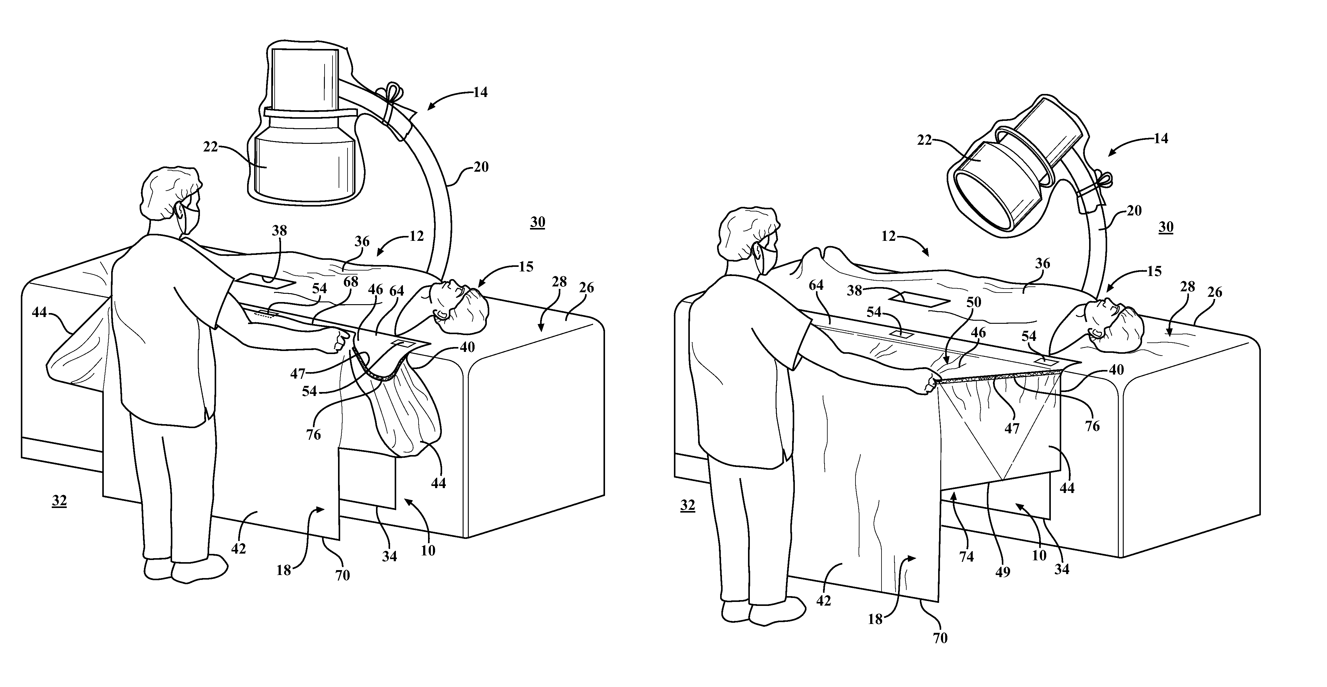
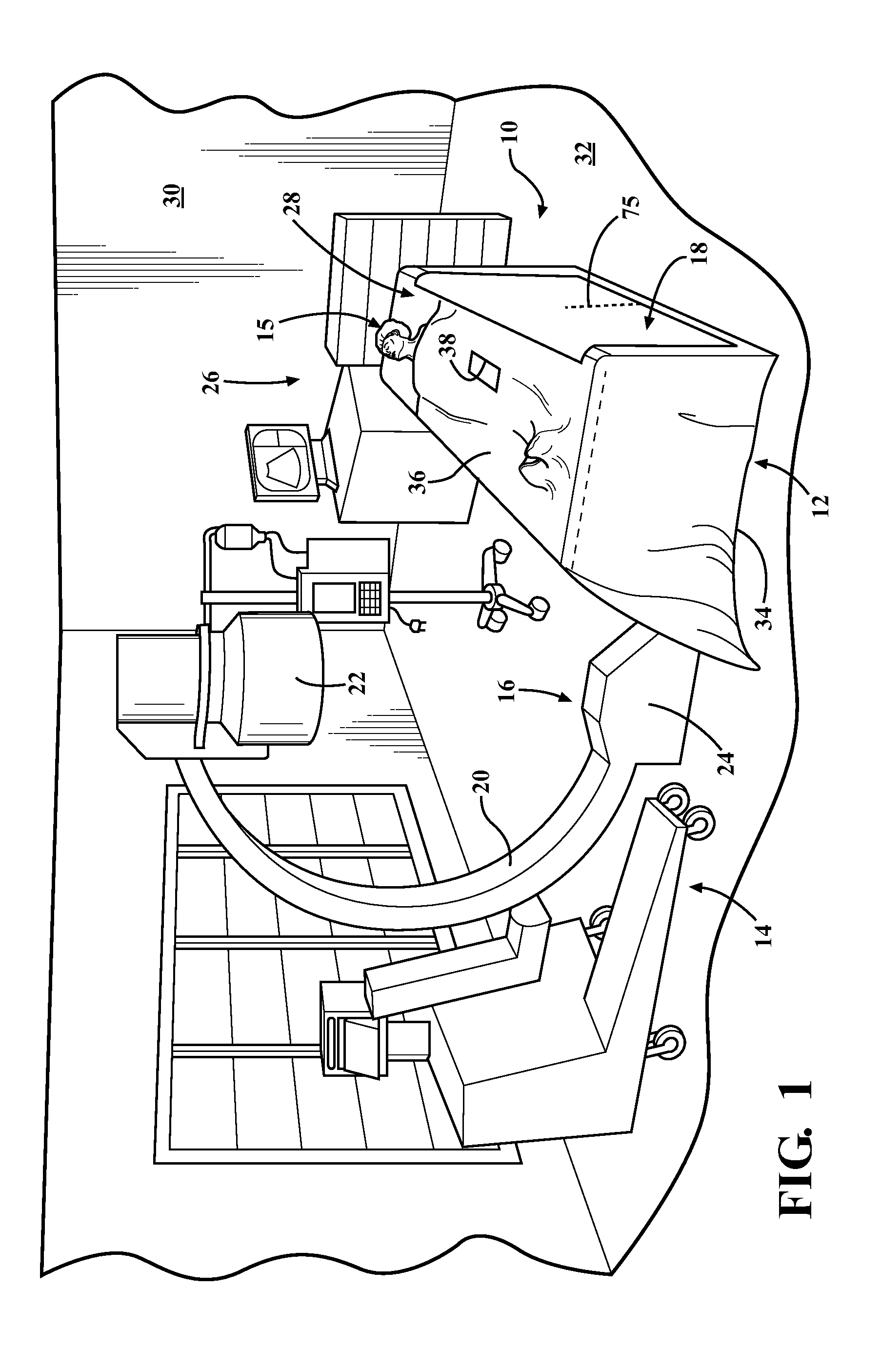
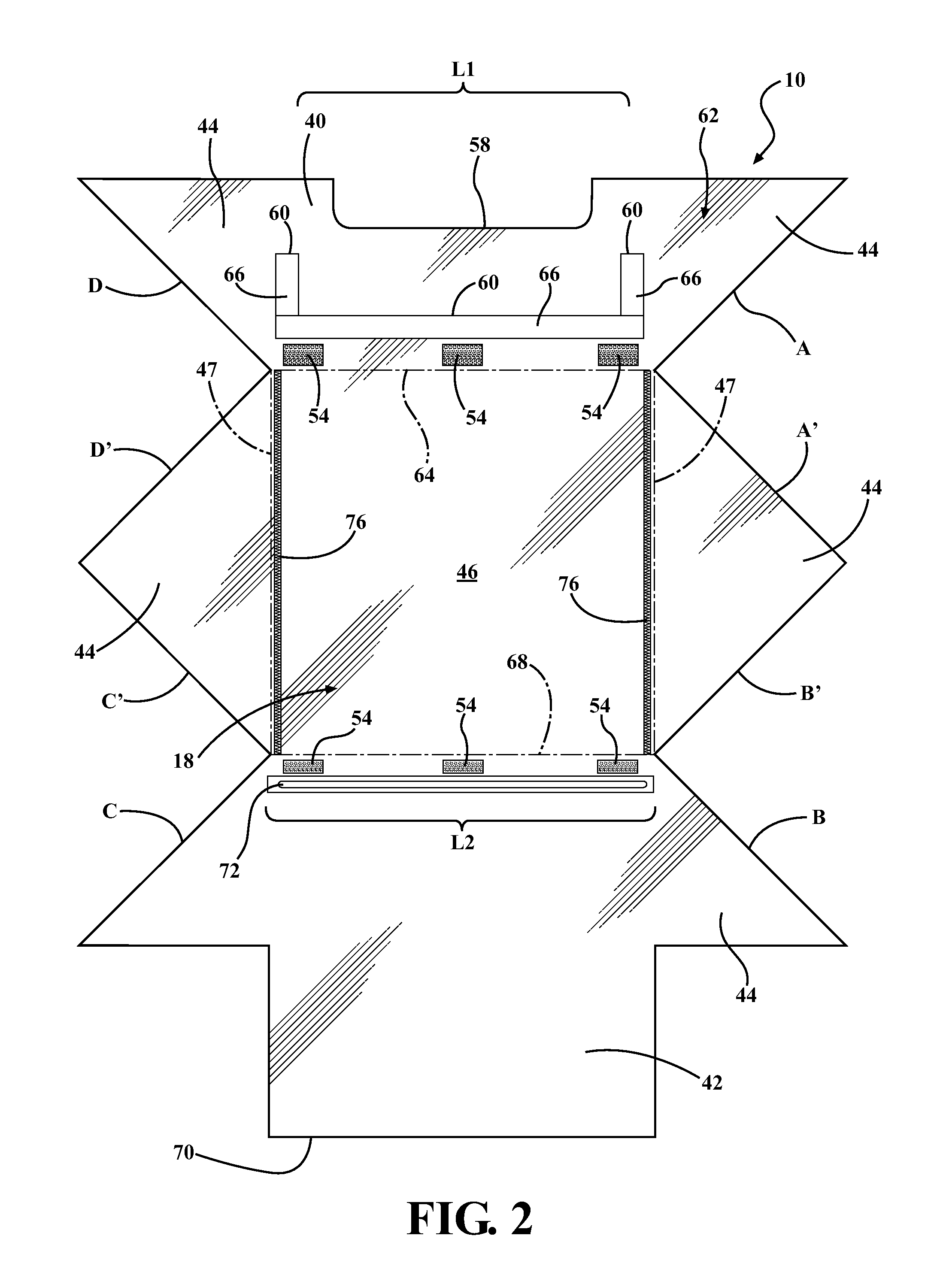
[0027]Referring in more detail to the drawings, FIG. 1 illustrates a sterile surgical C-arm radiological imaging unit drape, referred to hereafter as imaging drape 10, constructed in accordance with one aspect of the invention. The imaging drape 10 is shown attached, in a collapsed position, to a standard patient drape 12 to facilitate providing and maintaining a sterile outer surface about a portion of a C-arm type radiological imaging unit 14, and particularly about a distal end portion 16 of the imaging unit 14. The imaging drape 10 has a wall 18 providing an enclosure constructed of a flexible, tear and puncture-resistant material, such as a clear plastic material, for example, wherein the imaging drape 10 is economical in construction, and thus, is well suited to be disposed after use. The imaging drape 10 provides a quick and easy mechanism in which to reliably ensure a sterile surgical theater is maintained throughout a surgical procedure, at least with regard to a potential ...


PUM


Abstract

A surgical radiological C-arm imaging unit drape configured to provide a sterile outer surface about an end portion of a C-arm imaging unit and method of providing a sterile surface about an end of a C-arm imaging unit is provided. The drape includes a flexible enclosure having an upper wall with a pair of sidewalls and a rear side extending downwardly from the upper wall. The upper wall and the side walls are extendible between expanded and collapsed positions. When in the expanded position, a pocket sized for receipt of the end portion of the imaging unit beneath the upper wall is formed. The upper wall has a sterile outer surface configured to face away from and overlie the end portion of the imaging unit to maintain sterility in a sterile zone above an operating table. When in the collapsed position, the sterile outer surface shielded from external contamination.

Description

CROSS-REFERENCE TO RELATED APPLICATIONS[0001]This application is a continuation-in-part of U.S. patent application Ser. No. 12 / 248,291, filed Oct. 9, 2008 now U.S. Pat. No. 8,042,549, which claims the benefit of U.S. Provisional Application Ser. No. 61 / 043,644, filed Apr. 9, 2008, and this application further claims the benefit of U.S. Provisional Application Ser. No. 61 / 225,134, filed Jul. 13, 2009, all of which are incorporated herein by reference in their entirety.BACKGROUND OF THE INVENTION[0002]1. Technical Field[0003]This invention relates generally to surgical drapes, and more particularly to surgical drapes used to cover radiological imaging equipment to maintain sterility in a surgical theatre.[0004]2. Related Art[0005]The need to provide a sterile environment in a surgical theatre is directly associated with the known risk of infection that can be caused by bacteria, such as from a person or surgical equipment, in the surgical theatre. As such, it is known that in order to...

Claims


Application Information

Patent Timeline
Patent Type & Authority Patents(United States)
IPC IPC(8): A61B19/08H01J35/16A61B46/23
CPCA61B19/08A61B19/081A61B46/00A61B46/10
Inventor KASKA, SERGE
Owner TIDI PROD LLC
Why Eureka
Try Eureka