Method for measuring the area of a sample disposed within an analysis chamber

a technology of analysis chamber and sample, which is applied in the field of methods for analysis of biologic fluid samples, can solve the problem of difficulty in accurately dispensed sample amounts

Inactive Publication Date: 2012-12-04
ABBOTT POINT CARE
View PDF45 Cites 53 Cited by
  • Summary
  • Abstract
  • Description
  • Claims
  • Application Information

AI Technical Summary

Benefits of technology

[0009]An analysis chamber having a pair of transparent panels separated by a uniform height provides a convenient vehicle for the analysis of the sample. For many analyses, it is useful (or required) to know the volume of the sample being analyzed, but difficult to dispense an accurately known amount of sample. To avoid the problem of dispensing accurately known amounts of sample, it would be useful to measure the area of the film generated in the aforesaid analysis chamber and multiplying that area by the chamber height to determine the sample volume added. If the blood (or other biologic fluid) would distribute in geometrically perfect shapes, this would be relatively easy, but samples, as they spread through the chamber from their point of application, often trap air bubbles or have serpentiginous edges. The task of determining the area of a sample within a chamber is particularly problematic for blood samples having a low number of red blood cells, where the relatively clear plasma will separate from the majority of the sample, and this relatively colorless area may not be included in the area calculations if the coloration of the sample area is the only criteria used for its area calculation. The present method provides an accurate way of determining sample area and therefore volume in a chamber of uniform height, and thereby provides a solution to the problems associated with dispensing an accurate sample volume.

Problems solved by technology

For many analyses, it is useful (or required) to know the volume of the sample being analyzed, but difficult to dispense an accurately known amount of sample.
The task of determining the area of a sample within a chamber is particularly problematic for blood samples having a low number of red blood cells, where the relatively clear plasma will separate from the majority of the sample, and this relatively colorless area may not be included in the area calculations if the coloration of the sample area is the only criteria used for its area calculation.

Method used


Image

  • Method for measuring the area of a sample disposed within an analysis chamber
  • Method for measuring the area of a sample disposed within an analysis chamber
  • Method for measuring the area of a sample disposed within an analysis chamber

Examples


Embodiment Construction

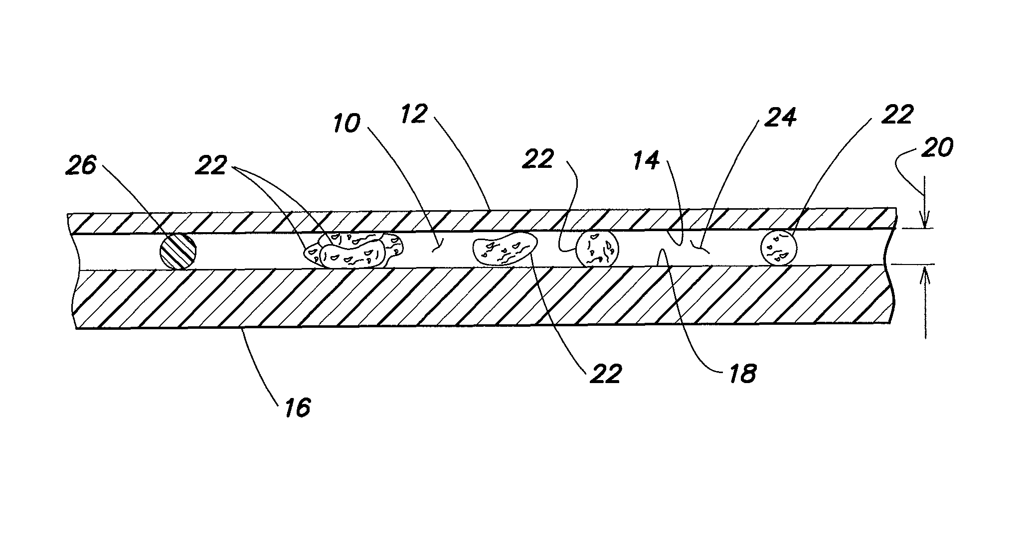
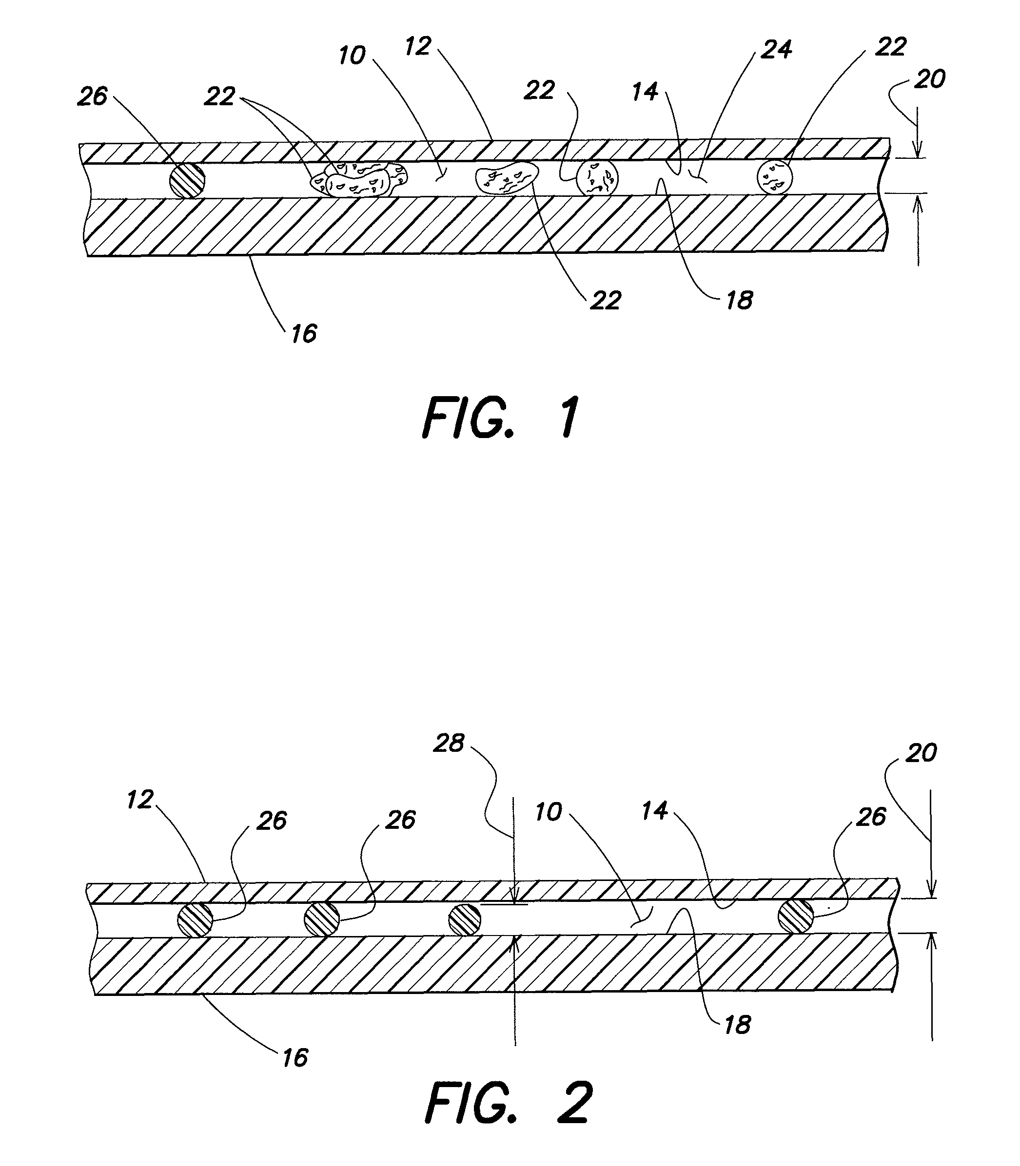
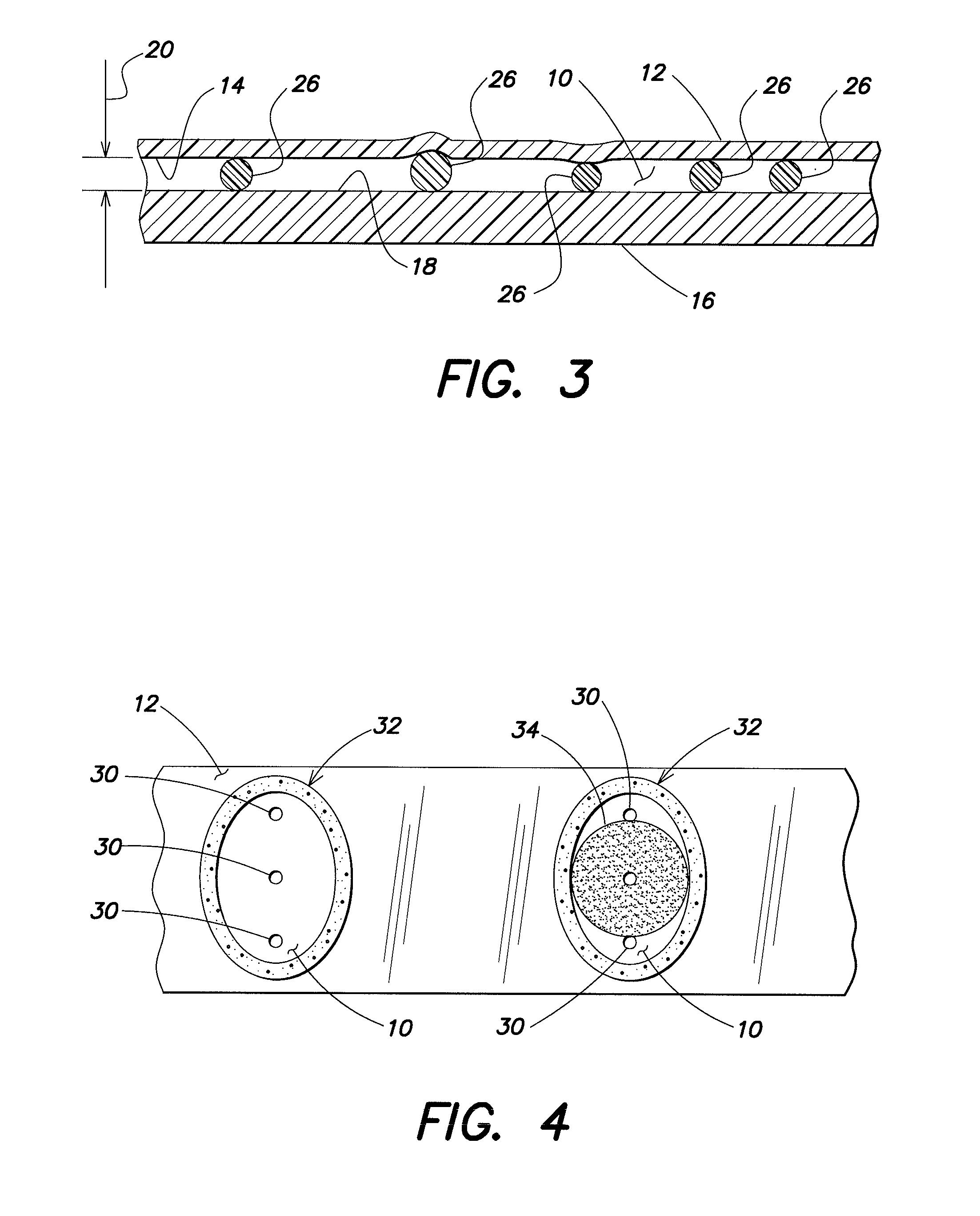
[0019]The present invention provides one or more methods for determining the area of a biological fluid sample quiescently residing within an analysis chamber. Depending on the analysis, the sample area itself can provide desirable, useful information. In other analyses, the area can be used to determine the volume of the sample within the chamber; e.g., where the chamber height is known or is determinable, the sample volume sample can be determined using the area and the known or determinable height of the chamber.

[0020]The present method(s) can be performed with an analysis chamber that is operable to quiescently hold a biological fluid sample (e.g., a sample of substantially undiluted anticoagulated whole blood) for analysis. The chamber is typically sized to hold about 0.2 μl to 1.0 μl of sample, but the method is not limited to use with any particular chamber volume, and the chamber volume can vary to suit the analysis application. In those instances when the present method is ...


PUM


Abstract

A method for determining the area of an analysis chamber covered by a biologic fluid sample quiescently residing within the chamber is provided. The chamber has a first panel with an interior surface, and a second panel with an interior surface, both of which panels are transparent. The method includes the steps of: a) illuminating the sample residing within the analysis chamber at one or more wavelengths operable to highlight interfaces between the sample and air, and to highlight a constituent within the sample; b) imaging the sample along the one or more wavelengths, and producing image signals representative of the interaction of the one or more wavelengths with the sample; c) determining a location of at least one interface between the sample and air, using the image signals; d) determining a location of one or more constituents within the sample relative to the at least one sample-air interface using the image signals; and e) determining an area of the chamber containing the sample, using the location of the one or more constituents and the at least one sample-air interface.

Description

[0001]The present application is entitled to the benefit of and incorporates by reference essential subject matter disclosed in U.S. Provisional Patent Application Ser. No. 61 / 043,567, filed Apr. 9, 2008.BACKGROUND OF THE INVENTION[0002]1. Technical Field[0003]The present invention relates to methods for analysis of biologic fluid samples general, and apparatus and methods for determining the area filled by the sample within an analysis chamber in particular.[0004]2. Background Information[0005]Physicians, veterinarians and scientists have examined human and animals' biologic fluids, especially blood, in order to determine constituent quantities as well as to identify the presence of unusual particulates not seen in healthy subjects. The constituents generally measured, quantified and identified include red blood cells (RBCs), white blood cells (WBCs), and platelets.[0006]Known blood examination techniques, described in detailed medical texts such as Wintrobe's Clinical Hematology 1...

Claims


Application Information

Patent Timeline
Patent Type & Authority Patents(United States)
IPC IPC(8): G06K9/00
CPCG06T7/0012G06T7/602G01N21/17G01N15/05G06T2207/10056G01N2015/055G06T2207/30024G06T2207/10064G06T7/62Y10T436/11
Inventor LALPURIA, NITEN V.WARDLAW, STEPHEN C.
Owner ABBOTT POINT CARE
Why Eureka
Try Eureka