Delivery system and method of use for the eye

a delivery system and eye technology, applied in the field of human tissue treatment, can solve the problems of increasing intraocular pressure in the diseased eye, damage to the optic nerve and blindness, and increased intraocular pressure in the eye, so as to improve the treatment of glaucoma, improve fluid flow, and reduce the effect of elevated intraocular pressur

Inactive Publication Date: 2013-09-24
IVANTIS INC
View PDF7 Cites 105 Cited by
  • Summary
  • Abstract
  • Description
  • Claims
  • Application Information

AI Technical Summary

Benefits of technology

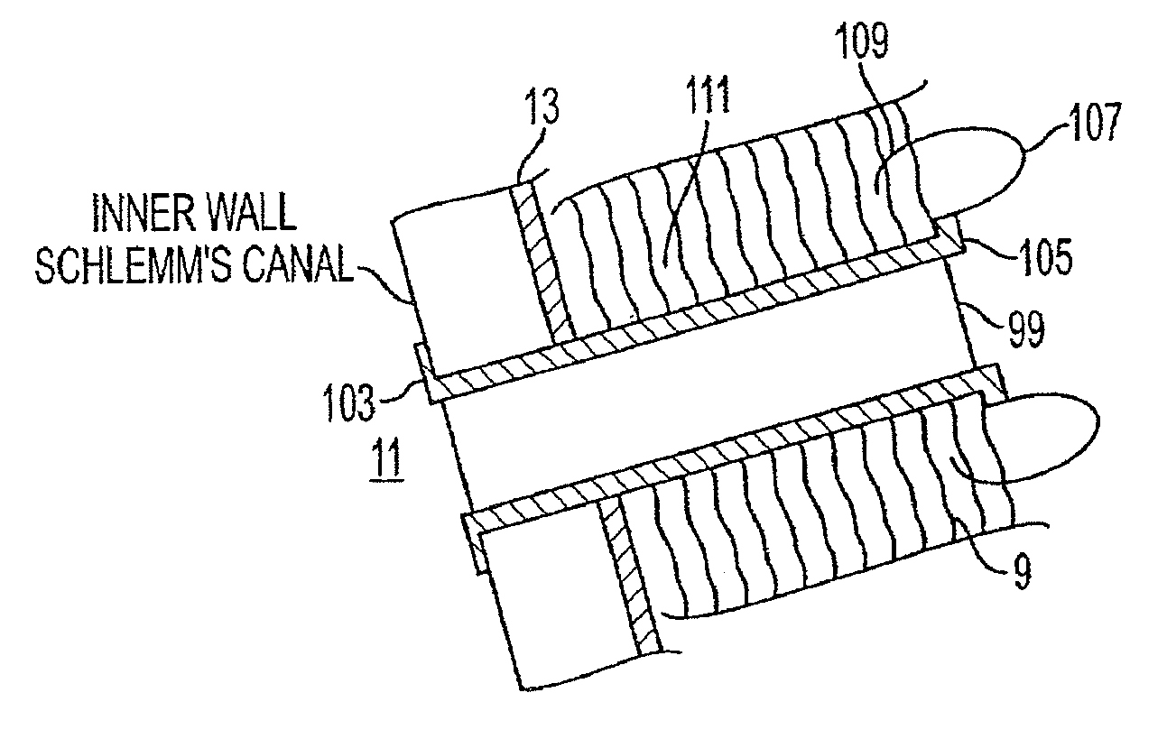
[0013]The present invention is an improved glaucoma treatment by providing a method and delivery system for creating an aqueous outflow pathway through the trabecular meshwork, juxtacanalicular trabecular meshwork and Schlemm's canal of an eye in order to reduce elevated intraocular pressure. The method includes the steps of introducing a fiber-optic probe between the outer surface of the eye and the anterior chamber until a distal end of the fiber-optic probe is in contact with or adjacent to a target site including the trabecular meshwork, the juxtacanalicular trabecular meshwork and Schlemm's canal distal to the meshwork. Pulsed laser radiation is delivered from the distal end of the fiber-optic probe sufficient to cause photoablation of the juxtacanalicular trabecular meshwork and an inner wall of Schlemm's canal in the target tissues. The fiber-optic probe may be stationery or advanced creating an aperture through these tissues to enable and improve fluid flow from the anterior chamber of the eye. The pulsed radiation is delivered in wavelengths, pulse durations and fluences to cause a minimal thermal effect on the tissue while removing and modifying tissue.
[0014]In a second aspect of the invention, a method of controlling an interior anatomy of an eye during an intraocular procedure includes the steps of creating an opening in the eye, and filling the anterior chamber of the eye through the opening with a viscoelastic material. The interior pressure within the eye may be sensed with a pressure sensor. The interior pressure may be adjusted by controlling the amount of viscoelastic material so as to compress or decompress the interior anatomy of the eye at a predetermined target anatomy site. In one aspect, the interior anatomy includes the trabecular meshwork. Viscoelastic materials of various viscosities and other protective agents placed within structures enable micro-manipulation of such structures for surgical modification while protecting adjacent structures from possible damage. Schlemm's canal may be inflated to enable perforation of the inner wall while protecting the outer wall structures.

Problems solved by technology

Glaucoma, a serious long-term health care problem, is a disorder of the eye in which elevated intraocular pressure ultimately leads to damage to the optic nerve and to blindness.
Because aqueous humor is constantly produced by the eye, any obstruction in the trabecular meshwork, the juxtacanalicular trabecular meshwork or in Schlemm's canal prevents the aqueous humor from readily escaping from the anterior eye chamber which results in an elevation of intraocular pressure within the eye.
While these treatments may be effective for a period of time, the intraocular pressure in the diseased eyes often increases in many patients.
The most frequent problems result from patients failing to follow their treatment regimen thus causing inadequately controlled glaucoma, which results in irreversible damage to the optic nerve that ultimately results in vision loss.
The human eye is a particularly challenging target for corrective surgery because of the size, fragility, distribution and characteristics of interior tissues.
The surgical therapies in current use, however, do not address the location of the outflow obstruction that is recognized for causing the elevated intraocular pressure.
Significantly, these techniques have been found to be unsuccessful for long term intraocular pressure control.
This procedure, however, has many complications including leaks, infections, hypotony (e.g., low eye pressure), and requirements for post-operative needling, undesirable antimetabolite use, a need for flap suture adjustment to maintain the function of the opening and a need for long-term monitoring to avoid late complications.
Another procedure, called deep sclerectomy, attempts to create an intrascleral filtration pocket, but does not alter anatomic relationships and does not treat the region of outflow obstruction.
Although attempting to address the outflow obstruction that causes the increased intraocular pressure, viscoanalostomy has not been shown to be successful.
First, they have limited ability to lower the intraocular pressure to a desirable level.
Second, while most found initial success in creating a puncture through the meshwork, the short duration of the reduced intraocular pressure proved to be ineffective in treating the long term effects of glaucoma.
As a result, patients suffered undesirable additional post operative procedures to lower the intraocular pressure and required continuous long-term monitoring.
These early laser procedures failed in that no consideration was given to the size of the openings in the trabecular meshwork.
In addition, these procedures also failed to recognize the importance of reducing collateral tissue damage surrounding the created hole.
The actual and potential thermal effect produced by the lasers is a significant contributing factor to the resultant tissue damage.

Method used


Image

  • Delivery system and method of use for the eye
  • Delivery system and method of use for the eye
  • Delivery system and method of use for the eye

Examples


Embodiment Construction

[0040]FIGS. 3-12 illustrate an embodiment of a laser delivery system 21 for micromachining, microscupting, or microshaping the interior anatomy of an eye. As shown in FIG. 4, a laser delivery system 21 may be operated to reduce the thermal component of laser energy contributing to collateral tissue damage. As further shown in FIG. 4, laser delivery system 21 includes a fiber-optic probe 23 for entering the eye and removal or manipulation of eye tissue. Fiber-optic probe 23 is mechanically and electrically coupled to a probe handset 25. Probe handset 25 includes controls for manipulating and operating the fiber-optic probe. A servo device 27 is connected to fiber-optic probe 23 for automatically controlling pressure within the eye during an intraocular procedure. If desired, a motion controller 29 may selectively automate transocular movement of fiber-optic probe 23 into a desired site for tissue removal and / or manipulation. A laser unit 31 provides laser energy in the form of wavele...


PUM


Abstract

A method and delivery system are disclosed for creating an aqueous flow pathway in the trabecular meshwork, juxtacanalicular trabecular meshwork and Schlemm's canal of an eye for reducing elevated intraocular pressure. Pulsed laser radiation is delivered from the distal end of a fiber-optic probe sufficient to cause photoablation of selected portions of the trabecular meshwork, the juxtacanalicular trabecular meshwork and an inner wall of Schlemm's canal in the target site. The fiber-optic probe may be advanced so as to create an aperture in the inner wall of Schlemm's canal in which fluid from the anterior chamber of the eye flows. The method and delivery system may further be used on any tissue types in the body.

Description

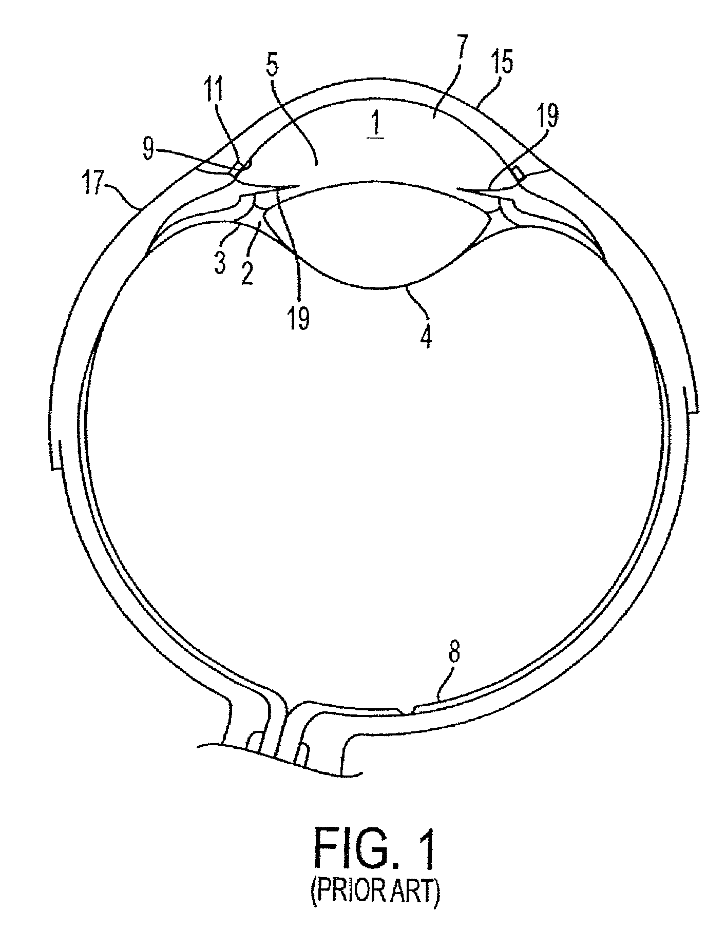
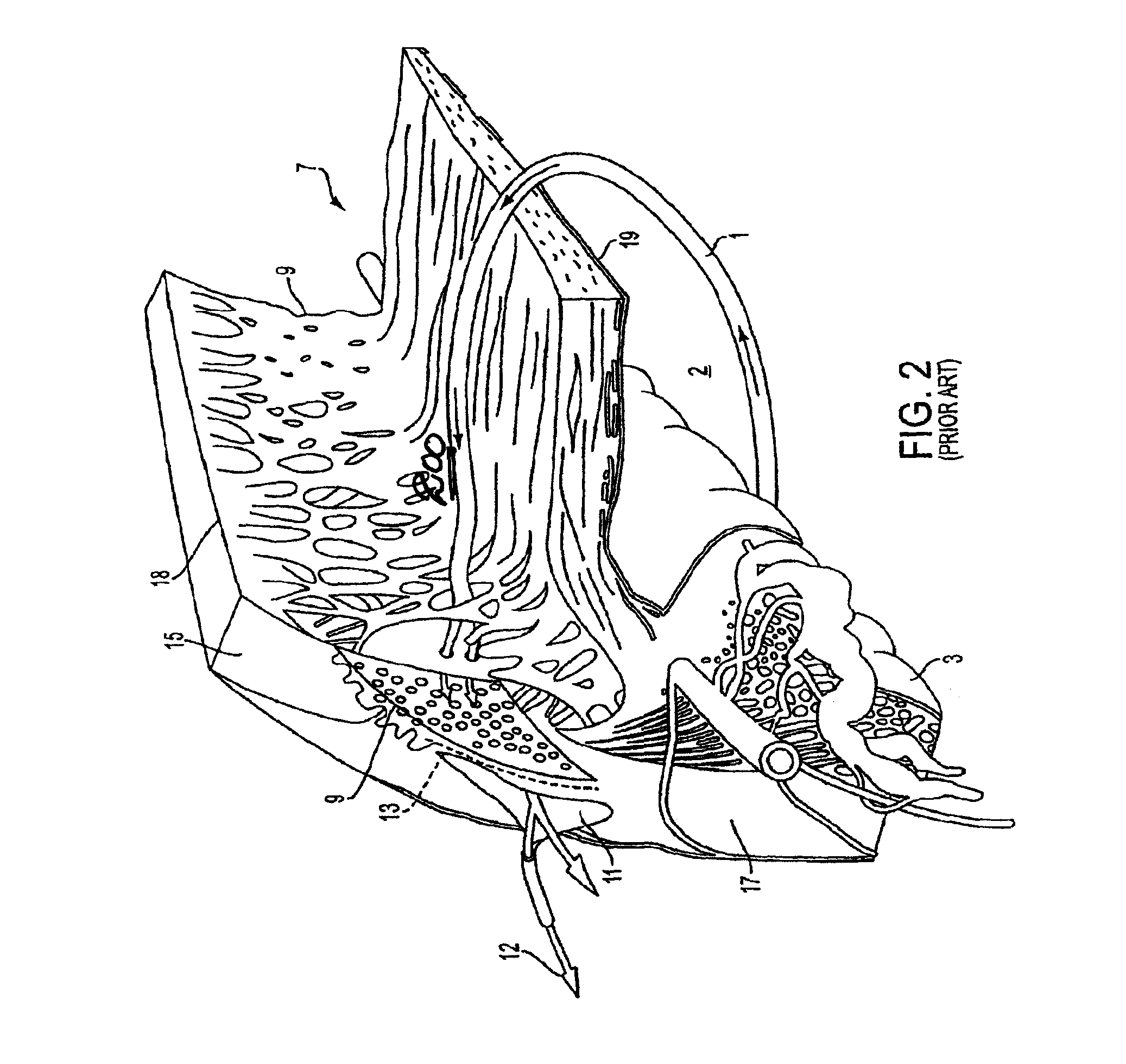
RELATED APPLICATIONS[0001]This is a divisional of U.S. patent application Ser. No. 09 / 860,842, filed May 21, 2001, which claims the benefit of U.S. Provisional Application No. 60 / 205,846, filed May 19, 2000, both of which are incorporated herein by reference.FIELD OF THE INVENTIONS[0002]This invention relates to devices and methods for treatment of human tissues, especially interior human tissue structures in the eye for restructuring, and more particularly to treatment of glaucoma.BACKGROUND[0003]Glaucoma, a serious long-term health care problem, is a disorder of the eye in which elevated intraocular pressure ultimately leads to damage to the optic nerve and to blindness. Glaucoma has been cited as the second most common cause of blindness affecting several million people in the United States alone.[0004]In order to fully appreciate the present invention, a brief overview of the anatomy of the eye is provided. As schematically shown in FIG. 1, the outer layer of the eye includes a ...

Claims


Application Information

Patent Timeline
Patent Type & Authority Patents(United States)
IPC IPC(8): A61M5/00A61B17/00A61B18/00A61B18/22A61F2/14A61F9/007A61F9/008A61F9/011
CPCA61F9/00802A61B2017/00039A61B2017/00057A61B2017/00092A61B2017/00119A61B2018/00011A61B2018/00982A61B2018/2255A61F2/14A61F9/00781A61F9/008A61F2009/00844A61F2009/00868A61F9/00A61B18/22A61B2017/00022A61B2017/00026A61B17/32A61B2017/00066A61B2017/00017A61B2018/00648A61B2018/00321A61B2018/00196A61B2018/00577A61B2018/00904A61F2009/00891A61F2/15
Inventor BERLIN, MICHAEL S.
Owner IVANTIS INC
Why Eureka
Try Eureka