Biomarkers for pancreatic cancer and diagnostic methods

a pancreatic cancer and biomarker technology, applied in the field of molecular biology and medicine, can solve the problems of high cost, significant interpretation expertise, and insufficient performance of single marker tests, and achieve the effect of improving performan

Active Publication Date: 2014-01-21
UNIVERSITY OF PITTSBURGH +1
View PDF17 Cites 7 Cited by
  • Summary
  • Abstract
  • Description
  • Claims
  • Application Information

AI Technical Summary

Problems solved by technology

These methods are useful for identifying pancreatic abnormalities and rendering an accurate diagnosis in many cases, but they come with high cost, significant expertise required for interpretation, and inherent uncertainty.
The performance of tests based on single markers so far has not been good enough to be recommended for clinical application.
The challenge for pancreatic cancer diagnostics is to find the particular protein alterations or combinations of protein alterations that usually occur early in cancer development and that do not occur in benign conditions.
The current methods for analyzing glycans are either cumbersome or very low throughput and not reproducible enough for diagnostics research.
While these methods are useful for determining glycan structures, they are not suitable for studies requiring reproducible measurements over many different samples or proteins, or for determining variation between populations of samples.
Lectins and antibodies against carbohydrate epitopes have been used to identify cancer-associated glycosylation, although those methods do not identify which proteins are carrying the epitopes.

Method used


Image

  • Biomarkers for pancreatic cancer and diagnostic methods
  • Biomarkers for pancreatic cancer and diagnostic methods
  • Biomarkers for pancreatic cancer and diagnostic methods

Examples


example 1

Serum Samples

[0158]Serum samples were assembled from three sites. At Evanston Northwestern Healthcare (ENH, Evanston, Ill.), serum samples were collected from patients with pancreatic adenocarcinoma (stage I to stage IV), other gastro-intestinal malignancies (such as colon cancer, duodenal or esophageal cancer), or benign gastro-intestinal diseases (such as ampullary adenoma, pancreatitis, cystadenoma, pseudocyst or diverticulosis). The samples were collected prior to any invasive procedures, and the patient diagnoses were confirmed after subsequent procedures. Control samples also were collected at ENH from high-risk individuals from pancreatic cancer prone families undergoing surveillance with Endoscopic Ultrasound (EUS) or Endoscopic Retrograde Cholangiopancreatography (ERCP). The control subjects had no pancreatic lesions, and serum samples were collected using the same procedure as for the other patients at ENH. Additional samples from pancreatic cancer patients and healthy con...

example 2

Antibodies and ELISA

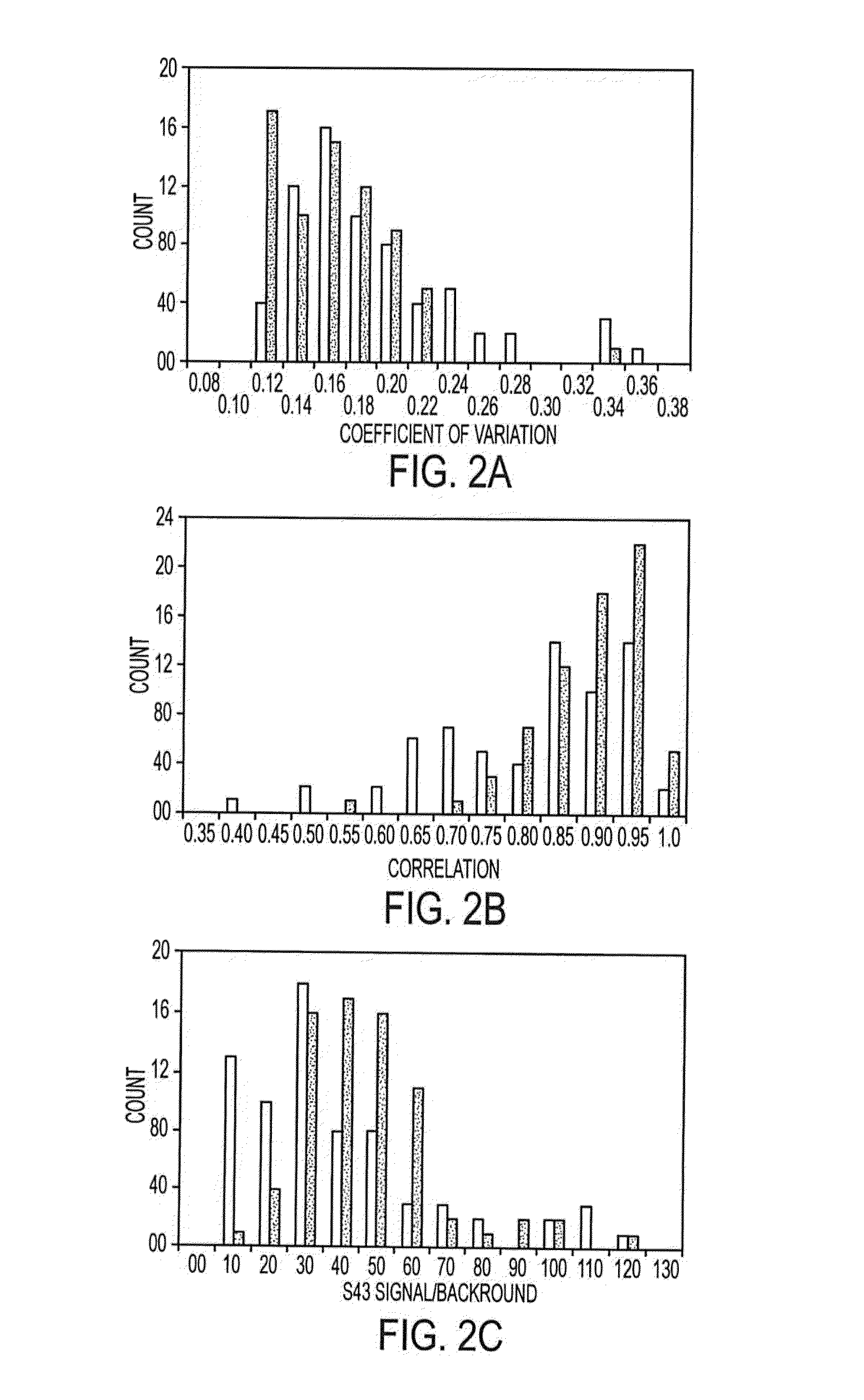
[0161]Antibodies were purchased from various sources. Table 1 shows the complete list of antibodies used, and performance characteristics. The average CVs, correlations, and average S / Bs (543 channel only) were calculated. The “correlation overlap” column refers to the number of samples that both gave measurable data in both sets one and two (out of 89 possible) or in both sets three and four (out of 138 possible). The “Control S / B / ” column gives the average S / B of each antibody in the negative control experiments, in which unlabeled sera were incubated on the arrays.

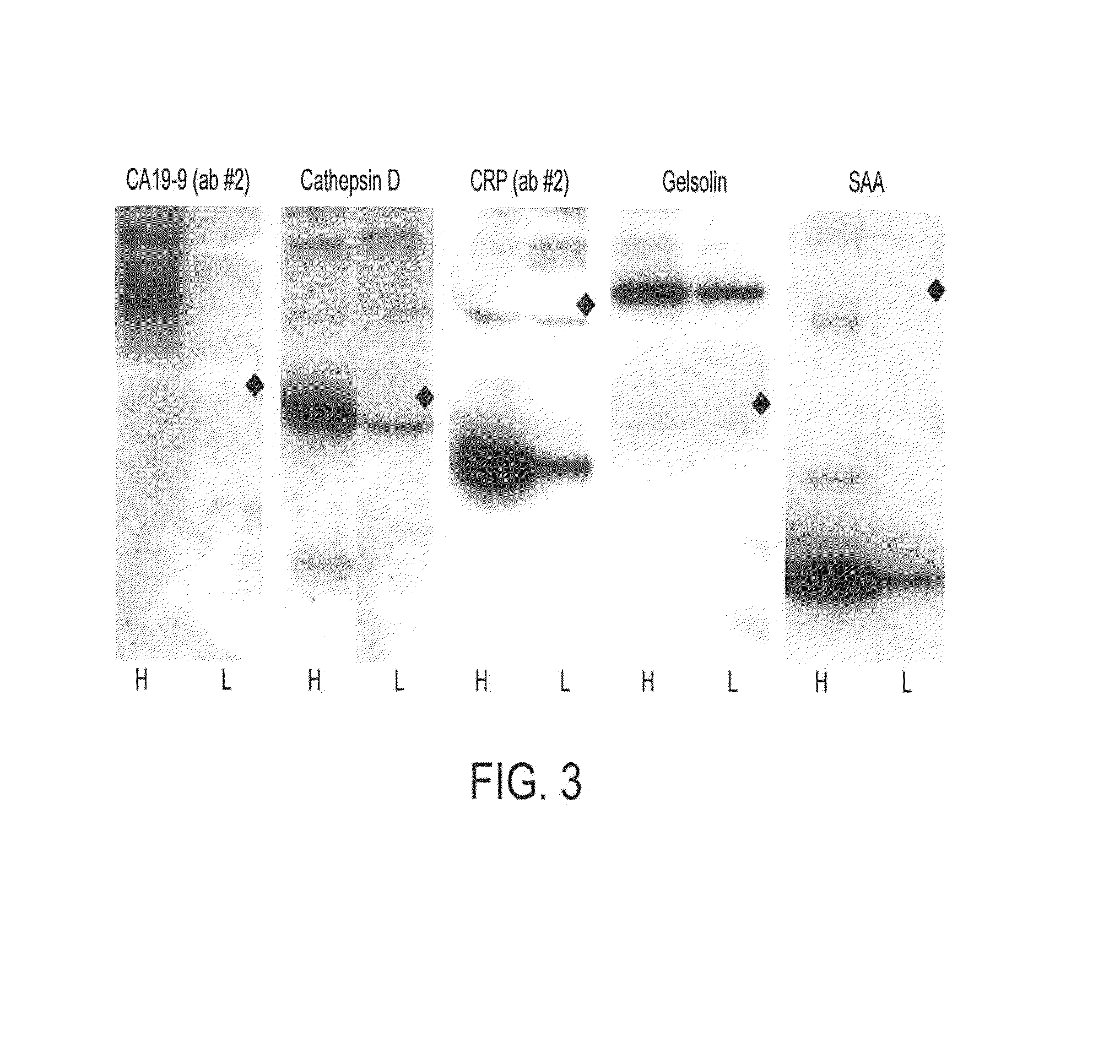
[0162]Antibodies that were supplied in ascites fluid, culture supernatant or antisera were purified using Protein A beads (Affi-gel Protein A MAPS kit, Bio-Rad) according to the manufacturer's protocol. The antibodies were prepared at concentrations of 200-2000 μg / ml (most at 500 μg / ml) in 10.1 mM Na2HPO4, 1.8 mM KH2PO4, 137 mM NaCl, 2.7 mM KCl, pH 7.5 (1×PBS) containing 0.02% NaN3. The integrity of ...

example 3

Fabrication of Antibody Microarray

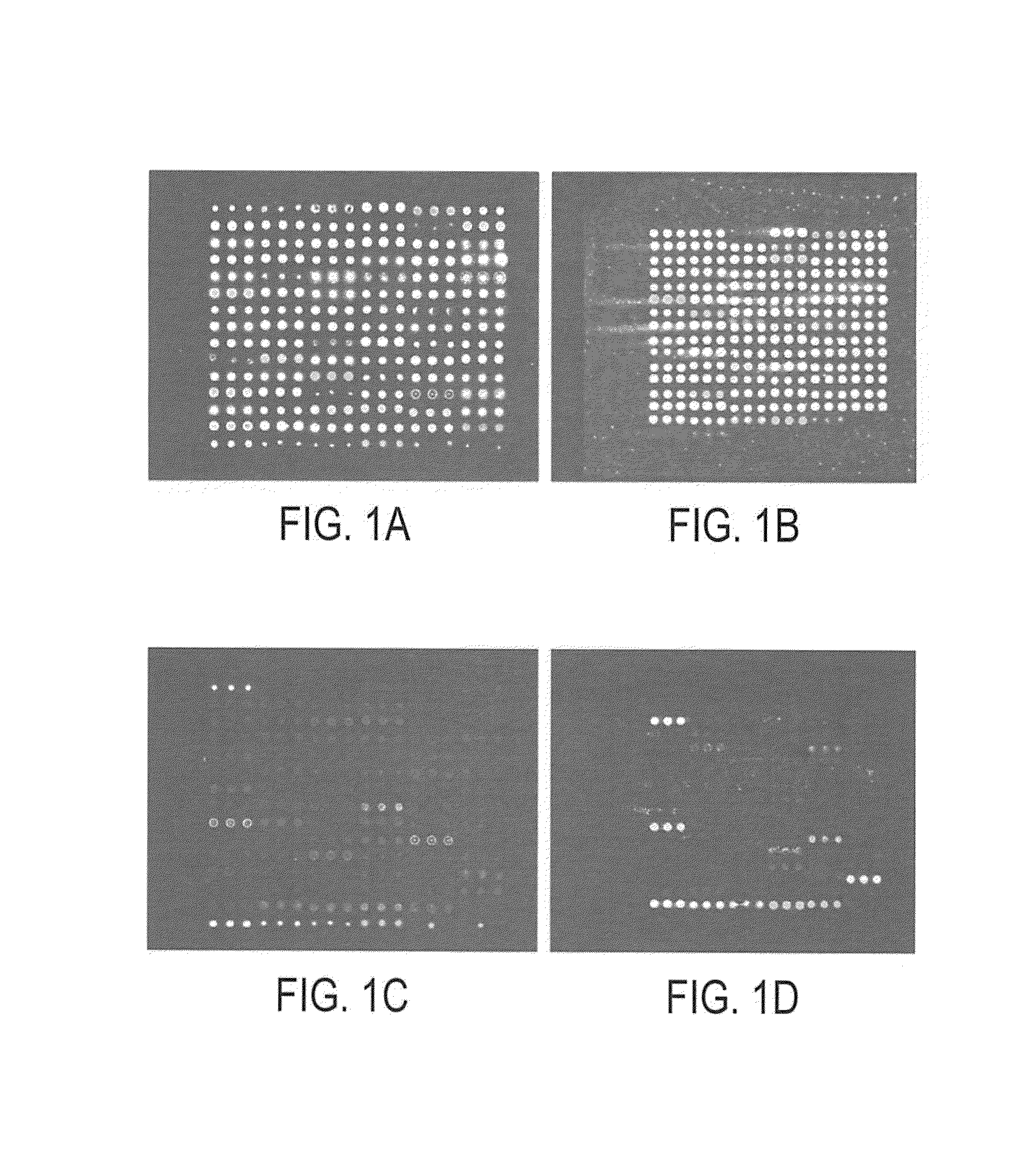
[0163]Microarrays were prepared as previously described by the inventor. Zhou, H., Bouwman, K., Schotanus, M., Verweij, C., Marrero, J. A., Dillon, D., Costa, J., Lizardi, P. M., and Haab, B. B. Two-color, rolling-circle amplification on antibody microarrays for sensitive, multiplexed serum-protein measurements. Genome Biology, 5: R28, 2004. The antibody solutions were assembled in polypropylene 384-well microtiter plates (MJ Research), using 20 μl in each well. A piezoelectric non-contact printer (Biochip Arrayer, PerkinElmer Life Sciences) spotted approximately 350 μl of each antibody solution on the surfaces of either hydrogel-coated (OptArray slides, Accelr8 Technology Corp.) or ultra-thin-nitrocellulose-coated microscope slides (PATH slides, GenTel Biosurfaces). Twelve identical arrays were printed on each slide, with each array consisting of 88-90 antibodies and control proteins spotted in triplicate in an 18×15 array. The printed hydrogel sli...


PUM

PropertyMeasurementUnit
thresholdaaaaaaaaaa
molecular weightaaaaaaaaaa
concentrationsaaaaaaaaaa

Abstract

Methods and related kits for differentiating pancreatic cancer from a benign pancreatic disease. The method includes assaying a patient biological sample for a total level of CA 19-9 antigen and for a glycan level in specific mucin(s), and comparing the total level of CA 19-9 antigen and the glycan level in the specific mucin(s) to statistically validated thresholds, wherein a different level of total CA 19-9 antigen in the patient biological sample as compared to a statistically validated threshold and a different level of glycan level in the specific mucin(s) as compared to statistically validated thresholds indicate pancreatic cancer in the patient rather than a benign pancreatic disease.

Description

CROSS-REFERENCE TO RELATED APPLICATIONS[0001]The present application is a continuation-in-part of U.S. application Ser. No. 11 / 375,756 filed Mar. 15, 2006, now U.S. Pat. No. 7,838,634, entitled METHODS FOR MEASURING GLYCAN LEVELS OF PROTEINS, which claims the benefit of provisional application Ser. No. 60 / 671,829 filed Apr. 15, 2005, entitled DCP BIOMARKER FOR PANCREATIC CANCER, the entire contents of which applications are incorporated herein in their entirety.[0002]This invention was made with government support under R33 CA122890 and R21 CA112153 awarded by the National institute of Health. The government has certain rights in the invention.FIELD OF THE INVENTION[0003]This invention relates to the field of molecular biology and medicine and specifically to biomarkers, kits, and methods for diagnosing pancreatic cancer.BACKGROUND OF THE INVENTION[0004]Methods to detect cancers more accurately or at earlier stages could lead to better outcomes for many cancer patients, including pa...

Claims


Application Information

Patent Timeline
Patent Type & Authority Patents(United States)
IPC IPC(8): C12Q1/00G01N33/53
CPCG01N33/6803G01N33/5308G01N33/57438
Inventor HAAB, BRIAN B.YUE, TINGTINGBRAND, RANDALL E.
Owner UNIVERSITY OF PITTSBURGH
Why Eureka
Try Eureka