Medical imaging apparatus

a medical imaging and apparatus technology, applied in the field of medical imaging apparatus, can solve the problem of difficulty in adjusting the same slice position for the operator, and achieve the effect of prolonging the examination tim

Active Publication Date: 2014-04-08
FUJIFILM HEALTHCARE CORP
View PDF3 Cites 5 Cited by
  • Summary
  • Abstract
  • Description
  • Claims
  • Application Information

AI Technical Summary

Benefits of technology

[0014]According to the present invention, in a medical imaging apparatus in which an arbitrary slice in a three-dimensional space can be set as a slice position, such as the aforementioned MRI apparatuses, a slice position can be automatically calculated, and an extracting slice can be automatically calculated in MPR without changing the conventional examination flow and without prolonging the examination time.

Problems solved by technology

Further, when imaging the same slice position repeatedly, it is difficult for the operator to set the same slice position for every imaging by a manual operation.
In any case, the same operation for setting the slice position must be repeated for every imaging, and it is mentioned as one of monotonous troublesome operations.

Method used


Image

  • Medical imaging apparatus
  • Medical imaging apparatus
  • Medical imaging apparatus

Examples


first embodiment

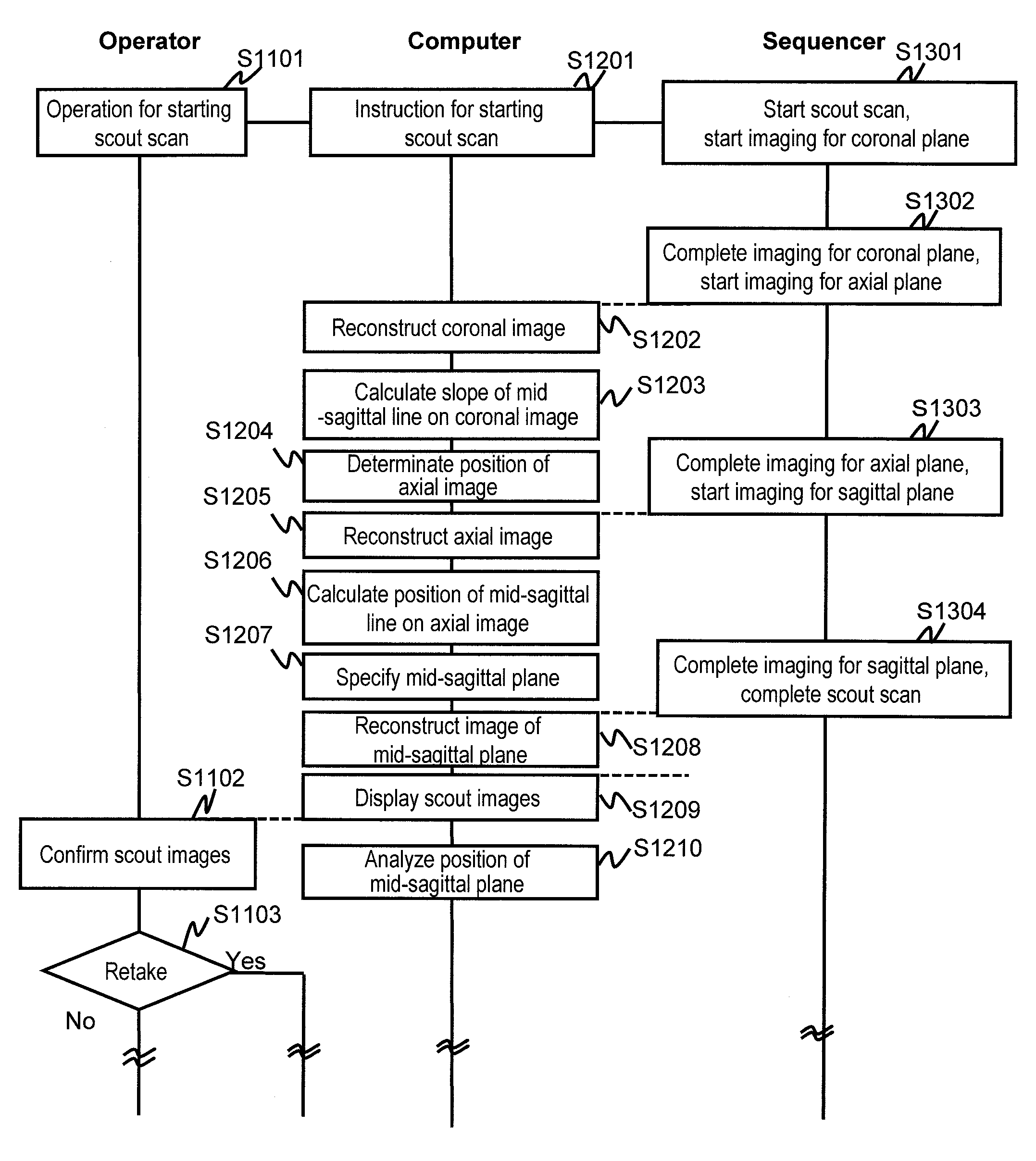
[0043]Hereafter, the first embodiment of the present invention will be explained. In all of the drawings for explaining the embodiments of the present invention, elements having the same function are indicated with the same numerals or symbols, and repetitive explanations thereof are omitted.

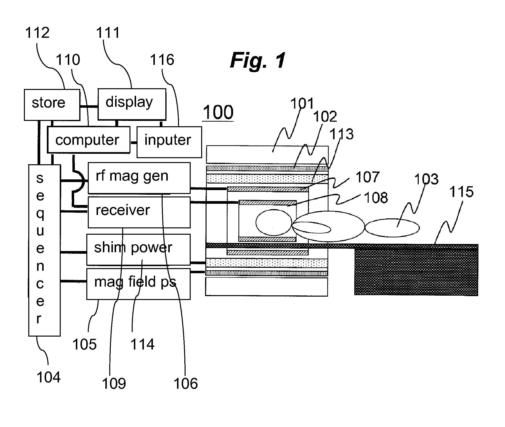
[0044]First, a magnetic resonance imaging (MRI) apparatus of this embodiment will be explained. An MRI apparatus 100 of this embodiment applies a radio frequency magnetic field to a subject 103 placed in a static magnetic field to excite nuclear magnetization in the subject 103, and measures generated magnetic resonance signals (echo signals), as described above. In this operation, a gradient magnetic field is applied to add positional information to the magnetic resonance signals to be measured for obtaining an image (imaging). FIG. 1 is a block diagram showing a typical configuration of the MRI apparatus 100 according to this embodiment, which implements the above operation. The MRI apparatus ...

second embodiment

[0158]Hereafter, the second embodiment of the present invention will be explained. The MRI apparatus according to this embodiment has basically the same configurations as those of the first embodiment. However, in this embodiment, there is provided a function of memorizing amount of adjustment received from the operator as learning data after a recommended slice position or a recommended slice for scout scan is displayed, and reflecting it in the subsequent processings. Hereafter, explanation will be made with focusing on the configurations different from those of the first embodiment.

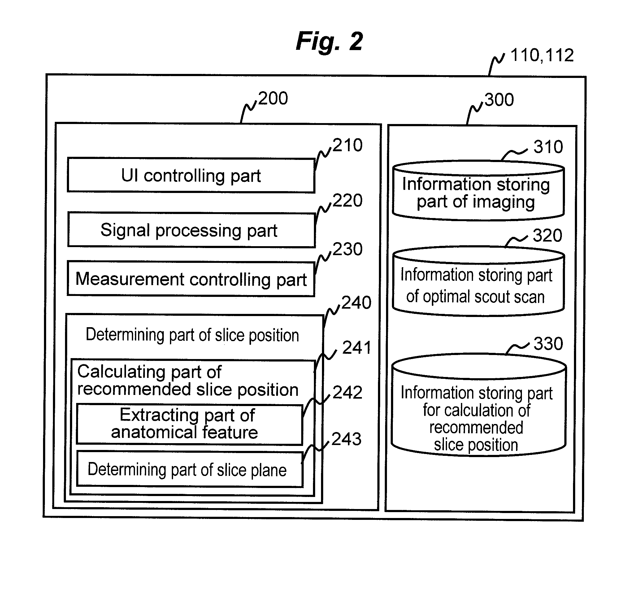
[0159]FIG. 14 is a functional block diagram of an information processor constituted by a computer 110A and a storage device 112A according to this embodiment. In the information processor according to this embodiment, in addition to the same configurations as those of the first embodiment, the determining part of slice position 240 of a control part 200A is provided with a learning function part 244, a...

third embodiment

[0170]Hereafter, the third embodiment of the present invention will be explained. The MRI apparatus according to this embodiment has basically the same configurations as those of either one of the aforementioned embodiments. However, in this embodiment, the calculating part of recommended slice position used in the first embodiment and the second embodiment is used for MPR processing. Hereafter, this embodiment will be explained with focusing on the configurations different from those of the embodiments described above.

[0171]FIG. 15 is a functional block diagram of an information processor constituted by a computer 110B and a storage device 112B according to this embodiment. The information processor according to this embodiment is basically the same as that of either one of the information processors of the aforementioned embodiments, but in the information processor of this embodiment, in addition to the same configurations as those of the aforementioned embodiments, the control p...


PUM


Abstract

There is provided a technique for, in a medical imaging apparatus enabling imaging of an arbitrary plane in a three-dimensional space, enabling automatic calculation of a slice position and automatic calculation of an extracting slice in MPR, without prolonging examination time. Two-dimensional scout scan similar to that used for manual setting of a slice position is performed, and the obtained scout images are processed to calculate a recommended slice position. Algorithms for the processing and various image processing procedures used for the processing are stored beforehand for every type of imaging region and every type of examination.

Description

TECHNICAL FIELD[0001]The present invention relates to a technique for determining a position of a slice position and a technique for extracting a diagnostic image for use in an examination using a medical imaging apparatus such as a magnetic resonance imaging apparatus.BACKGROUND ART[0002]There are medical imaging apparatuses enabling imaging of a slice of desired position and orientation in a three-dimensional space for use of the obtained image in diagnosis. In particular, magnetic resonance imaging (MRI) apparatuses are medical diagnostic imaging apparatuses utilizing the nuclear magnetic resonance phenomenon of, mainly, proton, which apparatuses apply a radio frequency magnetic field to a subject placed in a static magnetic field to excite nuclear magnetization, and construct images from measured magnetic resonance signals. The MRI apparatuses enable noninvasive imaging of an arbitrary slice without any restriction concerning imaging region.[0003]In the MRI apparatuses, in gener...

Claims


Application Information

Patent Timeline
Patent Type & Authority Patents(United States)
IPC IPC(8): G06K9/00
CPCA61B5/4528G01R33/4833A61B5/0037A61B5/055G01R33/546A61B5/4533A61B2576/026A61B5/0042G01R33/543G16H30/40
Inventor YOKOSAWA, SUGURUTANIGUCHI, YOBITO, YOSHITAKA
Owner FUJIFILM HEALTHCARE CORP
Why Eureka
Try Eureka