Wireless recording and stimulation of brain activity

a brain activity and wireless technology, applied in the field of wireless brain activity recording devices and methods, can solve the problems of increasing the likelihood of serious infection, long hospital stay, and difficulty in coping with patients, so as to reduce morbidity or mortality, avoid long and costly hospitalization, and eliminate the risk of dislodging the implanted array

Inactive Publication Date: 2014-09-30
SIGENICS +2
View PDF5 Cites 22 Cited by
  • Summary
  • Abstract
  • Description
  • Claims
  • Application Information

AI Technical Summary

Benefits of technology

[0008]In various embodiments, the present invention is directed to the use of subdural arrays that transmit ECoG recordings wirelessly. The ECoG waveforms are transmitted across the patient's skull, allowing the craniotomy used for surgical placement of the arrays to be completely closed. The patient may then wear a receiver incorporated into a skullcap, or similar headwear that collects the transmitted ECoG recordings. The receiver in the headwear is connected to external hardware that stores the recordings. The patient is then free to remove and replace the headwear for showering or other daily activities. Wireless ECoG recording has many advantages: (a) patients may be discharged from the hospital for the monitoring period, avoiding lengthy and costly hospitalization; (b) ECoG recordings can be obtained at home, where sitters are not necessary; (c) patients may move about freely; (d) because no cables enter the head, morbidity or mortality from infections is reduced; (e) elimination of transcutaneous cables minimizes or eliminates the risk of dislodging the implanted arrays; and (f) continuous recordings for many weeks or even months is possible, greatly increasing the probability of recording a higher number of seizures and improving the accuracy of the epileptogenic localization.
[0009]Wireless ECoG arrays in accordance with embodiments of the present invention avoid recording of atypical seizure activity, which can result in false localization information, and provide an improved approach to locating epileptogenic foci. Recording ECoG activity can take place when the patient is taking his or her normal (or substantially normal) anti-seizure medications, and the monitoring period can continue until sufficient habitual seizure activity is observed to identify the pharmacologically intractable epileptogenic foci. This approach increases the accuracy of and confidence in the identification of epileptogenic zones and accordingly improves the outcome of epilepsy surgery.
[0017]Embodiments of the present invention involve a wireless subdural electrode array having a plurality of electrodes for recording electrical brain activity and for electrically stimulating the brain, wherein each electrode is capable of providing the recording or stimulation function. The recording function of the array may be managed by an electrical circuit either on or connected to the array and implanted within the cranium of the patient. The electrical circuit is desirably capable of autonomous operation, recording and transmitting ECoG waveforms via a wireless electromagnetic link whenever power is provided to the array. The same electrical circuit or a second electrical circuit, also implanted within the patient's cranium, can provide electrical stimulation pulses between electrodes on the array; the electrode selection, stimulus magnitude, frequency, duration and time of imposition of the stimulation may be controlled by the clinician via the wireless electromagnetic link. Power is provided to the subdural array and electrical circuit by a wireless electromagnetic link or by a power source connected to the array and located within the cranium of the patient. An advantage of certain embodiments of the invention, therefore, is the ability not only to record but to facilitate wireless electrical stimulation of the patient's brain in a manner that can be commanded wirelessly by the clinician.

Problems solved by technology

This invasive surgical procedure has drawbacks, including: (a) a lengthy and costly hospital stay is required; (b) a family member or sitter must typically be with the patient at all times; (c) patients may have difficulty coping with the need to remain in bed and tethered to the wall by cables; (d) long duration implantations increase the likelihood of serious infection; (e) there is a risk of intracerebral hemorrhage if the cables are accidentally pulled and the grids move; and (f) 10-20% of patients do not have enough seizures during their hospitalization to identify the epileptic zone with certainty.
These disadvantages of ECoG monitoring stem from the use of subdural arrays that are wired directly from within the patient's skull to external recording and data storage equipment.
The risk of infection, created by the cables exiting the patient's head and the prohibitive expense of prolonged hospitalization, are generally the principal factors that limit the duration of ECoG monitoring to 1-2 weeks.
The patient's usual or habitual seizure pattern may be replaced by more severe convulsive seizures that place the patient at risk for seizure-related morbidities of bodily trauma, aspiration, and hypoxia, as well as potentially fatal cardiac arrhythmias or cardio-respiratory arrest.
The recording of atypical seizure activity can result in false localization information.

Method used


Image

  • Wireless recording and stimulation of brain activity
  • Wireless recording and stimulation of brain activity
  • Wireless recording and stimulation of brain activity

Examples


Embodiment Construction

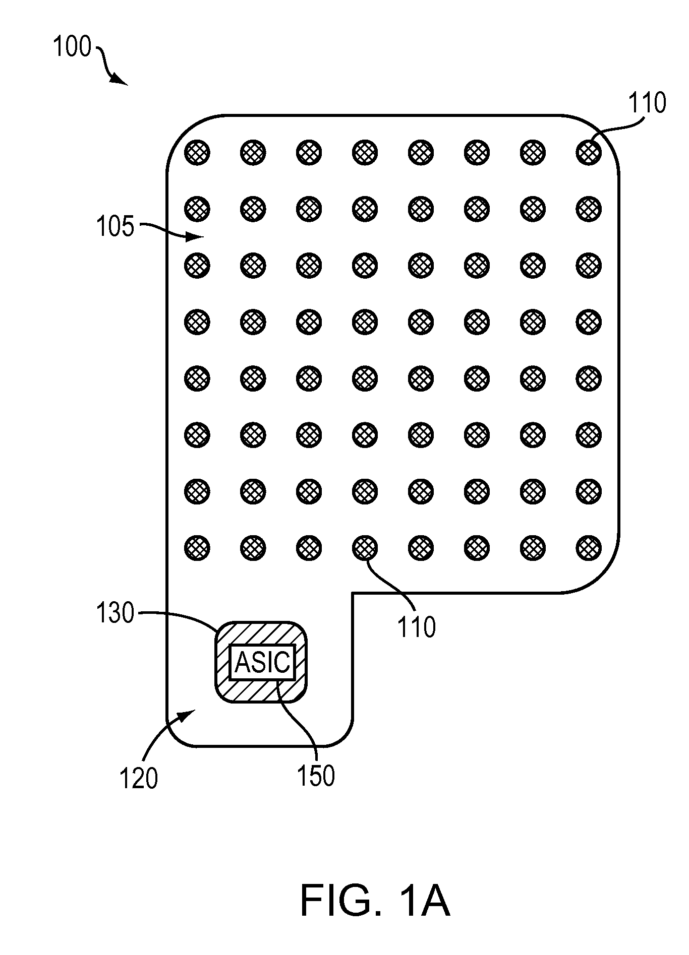
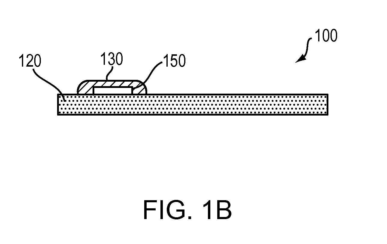
[0026]As used herein, the term “dura” refers to the fibrous membrane covering the brain and lining the inner surface of the skull; “electrocorticogram” (ECoG) refers to measurement of electrical brain activity from electrodes placed with the cranium and in close proximity to the cerebral cortex; “resection” means surgical removal of a part of the brain responsible for initiating or propagating seizure activity (and for purposes hereof, subpial transections are considered within the definition of resection); “subdural” refers to the volume within the cranium below the dura mater but above the arachnoid membrane of the meninges; “epileptogenic foci” refer to locations of epileptic abnormalities with the brain; and an “ASIC” means an application-specific integrated circuit, i.e., an electrical circuit device fabricated usually in silicon and designed for a specific application, or a general-purpose processor. The ASIC may be implemented in conjunction with, as appropriate, support circ...


PUM


Abstract

Subdural arrays transmit electrocorticogram recordings wirelessly, across the patient's skull, allowing the craniotomy used for surgical placement of the arrays to be completely closed. In various embodiments, the arrays also respond to commands, applying signal patterns to the patient's brain for diagnostic and treatment purposes.

Description

RELATED APPLICATION[0001]The present application claims priority to and the benefits of U.S. Provisional Application Ser. No. 61 / 144,929, filed on Jan. 15, 2009, the entire disclosure of which is hereby incorporated by reference.FIELD OF THE INVENTION[0002]This invention relates to improved devices and methods for recording electrical brain activity for the purpose of identifying regions in the brain exhibiting abnormal electrical activity, and more particularly to the identification of regions in the brain that originate or spread seizures associated with epilepsy and other brain dysfunctions.BACKGROUND[0003]Surgical resection of epileptogenic foci is a commonly practiced and often beneficial treatment for patients suffering debilitating seizures arising from otherwise intractable epilepsy. The success of this procedure depends on the ability of the medical team to precisely locate the epileptogenic zones in the patient's brain and to identify important cortical regions, such as el...

Claims


Application Information

Patent Timeline
Patent Type & Authority Patents(United States)
IPC IPC(8): A61B5/0478A61N1/05A61N1/36A61B5/00A61N1/02
CPCA61N1/0531A61N1/36082A61N1/0504A61B5/0006A61B5/0478A61N1/025A61B2562/125A61B5/4094A61B5/0031A61N1/36014A61B5/291A61B5/293A61N1/0529A61N1/36064A61N1/36139
Inventor COGAN, STUART F.TROYK, PHILIP R.EBERSOLE, JOHN S.TOWLE, VERNON L.
Owner SIGENICS
Why Eureka
Try Eureka