3D X-ray imaging of coronary vessels with ECG gating and motion correction

a three-dimensional imaging and motion correction technology, applied in the field of three-dimensional imaging of coronary vessels with ecg gating and motion correction, can solve the problems of insufficient period, poor motion estimation, and gaps in recording data, and achieve the effect of reducing anatomical nois

Inactive Publication Date: 2015-04-21
SIEMENS HEALTHCARE GMBH
View PDF13 Cites 4 Cited by
  • Summary
  • Abstract
  • Description
  • Claims
  • Application Information

AI Technical Summary

Benefits of technology

[0031]According to an embodiment of the invention a morphological filter is applied to the 2D projection data for the purpose of reducing anatomical noise.

Problems solved by technology

With long recording times the periodicity is no longer guaranteed due to arrhythmic heartbeat, respiration, patient motion.
Furthermore gaps in the recording data are produced as a result of ECG gating.
Estimating the motion from the recorded image data represents a challenge.
Motion estimation is a very poorly formulated problem.
The degrees of freedom of a general motion are very great and cannot be clearly determined from the measured recording data.
In the publication by C. Blondel et al [3] the motion estimation problem is limited based on the assumption that the motion is periodic, although this is not the case in reality.
The complexity of motion estimation is limited through use of an a priori 3D image.
With non-periodic motion, the image quality of the a priori 3D image and consequently also the image quality of the main reconstruction suffer.
The method has the disadvantage that if there is considerable movement no usable first preliminary 3D images can be generated.

Method used


Image

  • 3D X-ray imaging of coronary vessels with ECG gating and motion correction
  • 3D X-ray imaging of coronary vessels with ECG gating and motion correction
  • 3D X-ray imaging of coronary vessels with ECG gating and motion correction

Examples


Embodiment Construction

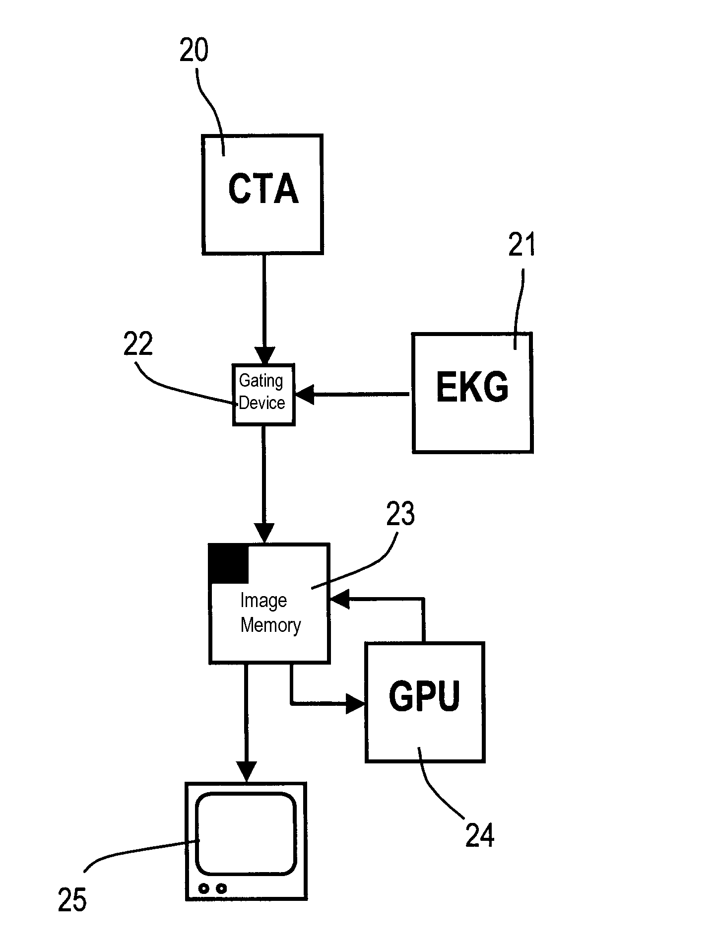
[0044]FIG. 3 shows an ACT X-ray system 20 for angiography CT and an ECG device 21 which controls the data flow of the ACT X-ray system 20 by means of a gating device 22. Connected to the gating device 22 is an image memory 23 in which initially a recorded 3D image data set is stored from which, following the acquisition of the complete 3D image data set, a 3D reconstruction image is computed by a GPU 24 and is then likewise stored in the image memory 23. The GPU 24 also initiates a motion estimation which will be described further below. The reconstructed and processed 3D images are displayed on the monitor 9 by means of a 3D playback device 25.

[0045]FIG. 4 shows method steps of an embodiment of the method according to the invention:

[0046]A first step (G1), recording of a projection image data set and e.g. an ECG; a second step (G2), calculation of a reference image with ECG gating and affine motion correction; and a third step (G3), 3D image reconstruction through correction of the...


PUM


Abstract

A method for three-dimensional visualization of a moving structure by a rotation angiography method is described. A series of projection images is recorded by an image acquisition unit from different recording angles during a rotation cycle. A three-dimensional image data can be reconstructed from the projection images. A continuous rotation cycle is proposed to be performed with simultaneous recording of at least one ECG. A three-dimensional reconstructed reference image is generated through a first correction of the motion of the moving structure by affine transformations. A three-dimensional image data of the moving structure is reconstructed from the data acquired in the continuous rotation cycle when using the reconstructed reference image while performing an estimation and correction of the motion by elastic deformations.

Description

CROSS REFERENCE TO RELATED APPLICATIONS[0001]This application claims priority of German application No. 10 2010 022 791.9 filed Jun. 4, 2010, which is incorporated by reference herein in its entirety.FIELD OF THE INVENTION[0002]The invention relates to a method for three-dimensional imaging of coronary vessels with ECG gating and motion correction.BACKGROUND OF THE INVENTION[0003]Three-dimensional rotation angiography (3D rotation angiography) is a standard method used for estimating the vascular anatomy before and during interventions. The reconstruction algorithms used for 3D reconstruction of the cardiac blood vessels by means of rotation angiography are heavily dependent on the periodicity of the cardiac motion. In the interventional environment patients often have arrhythmic heart signals or cannot hold their breath during a complete acquisition.[0004]For diagnostic examination purposes and for interventional procedures in for example cardiology, radiology and neurosurgery, int...

Claims


Application Information

Patent Timeline
Patent Type & Authority Patents(United States)
IPC IPC(8): G06T15/00A61B6/00G06T11/00
CPCA61B6/504A61B6/5264A61B6/5288A61B6/541G06T11/005A61B6/4441G06T2211/412G06T2211/424A61B6/4458
Inventor LAURITSCH, GUNTERROHKOHL, CHRISTOPHER
Owner SIEMENS HEALTHCARE GMBH
Why Eureka
Try Eureka