Analyte detection method

a technology of analyte and detection method, applied in the direction of material testing goods, biochemistry apparatus and processes, immunoassays, etc., can solve the problems of difficult detection of small molecules in biological samples, many compelling needs remain unmet, and even greater challenges

Active Publication Date: 2015-06-30
SPHERE MEDICAL
View PDF7 Cites 1 Cited by
  • Summary
  • Abstract
  • Description
  • Claims
  • Application Information

AI Technical Summary

Benefits of technology

[0013]The present invention seeks to provide a method of improving the lower limit of detection and / or concentration measurement of analytes, in particular propofol, in situations where a reaction step is required to generate a reaction product which is subsequently detected in order to qualitatively or quantitatively assess the presence of that analyte.
[0014]In accordance with a first aspect of the present invention, there is provided a method of determining the presence of an analyte of interest by means of a detection of a reaction product between the analyte of interest and a reactant, the method comprising performing a background measurement on the initial reaction mixture comprising at most a negligible concentration of the reaction product, wherein the reaction conditions present in said initial reaction mixture at least reduce the reaction rate of the formation of the reaction product such that the background measurement can be performed without a measurable change in said negligible concentration; altering the reaction conditions in the initial reaction mixture to accelerate said reaction rate; continuing said reaction until the concentration of the reaction product has stabilized; performing a second measurement on the resultant reaction mixture to obtain a signal correlated to said concentration; and determining the presence of the analyte of interest from a difference between the background measurement and the second measurement.
[0015]In accordance with the present invention, the rate of formation of the reaction product, e.g. the Gibbs-indophenol reaction product, is initially stopped or slowed down using appropriate measures, including, but not limited to, temperature, pH, slow mixing, the absence or removal of one or more reactant(s) and / or the addition of an inhibitor, in order to enable a reference measurement of the sample to be taken. The absence of the reaction or lower rate of reaction allows enough time to measure the level of the background “non-analyte” signal. Following the completion of the reference measurement, the reaction is allowed to proceed. Once the concentration of the target reaction product has stabilised, a second measurement, the sample measurement, is taken. In order to take account of the presence of the interfering species, the reference measurement is subsequently subtracted from the sample measurement in order to obtain an accurate estimation for the concentration of the target analyte.

Problems solved by technology

Despite recent advances in PoC testing, several compelling needs remain unmet.
For example, the detection of small molecules in biological samples is often very challenging, especially when no suitable receptor (e.g. enzyme, antibody, aptamer) with an appropriate specificity exists.
The challenge is even greater when the molecule is lipophilic and a large proportion of the analyte is unavailable for analysis due to its association with hydrophobic components of the sample matrix, such as cells, lipids and proteins.
The detection of small molecules in complex media (e.g. blood, plasma, saliva, urine, waste water and their extracts) is often difficult due to the association of the analyte with components of the sample matrix (e.g. plasma proteins and lipid membranes).
The complex and time-consuming nature of HPLC assays for small molecules in complex samples means that they are routinely performed by a very small number of specialist laboratories; for this reason the utility of these assays is rather limited.
In this case, the formation of the indophenol product from the reaction between the activated Gibbs reagent and propofol is the rate limiting step.
However, the optical measurement of propofol in whole blood below 1 μg / ml is limited by the presence of species in the blood sample that bind to and co-elute from the SPE column and absorb in the region of the indophenol signal.
In addition, insoluble aggregates can also be present and can scatter the light, thereby reducing the measured intensity at the detector.
These species contribute to the measured absorbance spectrum in the region of the indophenol signal at 595 nm, causing an offset in the measured absorbance at this wavelength and limiting the ability of the instrument to measure accurately the propofol concentration in blood samples containing less than 1 μg / ml of propofol.
As this non-propofol signal varies for different blood samples, this offset due to interfering species cannot be corrected for by applying, for example, correlation factors to the data.
However, it would necessitate the use of expensive spectrometers to measure the absorbance signal either side of the absorbance peak of interest and interpolating the signal in order to subtract the background contribution to the peak.
This, in turn, will increase the cost and complexity of the device.

Method used


Image

  • Analyte detection method
  • Analyte detection method
  • Analyte detection method

Examples


example

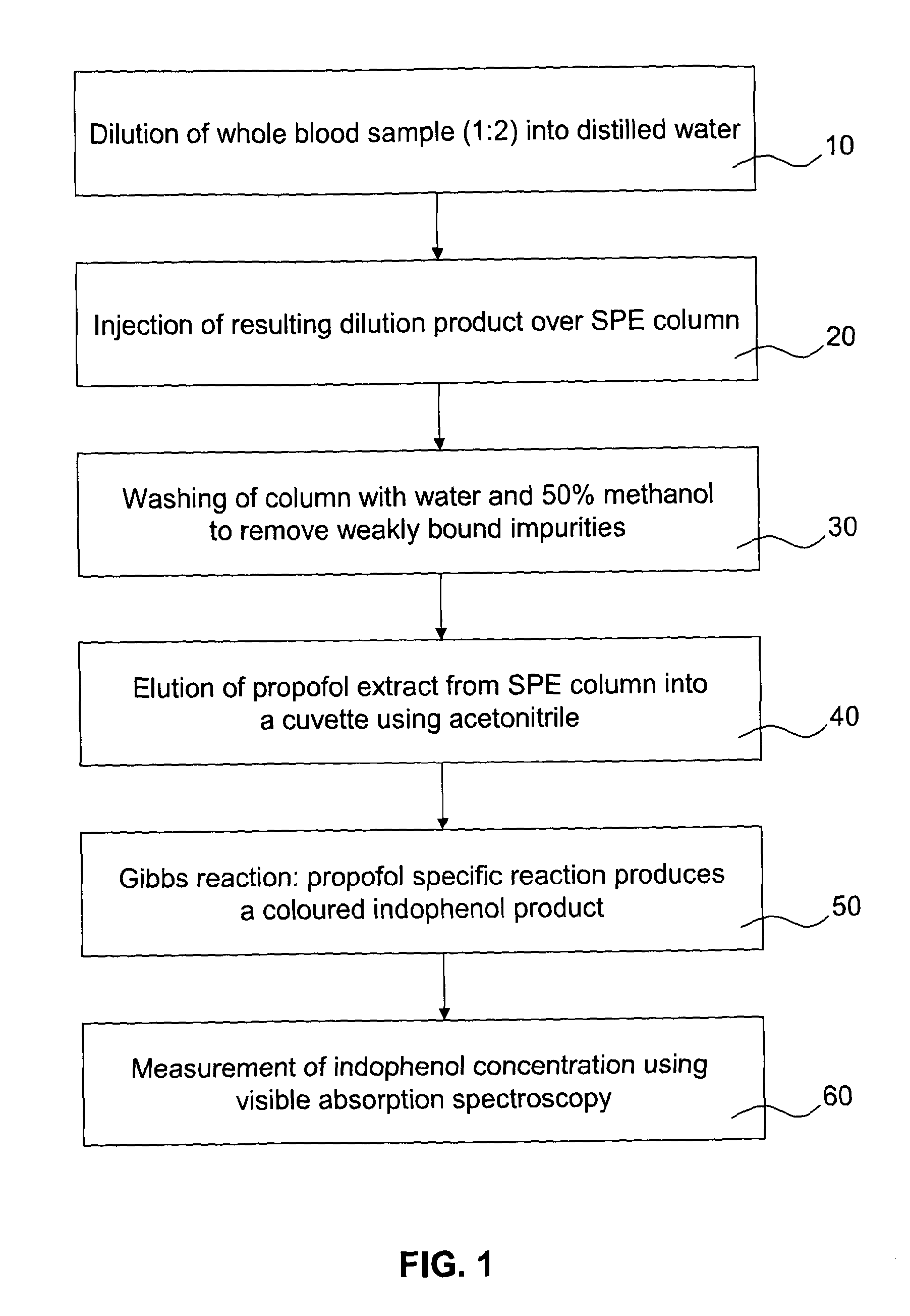
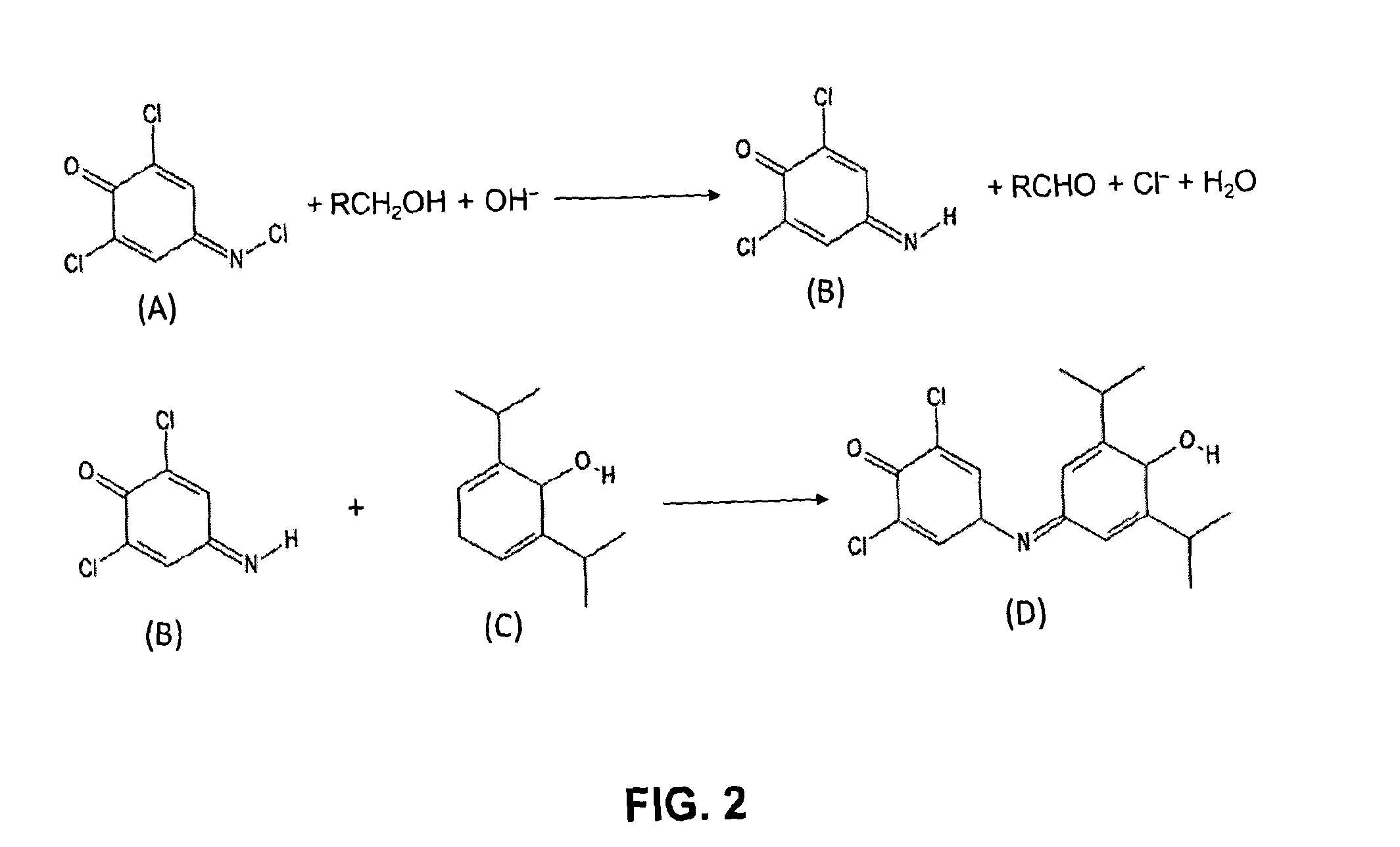
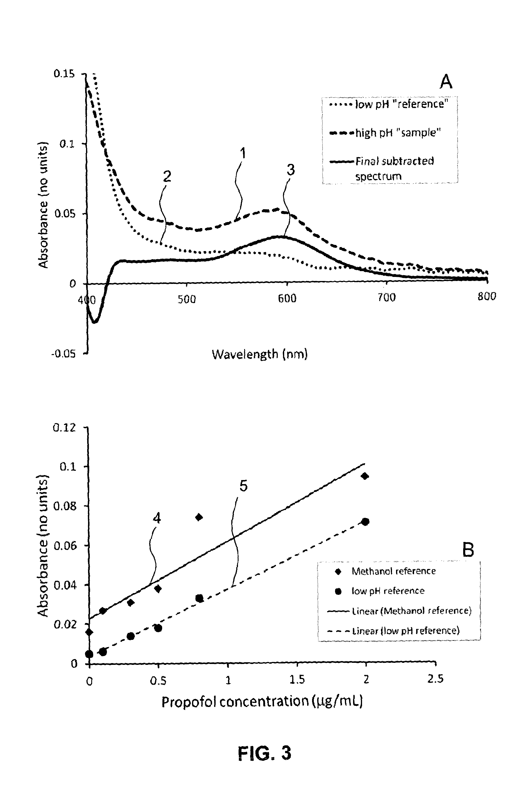
[0050]The present invention will be described in further detail by way of the following non-limiting example. In this example, the anaesthetic drug propofol is detected from whole blood using solid phase extraction (SPE) followed by a Gibbs reaction and detection by absorption spectroscopy. Established measurement protocols for low propofol concentrations rely on time consuming and complex HPLC-based assays. The low complexity and cost of the technique described in this example enables a propofol assay that can be performed in a near patient setting.

[0051]A blood sample (preferably 1 ml), which contains a known concentration propofol, is diluted 1:2 into water and then a known volume of this diluted sample, preferably 1.5 ml, is applied to a reverse-phase SPE column. The column is washed with (preferably (1.5 ml) of deionised water and (preferably 0.75 ml) of a 1:1 mixture of water and methanol to remove weakly bound impurities. The propofol extract is then eluted from the SPE colum...


PUM

PropertyMeasurementUnit
second wavelengthaaaaaaaaaa
wavelengthaaaaaaaaaa

Abstract

Disclosed is a method of determining the presence of an analyte of interest by means of a detection of a reaction product between the analyte of interest and a reactant, the method comprising extracting the analyte of interest from a complex sample matrix; transferring the analyte of interest to an initial reaction mixture; performing a background measurement on the initial reaction mixture comprising at most a negligible concentration of the reaction product, wherein the reaction conditions present in said initial reaction mixture at least reduce the reaction rate of the formation of the reaction product such that the background measurement can be performed without a measurable change in said negligible concentration; altering the reaction conditions in the initial reaction mixture to accelerate said reaction rate; continuing said reaction until the concentration of the reaction product has stabilized; performing a second measurement on the resultant reaction mixture to obtain a signal correlated to said concentration; and determining the presence of the analyte of interest from a difference between the background measurement and the second measurement. In a preferred embodiment, the analyte of interest is Propofol (2,6-di-isopropylphenol), and the reactant is the activated Gibbs reagent (2,6-dichloroquinoneimine).

Description

FIELD OF THE INVENTION[0001]The present invention relates to a method for improving the detection of analytes in complex sample matrices, in situations where a reaction step is required to generate a product, which is subsequently detected. In particular, this invention provides a means for selectively measuring the concentration of the anaesthetic drug propofol in whole blood.BACKGROUND OF THE INVENTION[0002]Modern healthcare relies extensively on a range of chemical and biochemical analytical tests on a variety of body fluids to enable diagnosis, therapy and management of disease. Medical and technological advances have considerably expanded the scope of diagnostic testing over the past few decades. Moreover, an increasing understanding of the human body, together with the emergence of technologies, such as microsystems and nanotechnology, are expected to have a profound impact on diagnostic technology.[0003]Increasingly, diagnostic tests in hospitals are carried out at the point-...

Claims


Application Information

Patent Timeline
Patent Type & Authority Patents(United States)
IPC IPC(8): G01N33/94G01N33/557G01N33/52G01N33/50
CPCG01N33/948G01N33/52G01N33/557Y10T436/203332Y10T436/14
Inventor PETTIGREW, DAVID MICHAELLAITENBERGER, PETER GEORGLIU, BO
Owner SPHERE MEDICAL
Why Eureka
Try Eureka