High throughput cell-based HPV immunoassays for diagnosis and screening of HPV-associated cancers

a cell-based, high-throughput technology, applied in the direction of instruments, biochemistry apparatus and processes, material analysis, etc., can solve the problems of low sensitivity and high false positive rate, and the inability to detect high-grade cancers in cervical biopsies. the effect of 33% to 50% of high-grade diseas

Inactive Publication Date: 2015-09-08
HEER MEDICAL TECH DEV CO LTD
View PDF123 Cites 0 Cited by
  • Summary
  • Abstract
  • Description
  • Claims
  • Application Information

AI Technical Summary

Benefits of technology

[0104]Attempts to detect the presence of HPV related antibodies in a human subject by ELISA (enzyme linked immunoabsorbent assays) generally lead to extremely low assay sensitivity and thus cannot be developed into a commercially suitable diagnostic test. Nindl, I., Benitez-Bribiesca, L., Berumen, J., N, F., Fisher, S., Gross, G., Lopez-Carillo, L., Muller, M., Tommasino, M., Vazquez-Curiel, A., and Gissmann, L. (1994) Antibodies against Linear and Conformational Epitopes of the Human Papillomavirus (HPV) Type 16 E6 and E7 Oncoproteins in Sera of Cervical Cancer Patients, Arch. Virol. 137, 341-353. Most of these ELISA assays target a single viral protein or short peptide fragments, which were not able to interact well or bind strongly and specifically to antibodies from the human subject. The assay specificity and sensitivity are so low such that even using samples from patients confirmed with HPV associated invasive cervical cancer, only 53% of the patient samples were found positive for HPV infection. Thus, there is no successful ELISA assay available as a diagnostic tool for clinic al samples.
[0106]The present invention provides a novel approach to viral oncoprotein testing, using a novel combination of tools that include novel antibodies, recombinantly-expressed HPV proteins including but not limited to E6, E7, L1, L2 and a whole-cell assay. The present invention enables the first whole-cell ELISA for detecting intracellular HPV oncoprotein in clinical samples. This whole-cell ELISA enables the detection of HPV oncoproteins as biomarkers for true precancers in the diagnosis and screening of cervical cancer and other HPV associated cancers.
[0107]Recombinant HPV E6 and antibody development. Development of E6 antibodies have been hampered by the difficulty of obtaining E6 oncoprotein, which is prone to aggregation. Furthermore, detection of E6 protein in clinical samples is difficult due to sample collection conditions which produce a protein conformational change. We have overcome the technical barriers to produce HPV E6 recombinant protein in a non-denatured, soluble form and have developed highly specific antibodies against HPV E6 oncoproteins. The present invention provides pan anti-HPV E6 antibodies, i.e. single antibodies capable of detecting most prevalent high-risk HPV types in fixed cells. The same method was used to develop recombinant HPV E7 protein and antibody to obtain pan anti-HPV E7 antibodies. The same method was also used to develop recombinant HPV L1 protein and recombinant HPV L2 protein, and antibody to obtain pan anti-HPV L1 and pan anti-HPV L2 antibodies.
[0108]Whole cell assay approach. We have also developed IHC and ICC assays to detect E6, E7, and L1 proteins using these types of antibody in fixed clinical samples. Our IHC and ICC study on cervical tissues and exfoliated cervical cytological samples demonstrate the utility of these antibody types that are capable of detecting E6, E7, and L1 proteins in clinical samples. To avoid invasive procedure for biopsy, and to avoid the subjectivity and variability of interpretation of morphology based staining and IHC / ICC results, we developed an objective platform based on whole-cell ELISA, to provide a robust tool for cervical cancer screening. The HPV E6, E7, and L1 whole-cell ELISAs respectively measure intracellular E6, E7, and L1 proteins objectively in a high throughput assay and enable the detection of HPV oncoproteins as biomarkers for true precancers in the diagnosis and screening of cervical cancer and other HPV associated cancers. These assays require no cell lysis, avoid use of detergent that may result in a change of the protein conformation in the process of protein extraction, and enable direct detection of native E6, E7, and L1 proteins in cervical cells. The assays provide an objective test to identify patients with high-grade precursor, thus avoids unnecessary invasive procedure for colposcopy and biopsy. This ELISA format uses more specific and clinical relevant biomarkers to provide an objective, more sensitive, robust, and high throughput test that can be automated to be suitable for routine cervical cancer screening and will provide a binary (presence or absence) or semi-quantitative measurement of E6, E7, and L1 proteins.
[0109]At present, there are no commercially-available immunological assays to clinically measure the presence of HPV-associated proteins or antibodies. Embodiments of the invention thus provide a diagnostic tool useful for diagnosis of HPV infection, dysplasia, and HPV related cervical cancer. In addition, the results from the immunological assays as described herein can be used to compare with other commercially-available immunological assays specifically-designed for p53 and RB. It is known that infection high risk type HPVs, such as HPV-16 and HPV-18 may cause cervical cancer due to the expression of E6 and E7, the viral oncoproteins that induce cervical cell malignancy and alter / reduce the expression of p53 and RB endogenous proteins of the host cells, leading to cellular dysfunction and ultimately carcinoma. Thus, it is contemplated to compare the assays results on the levels of all of these proteins altered by HPV infection perform on clinical samples, e.g., cervical tissues, body fluids, serum, etc., from the same human subjects.

Problems solved by technology

The test is subjective with significant inter-observer variability, and is limited by low sensitivity and high false positive results.
Although colposcopic biopsy has historically been considered the gold standard, recent reports indicate that cervical biopsies may miss 33% to 50% of high-grade disease because of sampling or diagnostic errors.
It therefore may be difficult to differentiate between false positive cervical cytology results and false-negative biopsy results.
These lesions, preferentially observed in women aged 35-40 yrs, are associated with a high risk of progression toward invasive cervical cancer.
However, LSIL does not invariably progress to HSIL in women infected with a high-risk HPV strain.
Nucleic acid-based HPV detection assays have been developed, but are not ideal for prognosing disease risk, in view of high cost, assay operation procedures, the requirements for facility, equipment, and highly trained personnel, and low positive predictive value for CIN.
Current DNA-based assays cannot differentiate LSIL from HSIL, nor CIN lesions from non-transforming latent or remissive viral infection.
Known protocols for the production of monoclonal antibodies to HPV are generally unsuitable for the production of anti-HPV monoclonal antibodies and cannot be used in immunocytochemical diagnostic tests for screening general human population.
Other attempts to detect the presence of HPV related antibodies or viral proteins in a human subject by ELISA (enzyme linked immunoabsorbent assays) also generally lead to extremely low assay sensitivity and thus cannot be developed into a commercially suitable diagnostic test.
Most of these ELISA assays target a single viral protein or short peptide fragments, which are not able to interact well or bind strongly and specifically to antibodies from the human subject.
Given the testing populations come from general screening, with or without low grade, or precancerous lesions, the sensitivity of the assay will be too low to apply for clinical practice.
Thus, there is no successful ELISA assay available as a diagnostic tool for clinical samples.
However, some progressive cervical cancer cases are related to infection by low risk HPV types, while infection of some HPV types will never progress into cervical cancer.
However, prior art antibodies have limited utility for detecting E6 / E7 oncoprotein in situ.

Method used


Image

  • High throughput cell-based HPV immunoassays for diagnosis and screening of HPV-associated cancers
  • High throughput cell-based HPV immunoassays for diagnosis and screening of HPV-associated cancers
  • High throughput cell-based HPV immunoassays for diagnosis and screening of HPV-associated cancers

Examples


example 1

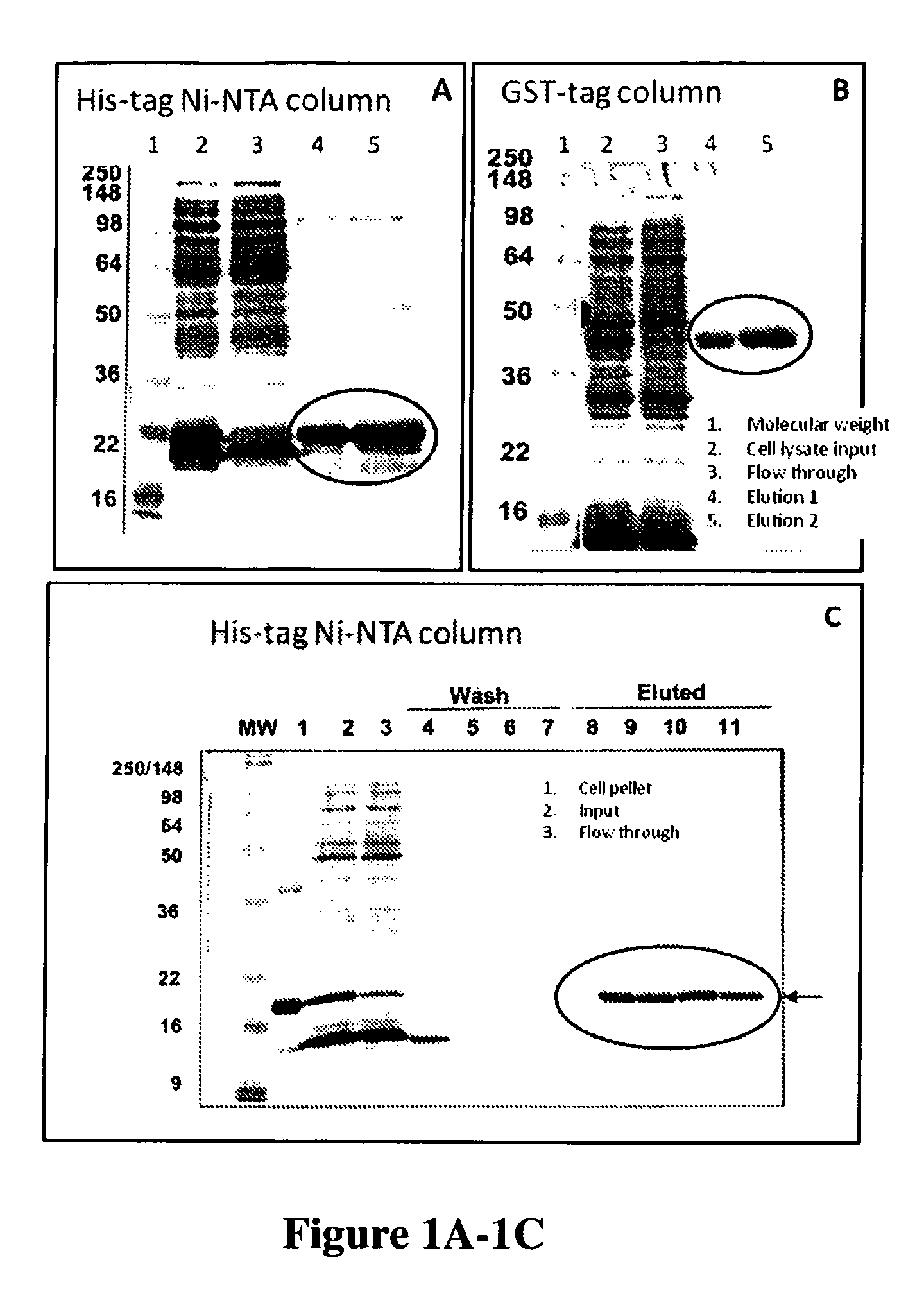
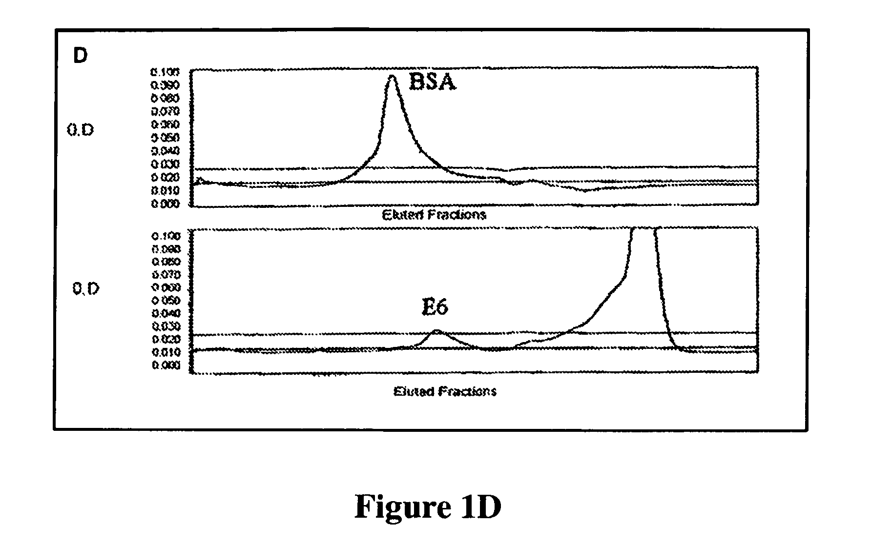
HPV Recombinant Protein Expression, Purification, and Preparation to be Used as Immunogens for Generating Antiserum, and Screening for Monoclonal Antibody from Hybridoma Cell Lines

[0158]HPV recombinant proteins can be any kinds of HPV proteins, HPV proteins of early genes and / or late genes, including, but not limited to, E2, E6, E7, L1, L2 and can be from various HPV types. One aspect of the invention provides recombinant proteins, such as recombinant hybrid proteins containing a partial sequence or a full length sequence of HPV oncogenic proteins. For example, full-length E6, E7, and / or L1 polypeptide sequence, which have been found very difficult to obtain and purify due to undesirable aggregation during protein purification, protein instability, low levels of expression, low immunogenic responses of purified proteins. For example, many early E6 oncoproteins contain many cysteine amino acids and thus the correct topography of the E6 oncoproteins requires formation of many disulfid...

example 2

Anti-HPV Antibody Preparation

[0167]Recombinant HPV proteins produced using the techniques as described in Example 1 were used as immunogens for generating antiserum, polyclonal antibodies, and monoclonal antibodies.

[0168]The basic techniques for cloning and for conducting the immunological assays can be found in “Antibodies: A Laboratory Manual”, Harlow and Lane, Cold Spring Harbor Laboratory, Cold Spring Harbor, N.Y. 1989; “Molecular Cloning”, A Laboratory Manual, eds. Sambrook, Fritsch and Maniatis, Cold Spring Harbor Laboratory Press, 1989, and others books and manuals known in the art. Details of our procedures for antibody production the characterization of the produced antibodies, and certain assays are described in co-owned U.S.

[0169]U.S. Pat. No. 8,865,162, filed on Jun. 10, 2009, titled “Novel Monoclonal Antibodies against HPV Protein,” U.S. Pat. No. 8,859,218, filed on Jun. 10, 2009, titled “In situ Detection of Early Stages and Late Stages HPV Infection,” U.S. Pre-Grant P...

example 2.6

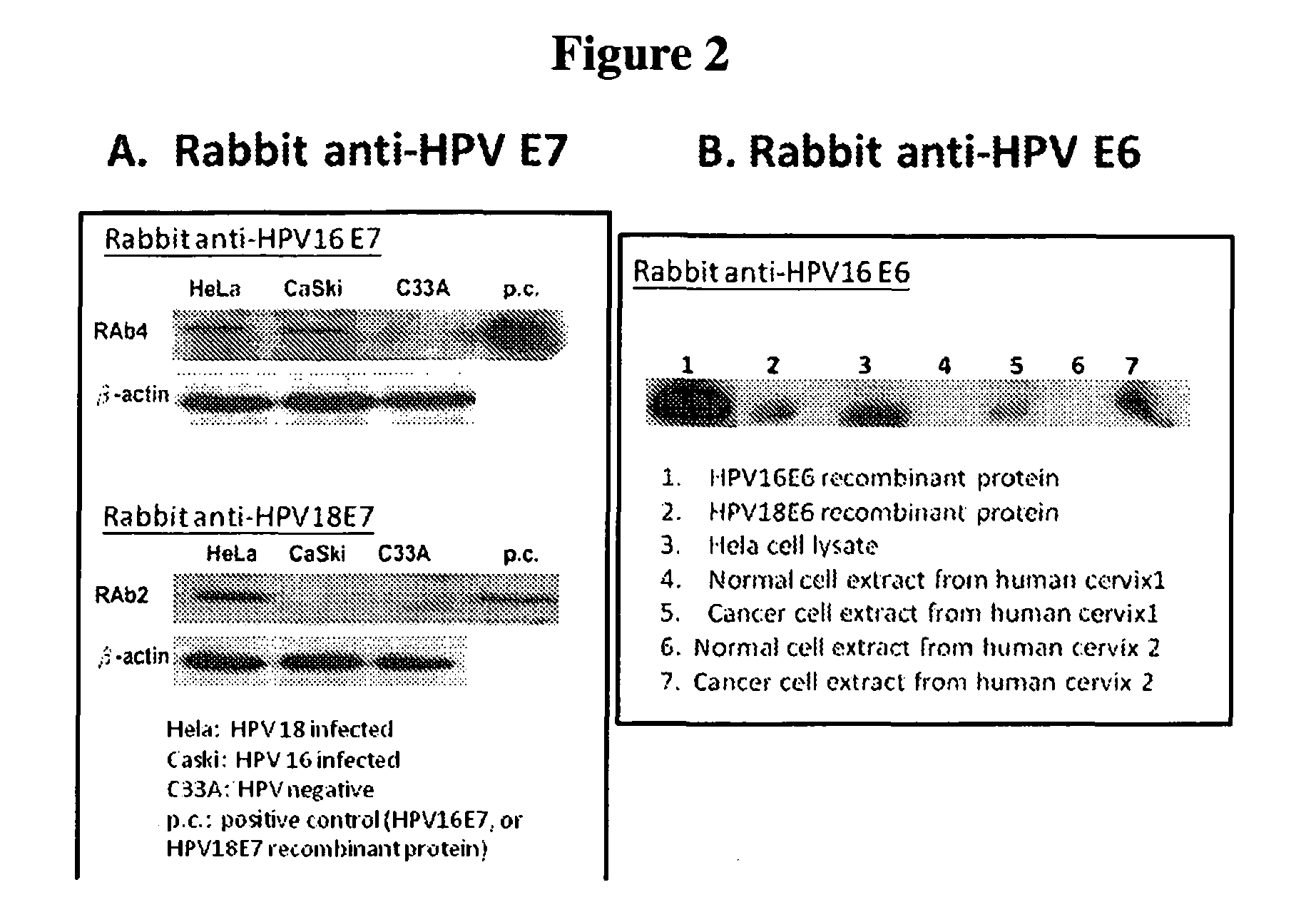
The Specificity of Anti-HPV Antibodies

[0171]One or more immunological assays can be used to test the specificity of the monoclonal antibodies generated by screening the hybridoma cell lines with two or more HPV recombinant proteins. EIA (Enzyme Immuno Assay) and / or Western blots were used as the assay format to test the specificity of the HPV antibodies described herein. Various purified recombinant HPV proteins, including the original screening proteins used for obtaining the anti-HPV antibodies and other proteins not used for screening, were used to coat on the microtiter plate to test the specificity of the obtained anti-HPV antibodies on EIA. Proteins in cell lysate from cervical cancer cell lines (with or without HPV infection) were also used to test the specificity of the anti-HPV antibodies by western blot. To confirm the binding and reactivity of the HPV antibodies with proteins from HPV infected cell lines, western blot is very useful to demonstrate specific protein bands c...


PUM

PropertyMeasurementUnit
pore sizeaaaaaaaaaa
dissociation constantaaaaaaaaaa
concentrationaaaaaaaaaa

Abstract

Methods for quantifying an HPV protein expression in a clinical sample are disclosed. The quantifying methods include the process for obtaining the clinical sample. Such a clinical sample is consisted of a population of cells that are susceptible to infection by an HPV. The quantifying methods also include the process for depositing the clinical sample into a container. The clinical sample is contacted with the first antibody that specifically binds to an HPV protein which is expressed by an HPV-infected cell under a condition that promotes specific binding of the first antibody to the HPV protein expressed by the population of cells. The methods further include the process for quantifying the specific binding of the first antibody and thereby quantifying the HPV protein expression in the clinical sample. The assay provides an objective test to identify patients with high-grade precursor from cytology samples before biopsy.

Description

CROSS-REFERENCE TO RELATED APPLICATIONS[0001]This application claims the benefit of U.S. Prov. App. Ser. No. 61 / 335,540, filed Jan. 8, 2010, titled “High throughput cell-based HPV immunoassays for diagnosis and screening of HPV associated cancers,” the contents of which are hereby incorporated by reference for all purposes.BACKGROUND OF THE INVENTION[0002]Cervical cancer is the second most common cause of cancer deaths in women worldwide, with an incidence about a half million new cases cumulatively resulting in about a quarter of a million deaths every year. In the US, cervical cancer mortality rate has decreased substantially as a result of cervical cancer screening programs that detect precancerous conditions so they can be treated before developing into cancer. The current paradigm for cervical cancer screening is based on the Pap test, a cytologically-based test of cells scraped from the cervix and examined microscopically to detect changes indicating dysplastic cell growth. Th...

Claims


Application Information

Patent Timeline
Patent Type & Authority Patents(United States)
IPC IPC(8): C12Q1/70G01N33/53G01N33/574G01N33/569
CPCG01N33/57411G01N33/56983G01N2333/025
Inventor CHENG, SHULING
Owner HEER MEDICAL TECH DEV CO LTD
Why Eureka
Try Eureka