

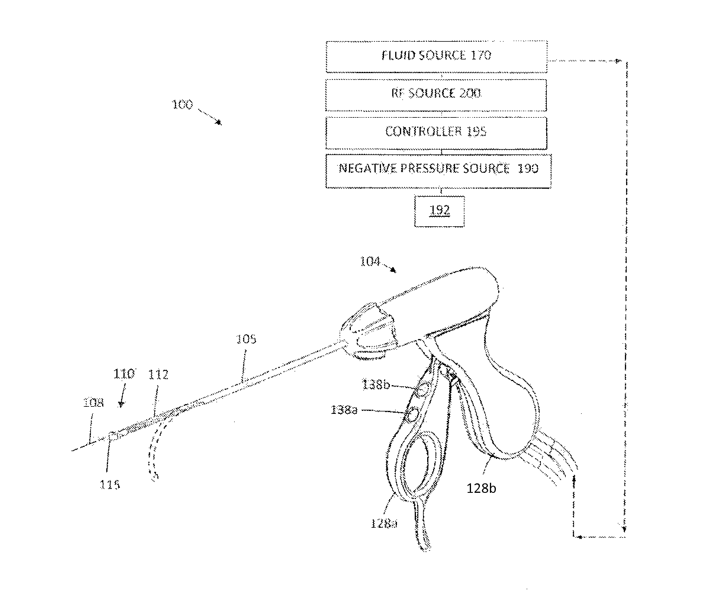
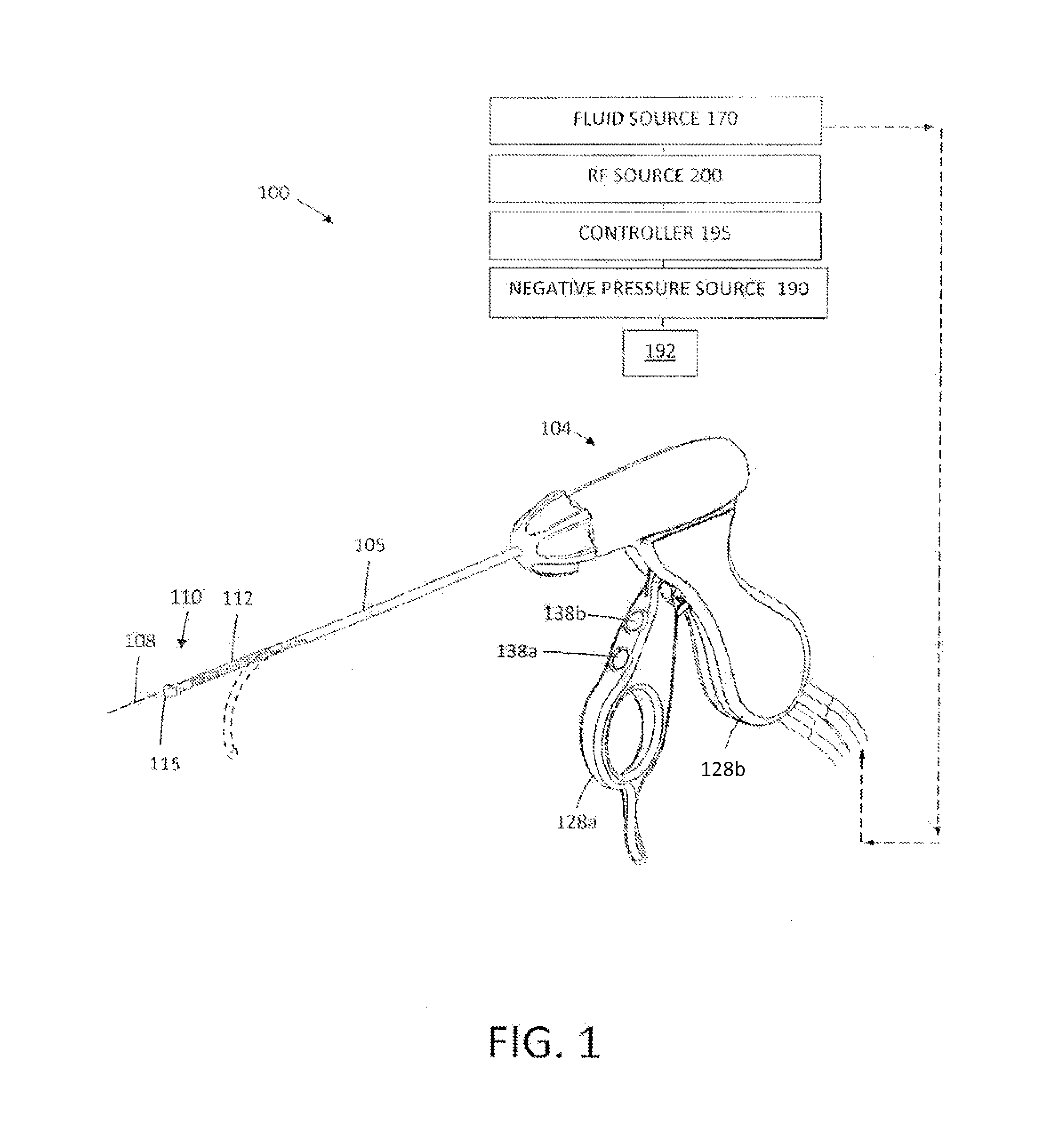
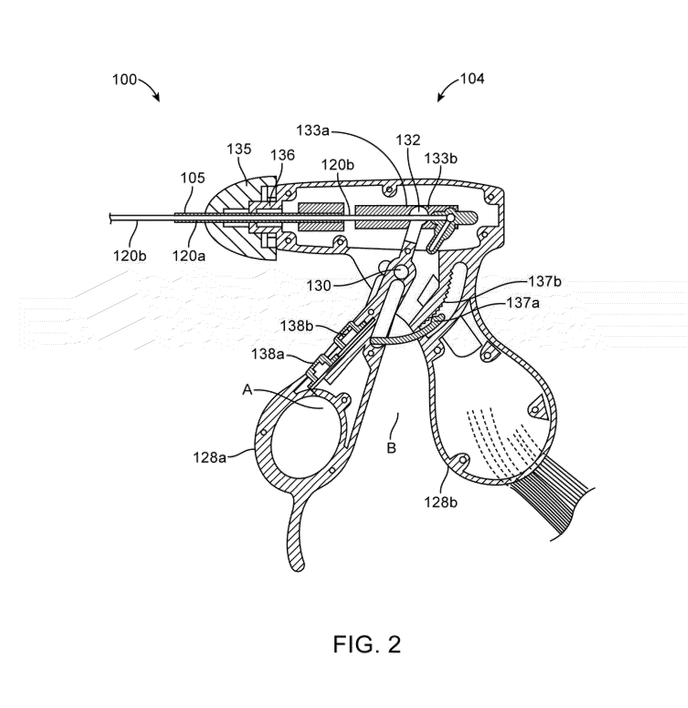
Medical ablation system and method of use
a technology of medical ablation and ablation surface, which is applied in the field of medical instruments and systems, can solve the problems of inability of rf and laser devices to provide smooth surfaces for cartilage ablating surfaces, and inability to provide cartilage tissue thermal damage,
- Summary
- Abstract
- Description
- Claims
- Application Information
AI Technical Summary
Benefits of technology
Problems solved by technology
Method used
Image

Examples
embodiment 680
[0081]FIG. 19 illustrates another system and working end embodiment 680 which is similar to that of FIGS. 16A-16B except that this embodiment includes a further inflow channel 682 for delivering a saline distention fluid through probe and outwardly from the working end. In this embodiment, another internal saline flow is provided in the paths as previously described circulating through inflow channel 622A, loop channel portion 630 and outflow channel 622B. The tissue extraction channel 640 is again as described previously. The variation in FIG. 19 has first and second negative pressure sources 635 and 635′ independently coupled to channel 622B and 640. The distension fluid channels 682 in FIG. 19 is operatively coupled to a second positive pressure source 625′ which provides the saline distention fluid. In the working end 680 of FIG. 19, the opposing polarity electrodes 650A and 685 are disposed entirely within the interior of the device. In one embodiment, the first polarity electr...
embodiment 800
[0089]FIG. 22 depicts another working end embodiment 800′ that similar to that of FIGS. 21A-21B except that electrode 810′ spans across the aspiration opening 644 and is fixed and not moveable. Again, the third electrode 810′ can cooperate with electrode 815 in disposed in the aspiration channel 640. It should be appreciated that the fixed electrode 810′ can comprise and single element or can comprise a plurality of electrode elements, a mesh or the like. As can be seen in FIG. 22, the system includes switch mechanisms 818 for operating the electrode arrangement to create the first plasma, the second plasma or both. In FIG. 22, the third electrode 810′ is somewhat recessed in port 644 but it should be appreciated that the electrode can be recessed in port 644, flush with the working surface or the electrode can project outward of the surface.
[0090]FIG. 23 illustrates another embodiment of working end 820 that is configured for providing first and second plasmas as described previous...
PUM
 Abstract
Description
Claims
Application Information