
Devices for securely closing tissue openings with minimized scarring

a tissue opening and tissue technology, applied in the field of tissue opening closure devices, can solve the problems of traumatizing and compromising the integrity of the tissue opening and the nutrient blood supply to the healing tissue edges, exposing the surgeon, the patient, and the surgeon, and achieving the effect of improving wound car

Active Publication Date: 2016-04-05
DERMACLIP US
View PDF58 Cites 17 Cited by
  • Summary
  • Abstract
  • Description
  • Claims
  • Application Information

AI Technical Summary

Benefits of technology

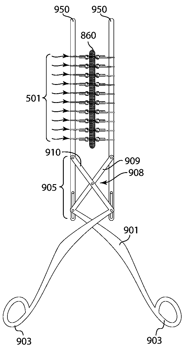
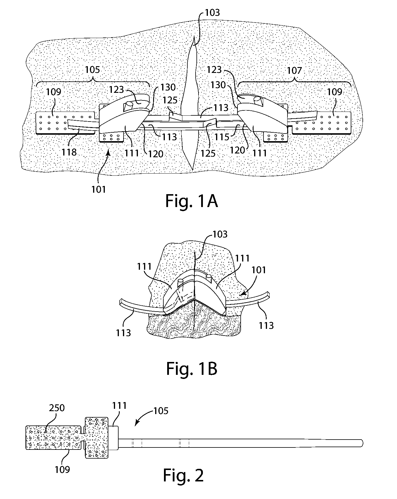
[0007]The invention is directed to devices, systems and methods for closing surgical incisions and non-surgical wounds that provide for improved wound care. In accordance with an embodiment of the invention, a tissue closure device for non-invasively closing a tissue opening includes an assembled pair of substantially identical closure components. Each component includes a tissue attachment base with an attachment mechanism on a first side thereof to affix the attachment base to the skin. A standoff assembly is mounted on a second side of the attachment base of each component. A toothed pull-tab, having first and second ends, is coupled to the attachment base of each component through the standoff assembly and defines a longitudinal axis. The standoff assembly has a forward face, to which the first end of the pull-tab is affixed, and an opposed rearward face, the forward face including a sloped portion that is sloped rearward as it approaches the attachment base. Each component further includes a female connective element, mounted on the second side of the attachment base, and which is built into the standoff assembly, that receives and engages the pull-tab from the other component and into which the pull-tab from the other component has been placed. Each pull-tab has a length sufficient to reach and be received by the female connective element of the other closure component. The second end of the pull-tab protrudes from the female connective element and defines an opening configured so as to releasably receive one of a corresponding pair of arms of an applicator tool, such that the device can be loaded onto the applicator tool in a position that straddles the arms of the applicator tool. When the loaded assembled pair is affixed to the skin with each of the attachment bases disposed on an opposite side of the tissue opening, the applicator tool can be manipulated to adjust the spacing between the arms and causes the second ends of the pull-tabs to be pulled away from each other. This brings the sloped portions of the forward faces of the components into substantial contact with one another at a location of contact, causing each standoff assembly to pivot about the location of contact, inducing eversion of tissue edges that defined the opening.
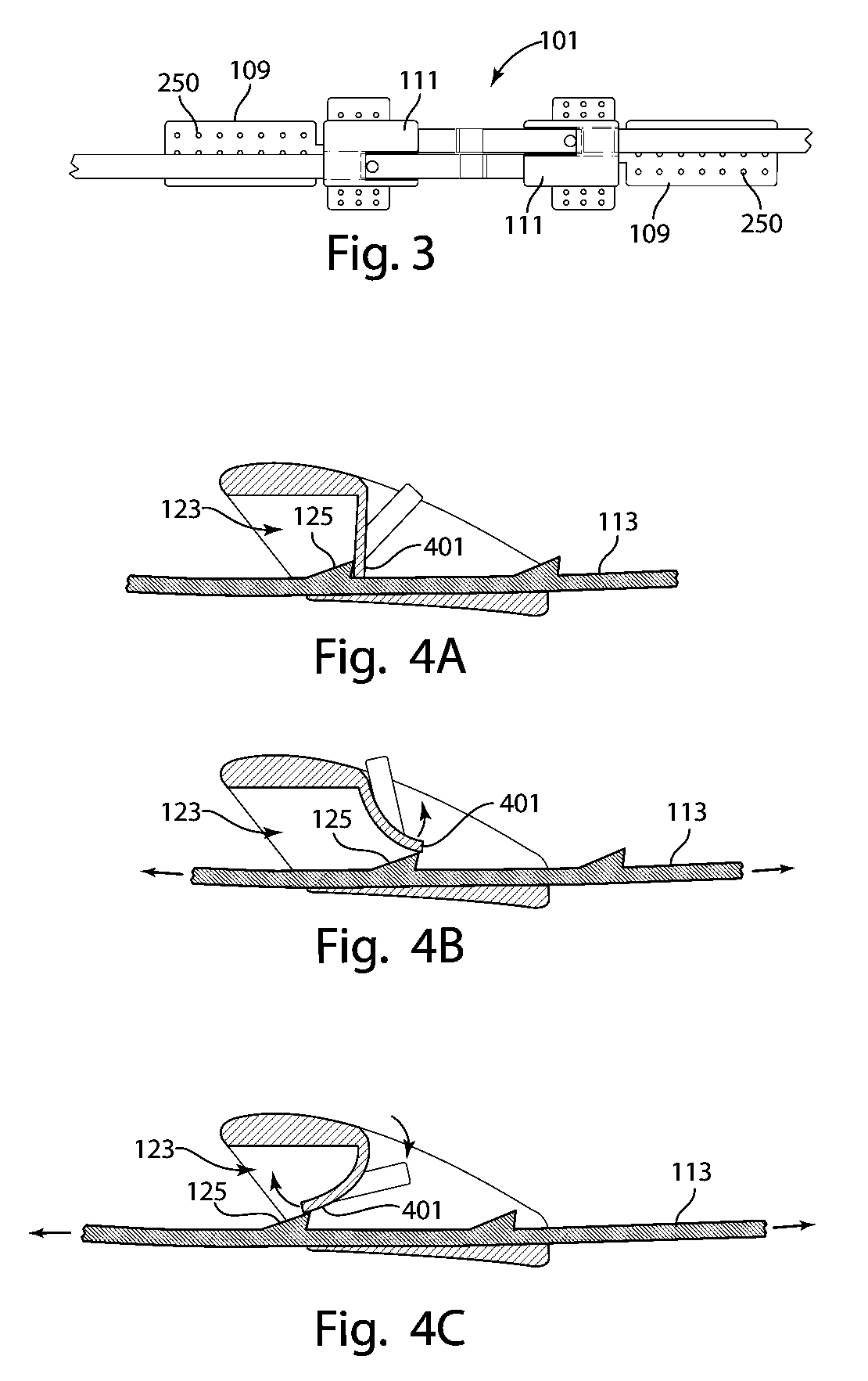
[0010]In accordance with further embodiments of the invention, a female connective element of a tissue closure device may have a releasable tab-locking mechanism configured to releasably lock the pull-tab in position. The releasable tab locking mechanism may include a releasable finger having a tip that engages against such pull-tab. The finger may have an unreleased position wherein the tip engages against such pull-tab and a released position wherein the pull-tab can be freely moved, the finger being biased in the unreleased position. In various embodiments, a ratchet / piston locking mechanism may be provided that allows for precise closure and locking of the paired components. By depressing the piston, the locking mechanism is securely unlatched. The releasable tab-locking mechanism may be manipulated by hand, or may require a tool, for example, tweezers or a needle. More particularly, in various embodiments, the tab-locking mechanism may include a hollow recess. A tool, for example a needle, can be used to unlock the locking mechanism, for example by engaging the end of the needle into the hollow recess of the locking mechanism, applying force to the locking mechanism through the tool, and with that force unlocking the locking mechanism. The releasable tab-locking mechanism allows for drainage of wound edema and / or the treatment of infection, without removal and reapplication of the device. Such a feature can also be helpful when a wound needs to be temporarily left open to facilitate healing.

Problems solved by technology

Both of these procedures are invasive, which can traumatize and compromise the integrity of the tissue opening and the nutrient blood supply to the healing tissue edges.
They cause pain, increase the possibility of infection, expose the surgeon, as well as the patient, to blood-borne disease, leave behind scars, and require a follow-up visit for suture or staple removal.
Surgical glue is also used, but has only been proven adequate for small wounds where skin edges are not widely separated or under tension during closure.
Suturing even by a skilled surgeon punctures and stresses tissue causing scarring.
Unfortunately, skin edge eversion and apposition is difficult to achieve with simple stitches or glues.
More complex stitches and devices that promote adequate eversion and apposition are time consuming to place and are used in less than one case in one hundred.
Additionally, conventional tissue closure devices for tissue do not allow for the breathing and the egress of tissue fluids.
Often, the durability of the applied tissue device is compromised by the impervious barrier to moisture and other substances beneath polymer components.
This may lead to dislodgement of the tissue device.

Method used


Image

  • Devices for securely closing tissue openings with minimized scarring
  • Devices for securely closing tissue openings with minimized scarring
  • Devices for securely closing tissue openings with minimized scarring

Examples


Embodiment Construction

[0007]The invention is directed to devices, systems and methods for closing surgical incisions and non-surgical wounds that provide for improved wound care. In accordance with an embodiment of the invention, a tissue closure device for non-invasively closing a tissue opening includes an assembled pair of substantially identical closure components. Each component includes a tissue attachment base with an attachment mechanism on a first side thereof to affix the attachment base to the skin. A standoff assembly is mounted on a second side of the attachment base of each component. A toothed pull-tab, having first and second ends, is coupled to the attachment base of each component through the standoff assembly and defines a longitudinal axis. The standoff assembly has a forward face, to which the first end of the pull-tab is affixed, and an opposed rearward face, the forward face including a sloped portion that is sloped rearward as it approaches the attachment base. Each component furt...


PUM


Abstract

Devices, tools, systems, kits and methods for closing a tissue opening non-invasively are presented. The devices include a tissue closure device that reliably induces eversion of tissue edges that define a tissue opening. The devices may be used in combination with an applicator tool to provide easy and uniform apposition of the tissue closure devices across the tissue opening. Further embodiments provide for releasable locking of the tissue closure devices, allowing for drainage of wound edema and / or treatment of infection, without removal and reapplication of the devices. Still further embodiments allow for the passing of moisture or other substances that may collect under a tissue closure device.

Description

CROSS-REFERENCE TO RELATED APPLICATIONS[0001]The present application is a continuation of Patent Cooperation Treaty Application PCT / US2012 / 036265, filed May 3, 2012, which in turn claims the benefit of U.S. Provisional Patent Application Ser. No. 61 / 481,986, entitled “Devices for Securely Closing Tissue Openings with No Scarring,” filed May 3, 2011, each of which are hereby incorporated herein by reference in its entirety.TECHNICAL FIELD[0002]This invention is directed to tissue closure devices, tools, systems, kits, and methods for tissue repair and closure. In particular, the invention is directed to devices and methods that safely and securely close wounded tissue openings and permit and encourage healing to occur.BACKGROUND ART[0003]Closure of tissue openings, such as, for example, for surgical incisions and accidental lacerations or wounds, is critical both to minimize the risk of infection and to promote optimal healing of the wound or incision. Both of these outcomes require ...

Claims


Application Information

Patent Timeline
Patent Type & Authority Patents(United States)
IPC IPC(8): A61B17/08
CPCA61B17/08A61B2017/081A61B2017/086A61B17/085A61B2017/00004
Inventor FOX, ANDREW D.
Owner DERMACLIP US
Learn More