Method and apparatus for minimally invasive treatment of unstable pelvic ring injuries combined with hip arthroplasty
a technology of unstable pelvis and hip joint, applied in the field of methods and equipment for the treatment of unstable pelvic fractures, can solve the problems of acute pelvic fractures that are potentially lethal, unstable pelvic fractures that are typically occurring, and the type of fractures tend to be more complex, so as to avoid any potential compressive complications
- Summary
- Abstract
- Description
- Claims
- Application Information
AI Technical Summary
Benefits of technology
Problems solved by technology
Method used
Image
Examples
first embodiment
Surgical Technique First Embodiment
[0034]The patient may be positioned in the supine position on a radiolucent table. The skin may be prepped and draped from above the umbilicus to the proximal thigh. The lower extremity may be prepped into the field as well to facilitate reduction techniques.
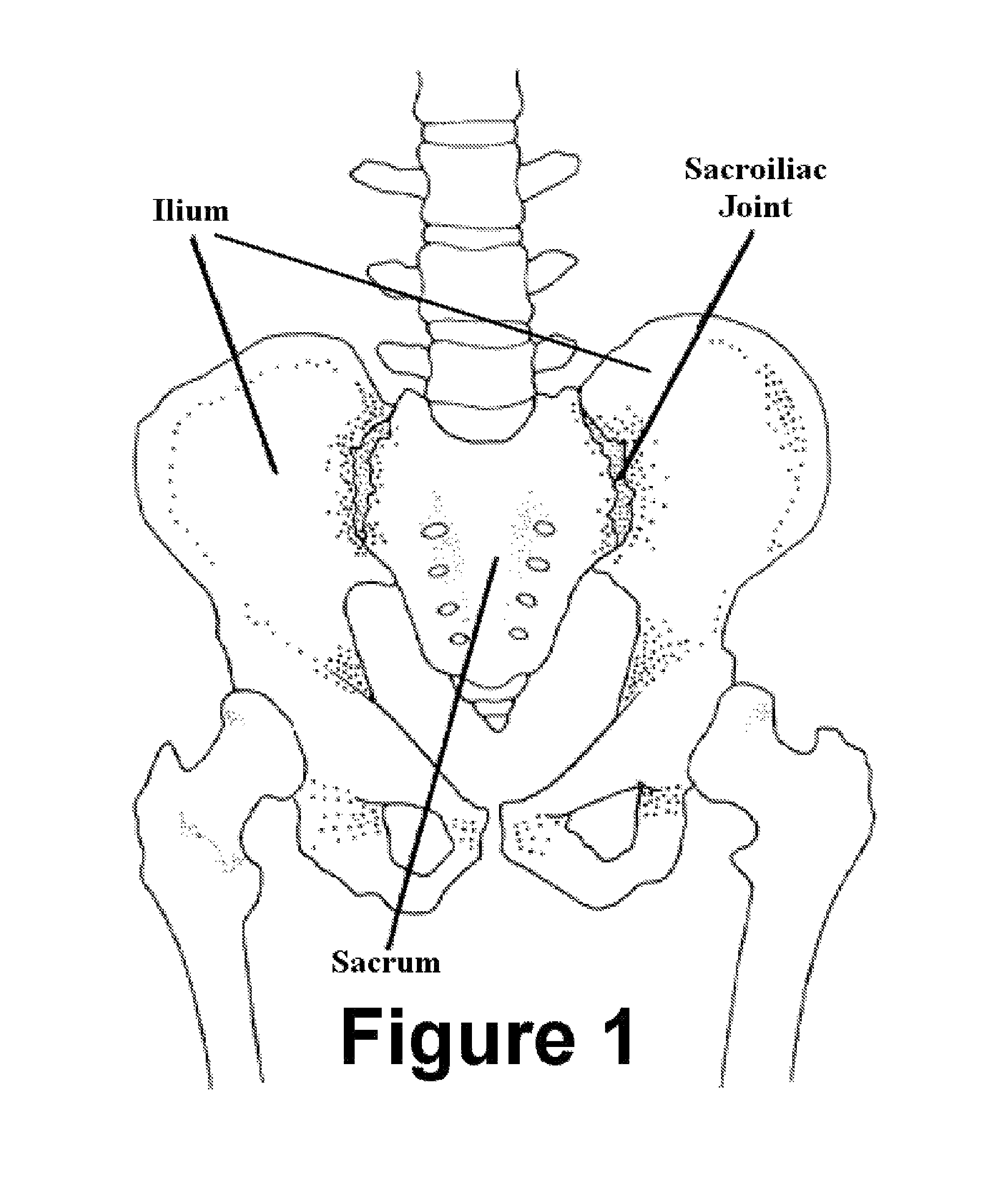
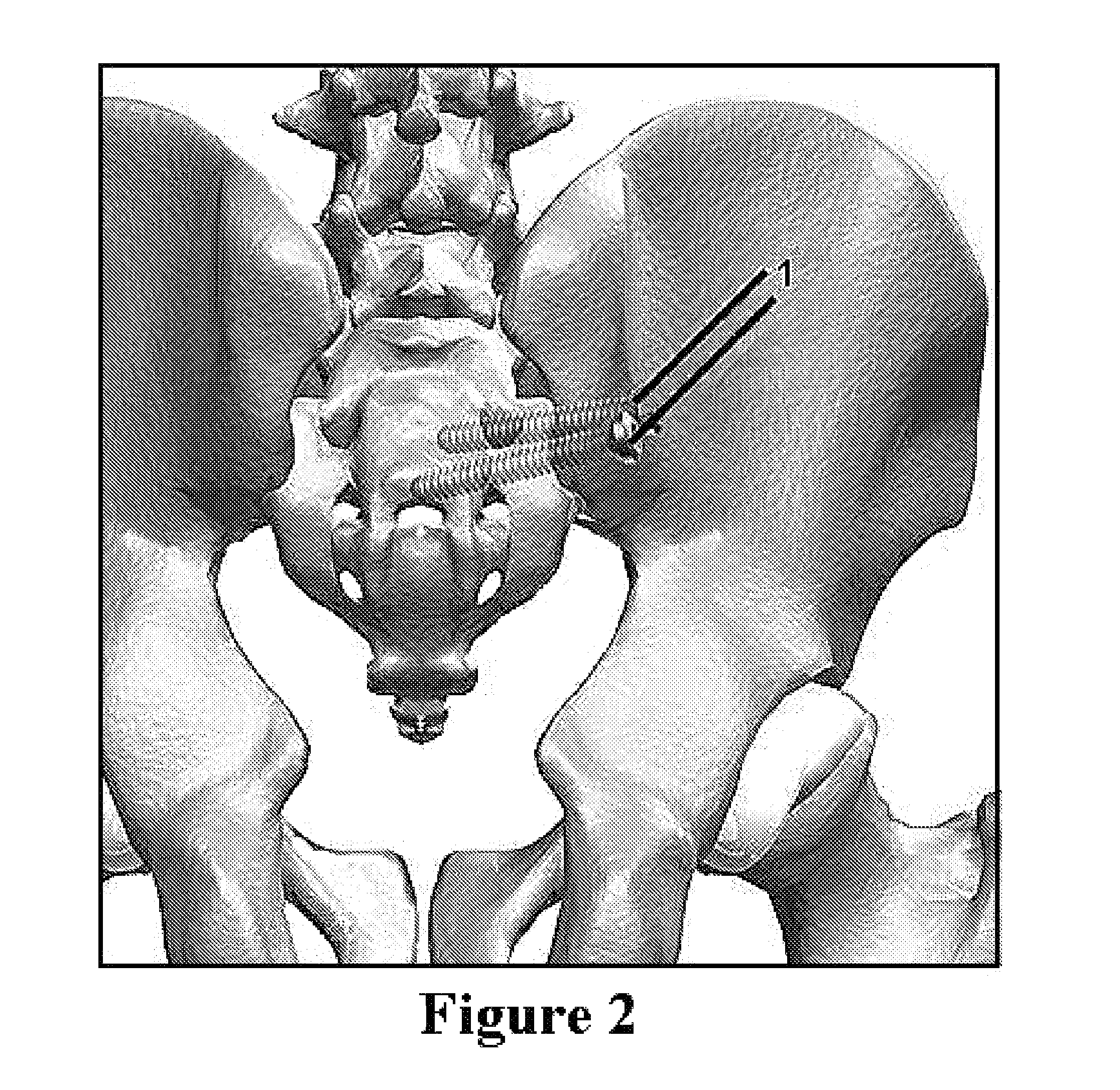
[0035]The posterior instability may be addressed first. The procedure for placement of iliosacral screws for posterior pelvic instability has been well described and will not be discussed here. See for example “CT-Guided Iliosacral Screw Placement: Technique and Clinical Experience” by Robert L. Sciulli, et al., American Journal of Roentgenology 2007; 188:W181-W192 (reproduced at http: / / www.ajronline.org / cgi / content / full / 188 / 2 / W181). FIG. 2 illustrates the way in which iliosacral screws 1 are inserted through the rear of the ilium and into the sacrum, thus stabilizing the posterior instability. FIG. 3 depicts typical iliosacral screws 1.
[0036]After stabilizing the posterior elements via the ili...
second embodiment
Surgical Technique Second Embodiment
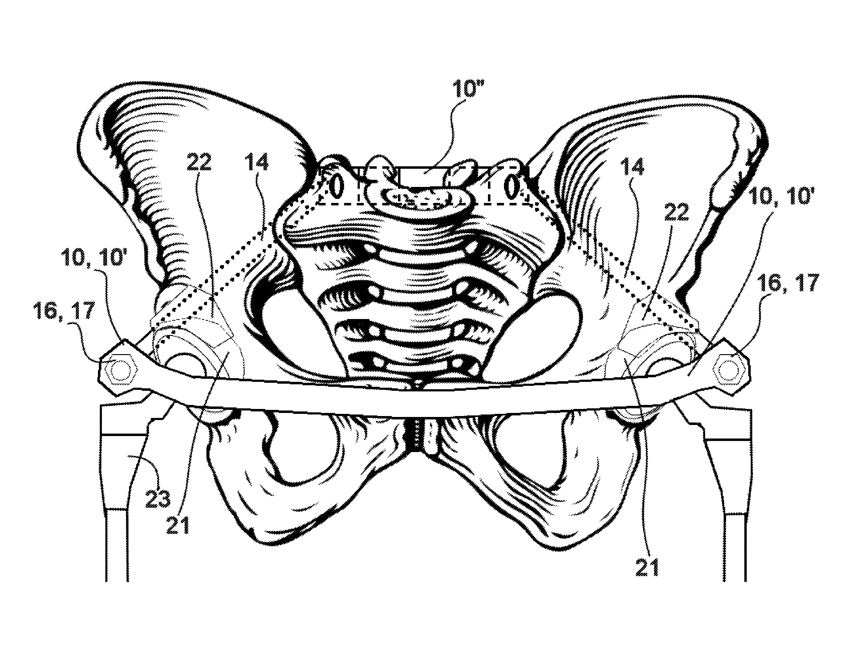
[0053]In an alternative embodiment, an anteriorly bowed subcutaneous elongated plate is used in place of the bowed rod. The elongated plate 10 is depicted in FIG. 8a. The plate 10 has at least one hole 11 in each end to accommodate the fixation means which affix the plate to the pelvis. Preferably the at least one hole 11 may be an elongated slot. More preferably the elongated slot is open on one end as shown in FIG. 8a. The elongated plate 10 may be bowed in a continuous arc or may have bends 13 (see FIG. 8b) to give the plate an approximation of an arc shape. The elongated plate 10 may be bent by the surgeon before insertion into the patient or may come pre-bent in a kit along with the fixation means. The pre-bent elongated plate 10 may come in a variety of lengths. It is believed that a most patients needs can be addressed using one size from a group of only three different sizes. The elongated plate 10 has a length much greater that its width ...
third embodiment
Surgical Technique Third Embodiment
[0062]In yet another embodiment, an anteriorly bowed subcutaneous elongated plate is used in place of the bowed rod and a second plate is used on the posterior in place of the iliosacral screws. The two plates are shown in FIGS. 11a-11d. The side view and top view of the posterior plate 10″ are depicted in FIGS. 11a and 11b. The posterior plate 10″ has at least two holes 11 in each end thereof to accommodate the fixation means which affix the plate to the pelvis and the sacrum. The outermost holes 11 will be used to affix the posterior plate 10″ to the ilia of the pelvis. The innermost holes 11 will be used to affix the posterior plate 10″ to the sacrum. The posterior plate 10″ has bends 13 to give the plate a shape that allows for fixation to the ilia of the pelvis and the sacrum, wile pressing against the posterior of the sacrum.
[0063]The side view and top view of the elongated anterior plate 10′ are depicted in FIGS. 11c and 11d. The elongated a...
PUM
 Abstract
Description
Claims
Application Information
 - R&D
- Intellectual Property
- Life Sciences
- Materials
- Tech Scout
- Unparalleled Data Quality
- Higher Quality Content
- 60% Fewer Hallucinations
Browse by: Latest US Patents, China's latest patents, Technical Efficacy Thesaurus, Application Domain, Technology Topic, Popular Technical Reports.
© 2025 PatSnap. All rights reserved.Legal|Privacy policy|Modern Slavery Act Transparency Statement|Sitemap|About US| Contact US: help@patsnap.com


