Composition for diagnosing liver cancer and methods of diagnosing liver cancer and obtaining information for diagnosing liver cancer
a liver cancer and information technology, applied in the field of liver cancer diagnosis and information retrieval, can solve the problems of difficult to use patients' afp levels to detect the progression from cirrhosis to liver cancer, and difficulty in diagnosing diseases such as cancer using biological fluids
- Summary
- Abstract
- Description
- Claims
- Application Information
AI Technical Summary
Benefits of technology
Problems solved by technology
Method used
Image
Examples
example 1
Selection of a Protein Marker Specific to Liver Cancer
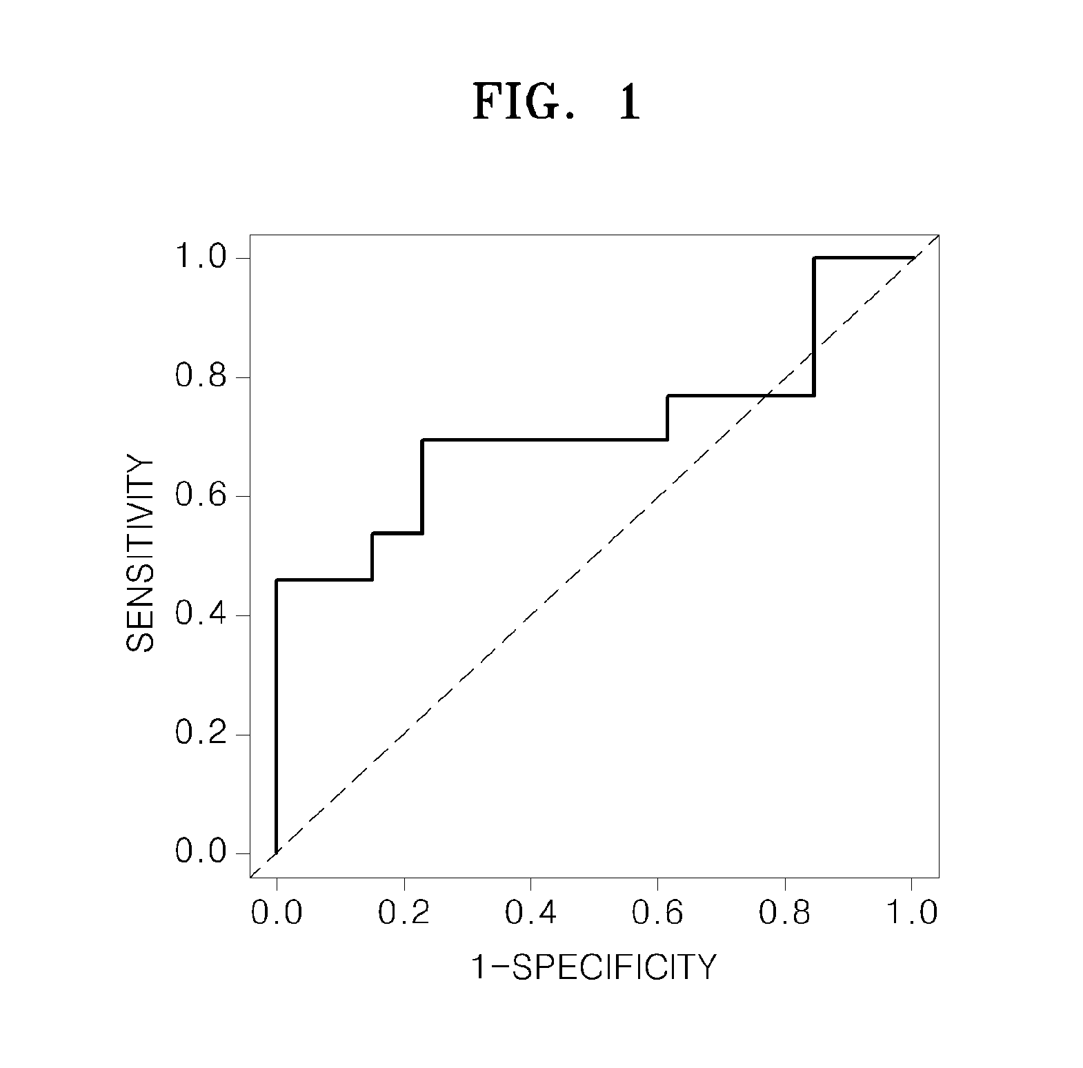
[0063]The presence of a marker specific to liver cancer contained in blood-derived microvesicles was confirmed using each of the samples derived from patients with cirrhosis and patients with liver cancer as explained below.
[0064]Blood samples in a range of 8 ml to 10 ml were each derived from 13 patients with cirrhosis without liver cancer and 13 patients with liver cancer without a cirrhosis, which are examined and confirmed by X-ray computed tomography (x-ray CT) scan and / or magnetic resonance imaging (MRI) scan, by using BD Vacutainer® Plus plastic whole blood tubes. X-ray CT is a technology that uses computer-processed x-rays to produce tomographic images (virtual “slices”) of specific areas of the scanned object, allowing the user to see what is inside it without cutting it open. MRI is a medical imaging technique used in radiology to investigate the anatomy and function of the body in both health and disease. Then, the blo...
example 2
Selection of Liver Cancer-Specific Protein and miRNA Marker
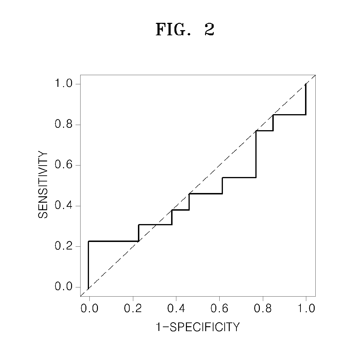
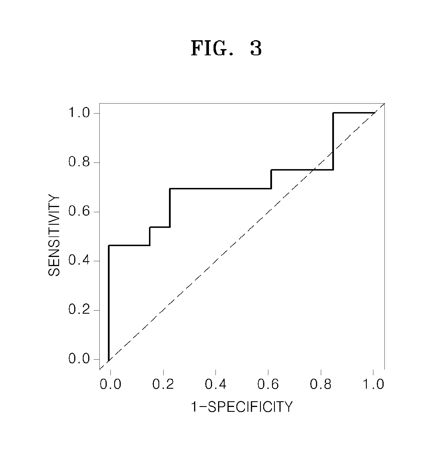
[0084]The presence of a liver cancer-specific protein and a miRNA marker in blood-derived microvesicles was confirmed using samples taken from a patient with cirrhosis and a patient with liver cancer.
[0085]Blood samples in a range of 8 ml to 10 ml were each derived from 13 patients with cirrhosis and 13 patients with liver cancer by using BD Vacutainer® Plus plastic whole blood tubes. Then, the blood samples were separately centrifuged at 1300×g at a temperature of 4° C. for 10 minutes, and accordingly plasma was separated therefrom. The separated plasma was then stored at a temperature of −80° C. A plasma sample was prepared by thawing the stored plasma followed by centrifuged at 3000×g at a temperature of 4° C. for 5 minutes so as to use the supernatants, and remove any precipitates.
[0086]Three hundred microliters of the plasma sample were mixed with 30 ul of beads (0.8 ug antibodies / bead ul) each coated with anti-CD9 anti...
PUM
| Property | Measurement | Unit |
|---|---|---|
| temperature | aaaaa | aaaaa |
| temperature | aaaaa | aaaaa |
| temperature | aaaaa | aaaaa |
Abstract
Description
Claims
Application Information
 - R&D
- Intellectual Property
- Life Sciences
- Materials
- Tech Scout
- Unparalleled Data Quality
- Higher Quality Content
- 60% Fewer Hallucinations
Browse by: Latest US Patents, China's latest patents, Technical Efficacy Thesaurus, Application Domain, Technology Topic, Popular Technical Reports.
© 2025 PatSnap. All rights reserved.Legal|Privacy policy|Modern Slavery Act Transparency Statement|Sitemap|About US| Contact US: help@patsnap.com


