Heart imaging method

a heart and heart valve technology, applied in the field of heart valve movement imaging, can solve the problems of limited diagnostic value, poor quality, and difficulty in assessing the heart and coronary arteries sufficiently to allow full evaluation

Active Publication Date: 2017-02-21
4DX LTD
View PDF6 Cites 11 Cited by
  • Summary
  • Abstract
  • Description
  • Claims
  • Application Information

AI Technical Summary

Benefits of technology

[0032]An object of the present invention is to provide improved images of the heart that are truer, more accurate multidimensional (that is, 2D, 3D and 4D) reconstructions.
[0033]An object of the present invention is to provide improved images of the heart in combination with the lungs that are truer multidimensional reconstructions.
[0034]A further object of the present invention is to provide an improved method of obtaining 2D, 3D and 4D reconstructions of the heart or heart / lung combination.

Problems solved by technology

Medical diagnosis is significantly based on echocardiograph (ECG) measurements but these have limited diagnostic value for many medical conditions and the quality can be poor.
One of the most significant problems associated with measurement of the cardiovascular system is that the constant motion of the heart makes it difficult to visualize the heart and coronary arteries sufficiently to allow full evaluation.
However, like other currently available techniques for flow field measurement in opaque vessels, MRI based techniques, suffer from poor spatial and temporal resolution, limiting the application of these techniques for in vivo flow analysis.
PIV techniques using visible light are limited to optically transparent sample.
These algorithms assume pulsed (instantaneous) illumination and zero out-of-plane flow gradients and therefore fail to take into account the 3D characteristics of imaging real flows using X-rays.
This results in a significant under estimation of flow velocity.
Single projection images are insufficient for evaluating three components of velocity.
However, any patient exposure to X-rays is a concern and there is an ongoing need to extract as much useful data as possible per exposure to X-rays or more preferably, reduced the amount of X-ray exposure without reducing the quality of data obtained.
However, proposed applications of EIT have not extended past monitoring of lung function, detection of cancer in the skin and breast and location of epileptic foci in the brain.
Imaging the lung or other areas where the water content was low, conventional MRI had proved inadequate.
In the past, MRI imaging has often proved inadequate in areas where water content is relatively low, such as the lung, however its application is substantially limited to areas of the body which can be permeated by helium gas.

Method used


Image

  • Heart imaging method
  • Heart imaging method
  • Heart imaging method

Examples


Embodiment Construction

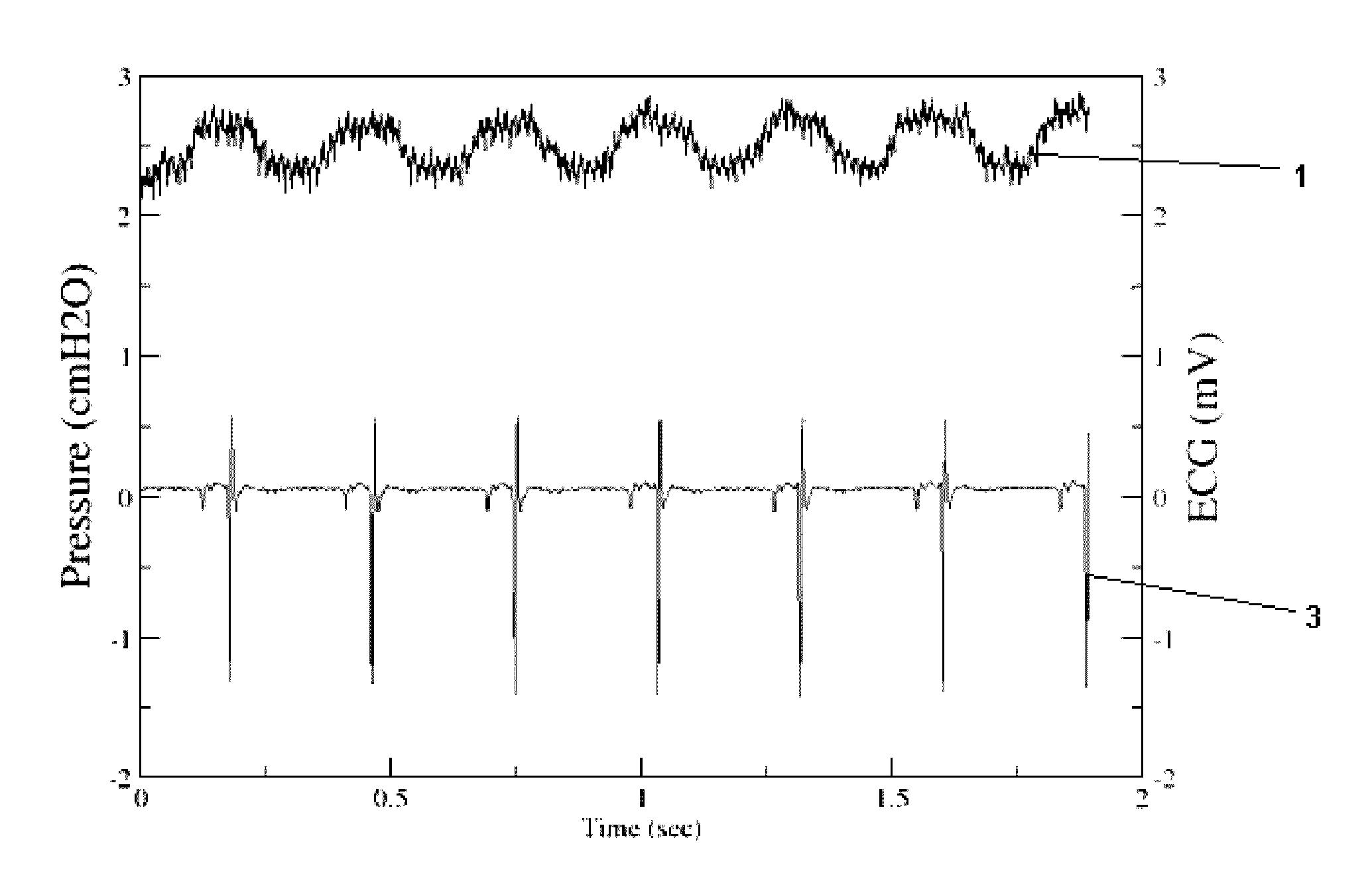
[0174]FIG. 1 is a plot comparing pressure oscillation (cm(H2O)) (1) with an electrocardiogram (ECG) trace (mV) (3) synchronised in time during expiratory breath hold. It illustrates typical prior art measurement of heart rate, heart function and the effect of the heart on the lungs.

[0175]An ECG is a commonly used prior art measure of heart rate and heart function. It is a measure of the electrical activity of the heart and is not a complete analysis of the cardiac cycle.

[0176]The measurement of pressure and gas content at the airway opening is a global measure and tells no spatial information. Specifically, a global measure of this type is indicative of activity in the lungs, but it is not a robust measure. A global measure is merely the sum of activity in all regions in the lungs and does not take into account destructive interference.

[0177]In a study by Lichtwarck-Aschoff (2003), cardiogenic oscillations on the pressure trace at the airway were used to show a relationship between ...


PUM


Abstract

The present invention relates to imaging of a human or animal heart, particularly imaging of movement of the heart and can be used for imaging function and form in a wide range of research, medical, veterinary and industrial applications. In particular, the present invention provides a method and apparatus for imaging a subject heart, the method including the steps of (1) recording at least one in vivo image of a lung of the subject in one or more regions; (2) applying said at least one in vivo image to a 2D or 3D heart model; and (3) reconstructing a 2D or 3D image field of the subject heart.

Description

CROSS REFERENCE TO RELATED APPLICATIONS[0001]This is a National Stage of International Application No. PCT / AU2012 / 001225 filed Oct. 10, 2012, claiming priority based on Australian Patent Application No. 2011904256 filed Oct. 10, 2011, the contents of all of which are incorporated herein by reference in their entirety.FIELD OF INVENTION[0002]The present invention relates to imaging of a human or animal heart, particularly imaging of movement of the heart[0003]In one aspect the present invention relates to the field of biomedical engineering, particularly in vivo or in vitro imaging of the heart.[0004]In another aspect, the invention relates to technology for imaging of function and form in a wide range of research, medical, veterinary and industrial applications.[0005]In a yet further aspect the present invention is suitable for use as a method and device for imaging the movement of living heart tissue.[0006]It will be convenient to hereinafter describe the invention in relation to i...

Claims


Application Information

Patent Timeline
Patent Type & Authority Patents(United States)
IPC IPC(8): G06T7/00G06T19/00A61B6/03A61B6/00
CPCG06T7/0012A61B6/032A61B6/481A61B6/486A61B6/50A61B6/503A61B6/507A61B6/5211A61B6/5288G06T19/00A61B6/508G06T2210/41G16H50/20
Inventor FOURAS, ANDREASDUBSKY, STEPHENTHURGOOD, JORDAN
Owner 4DX LTD
Why Eureka
Try Eureka