PME-1 as a biomarker to predict and diagnose an increased risk of endometrial cancer and gene silencing of PME-1 to inhibit epithelial to mesenchymal transition

a biomarker and gene silencing technology, applied in biochemistry, instruments, enzymology, etc., can solve the problems of ineffective pap smear in detecting endometrial cancer, inability to detect early endometrial cancer, and inability to use a method, so as to increase increase the risk of endometrial cancer. , the effect of increasing the expression level of pme-1

Inactive Publication Date: 2017-05-23
MEDICAL DIAGNOSTIC LAB
View PDF0 Cites 2 Cited by
  • Summary
  • Abstract
  • Description
  • Claims
  • Application Information

AI Technical Summary

Benefits of technology

[0019]In one aspect, the present invention provides a kit for detecting endometrial cancer in a human, comprising: a) a reagent for quantifying PME-1 expression level; and b) an instruction for use of said reagent in quantifying PME-1 protein expression level. An increased PME-1 protein expression level is indicative of an increased risk in endometrial cancer.

Problems solved by technology

Notably, Pap smear is ineffective in detecting endometrial cancer, while it is useful in screening for cervical cancer.
However, such procedure is not practical because it requires strict sterile environments and physician's competent skill, as well as the high associated costs.
However, the method is often not sensitive to detect early endometrial cancer.

Method used


Image

  • PME-1 as a biomarker to predict and diagnose an increased risk of endometrial cancer and gene silencing of PME-1 to inhibit epithelial to mesenchymal transition
  • PME-1 as a biomarker to predict and diagnose an increased risk of endometrial cancer and gene silencing of PME-1 to inhibit epithelial to mesenchymal transition
  • PME-1 as a biomarker to predict and diagnose an increased risk of endometrial cancer and gene silencing of PME-1 to inhibit epithelial to mesenchymal transition

Examples


example 1

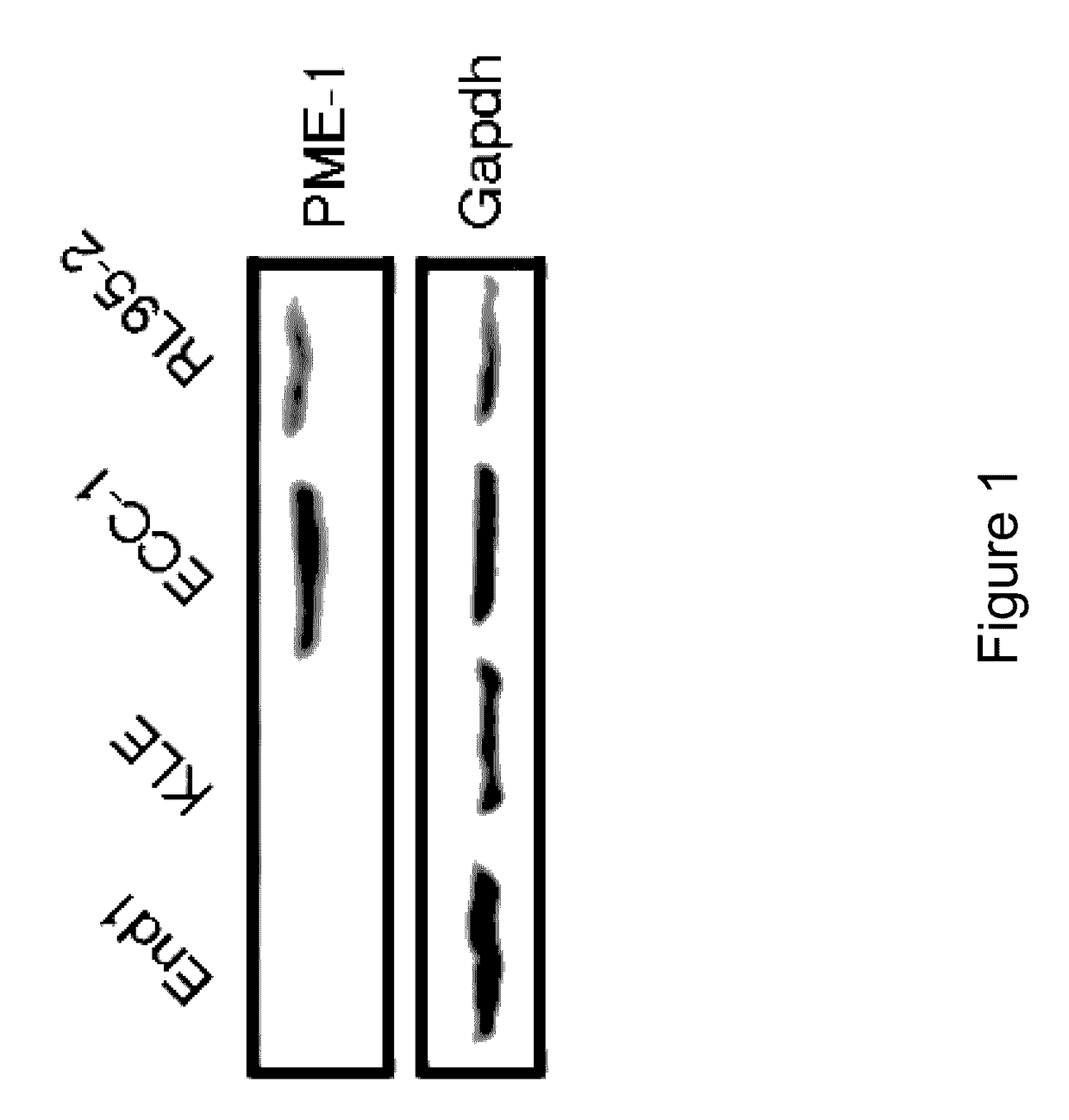
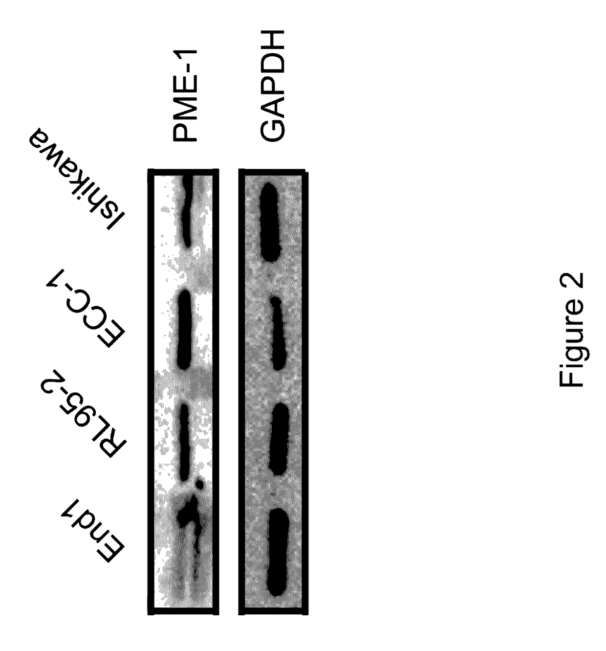
PME-1 Expression in Endometrial Cell Lines

[0214]In these initial studies, we sought to determine the expression levels of PME-1 in various human endometrial cell lines (EC). We examined the expression level of PME-1 protein using Western Blot analysis (See, Materials and Methods).

[0215]As shown in FIGS. 1 and 2, the more aggressive / metastatic EC cell lines (RL95-2, Ishikawa, and ECC-1) were found to express more abundant PME-1 protein as compared to the less aggressive / metastatic EC line (KLE) and the immortalized endocervical cell line, End1. GAPDH was used as a loading control in these studies. These data suggest that expression of PME-1 protein levels correlates with the degree of aggressiveness of EC; that is the more aggressive / metastatic EC, the higher the expression of PME-1 protein levels.

example 2

Western Blot Analysis of PME-1 Protein Expression in Tissue Samples from Patients with Endometrial Adenocarcinoma

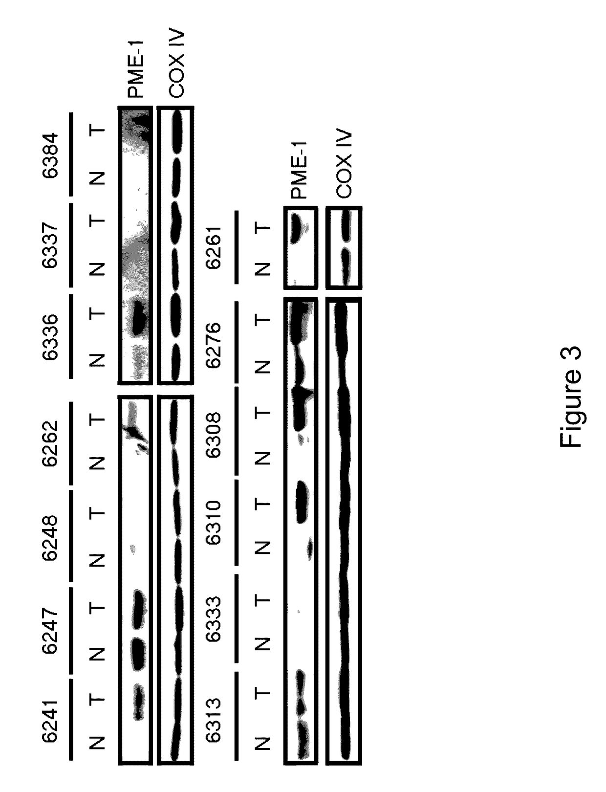
[0216]After establishing that the degree of aggressiveness and metastasis in EC cell lines indeed correlates with an increased level of PME-1 protein, we next determined whether the same trend might occur in tissue samples from patients.

[0217]We obtained 30 matched pairs of tissue samples harvested from patients with endometrial adenocarcinoma. The details of the relevant patient data are provided in Table 1. We used Western Blot analysis to monitor the expression of PME-1 protein levels in these tissue samples from patients (FIG. 3).

[0218]Representative Western blots are displayed for several stage I EC patient samples. All stage II and stage III tissue samples purchased are also shown in FIG. 3. The top left panel shows stage IB patient samples (patient #6241, 6247, 6248, 6262) except for #6262, which is a grade 1 tumor. The top right panel shows three (3) stage IC in w...

example 3

Immunohistochemistry Analysis

[0222]We confirmed the PME-1 protein expression data by evaluating the immunohistochemistry of the tissue samples from EC patients. This immunohistochemistry study provides the basis for our observation of an increased PME-1 expression level in the context of tissue morphology.

[0223]We performed H&E staining on the tissue samples (FIG. 5i-iv) and co-stained tissue samples with anti-PME-1 antibodies (FIG. 5v-viii). Normal tissue showed regular duct formation in the endometrium with light PME-1 expression around ducts within epithelial cells, as illustrated by the black arrows (FIG. 5i, v). In stage I EC, ducts became irregular and PME-1 localization becomes more punctuate and localized, as illustrated by the black arrows (FIG. 5ii, vi). Stage II EC was even less organized with increased PME-1 staining and more foci, as illustrated by the black arrows (FIG. 5iii, vii).

[0224]In stage III EC, PME-1 staining was darker and appeared to be limited to individual...


PUM

PropertyMeasurementUnit
incubation timeaaaaaaaaaa
temperatureaaaaaaaaaa

Abstract

Disclosed are methods of attenuating activity of the PME-1 gene. siRNAs or shRNAs are used to target against PME-1, thereby reducing the PME-1 mRNA. It is disclosed that the siRNAs or shRNAs targeted against PME-1 attenuate the epithelial to mesenchymal transition, thereby inhibit endometrial cancer development. A kit containing siRNA or shRNA reagents for attenuating the PME-1 gene expression is also disclosed.

Description

CROSS-REFERENCE TO RELATED APPLICATION[0001]This application claims the benefit under 35 U.S.C. §119(e) to U.S. Provisional Application No. 61 / 816,472 filed Apr. 26, 2013 and Provisional Application No. 61 / 905,947 filed Nov. 19, 2013, the content of which are incorporated by reference herein in their entirety.FIELD OF THE INVENTION[0002]The present invention generally relates to the discovery that PME-1 represents a novel endometrial cancer biomarker (PME-1 is a biomarker used to predict and diagnose an increased risk of endometrial cancer) and gene silencing of the PME-1 gene to inhibit the epithelial to mesenchymal transition and endometrial cancer development.BACKGROUND OF THE INVENTION[0003]Endometrial cancer refers to the malignancies of endometrium of the uterus. It is the most common gynecologic cancers in the United States, with over 35,000 women diagnosed each year. It represents the third most common cause of gynecologic cancer death, behind ovarian and cervical cancer (Si...

Claims


Application Information

Patent Timeline
Patent Type & Authority Patents(United States)
IPC IPC(8): C12Q1/68G01N33/574A61K31/713A61K31/7105C12N15/113C12N5/00
CPCC12Q1/6886A61K31/713A61K31/7105C12N15/1137G01N33/57442C12N2310/14C12N2310/531C12Q2600/158C12Y301/01089G01N2333/918
Inventor RICE, LYNDIPUSEY, MICHELLEWANDZIOCH, EWABAIL, SOPHIE MARIE GENEVIEVEWERDA, AMY LYNN
Owner MEDICAL DIAGNOSTIC LAB
Why Eureka
Try Eureka