Ventricular electrical activity indicator

a technology of electrical activity indicator and ventricular electrical activity, which is applied in the field of ventricular electrical activity indicator and operator interface in medical imaging system, can solve problems such as difficulty in distinguishing near-field atrial activity from far-field ventricular activity

Active Publication Date: 2017-07-18
BIOSENSE WEBSTER (ISRAEL) LTD
View PDF12 Cites 3 Cited by
  • Summary
  • Abstract
  • Description
  • Claims
  • Application Information

AI Technical Summary

Benefits of technology

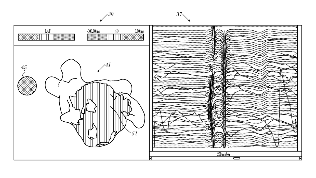
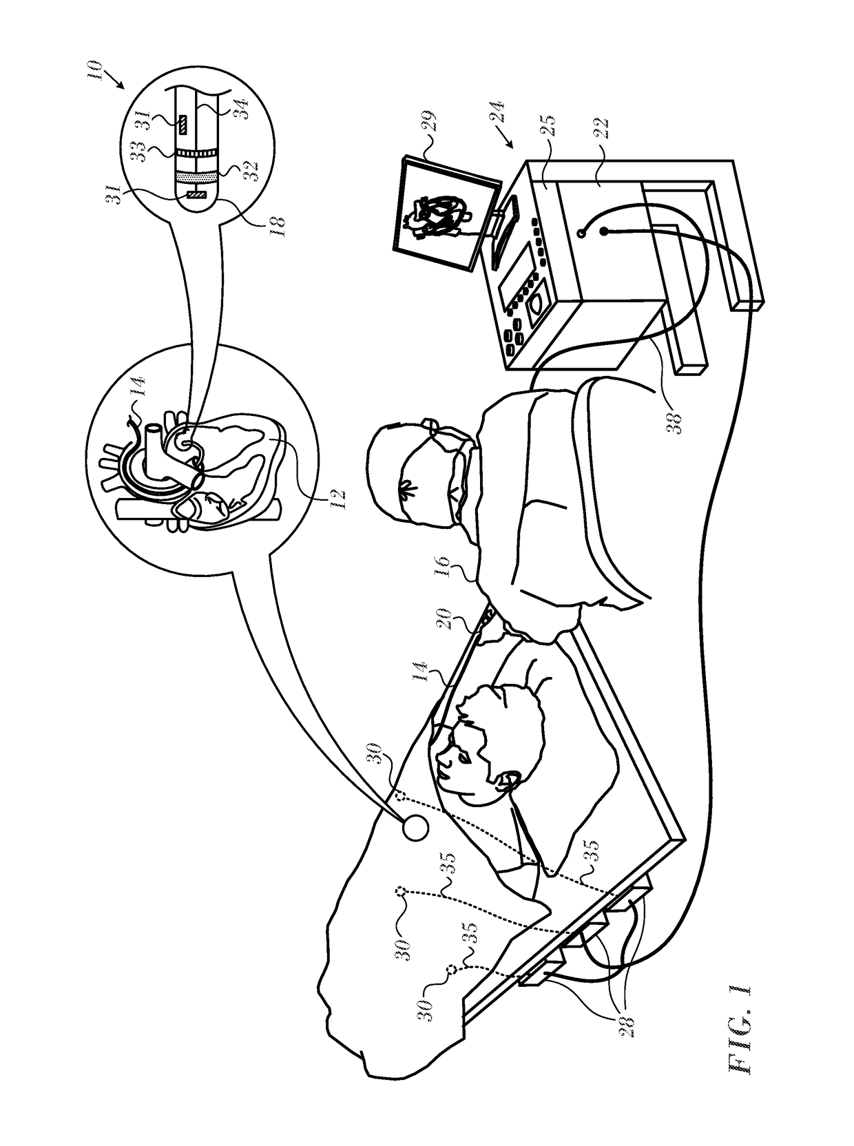
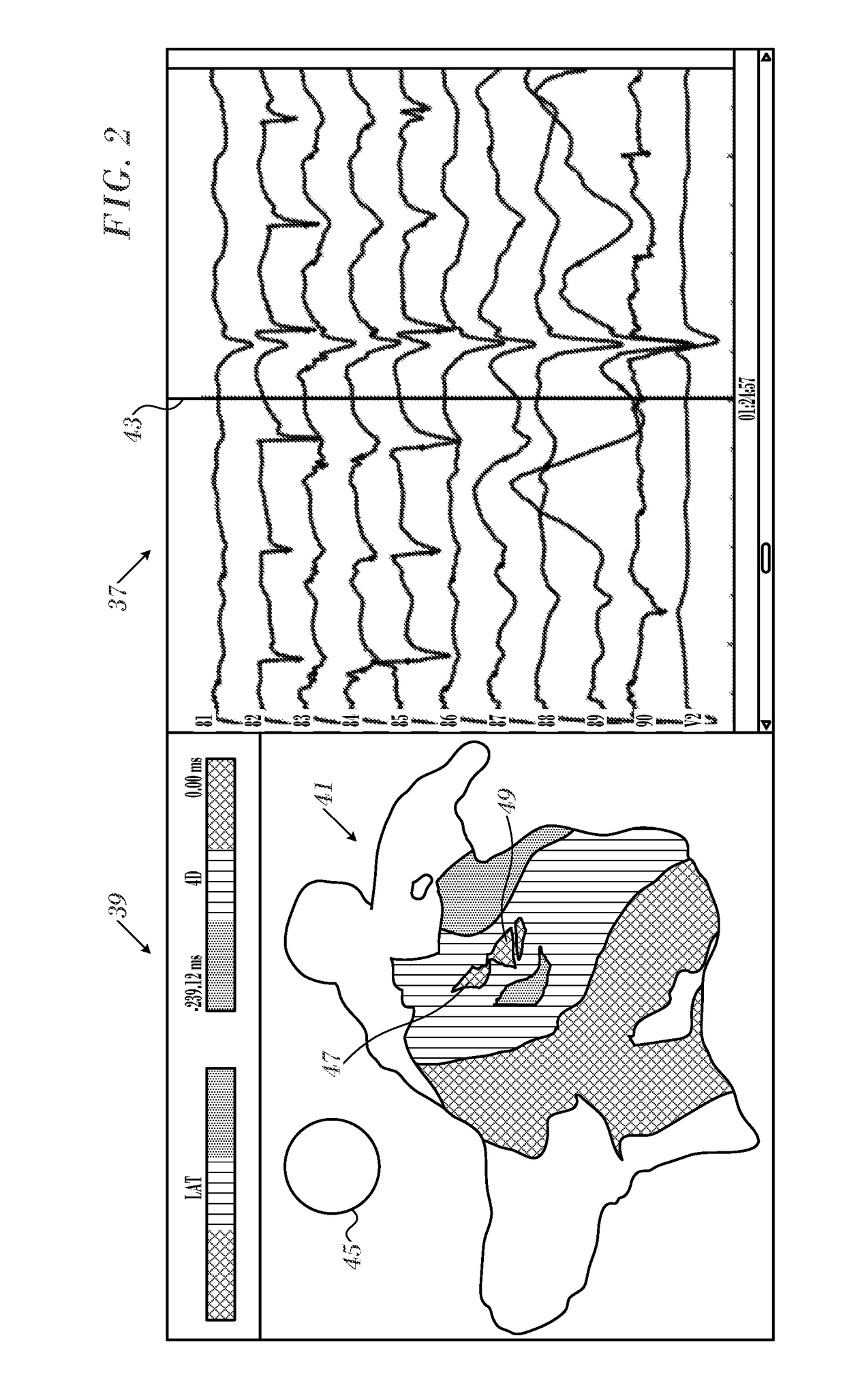
[0014]There is further provided according to embodiments of the invention an apparatus, including a processor connectable to an electrocardiographic sensor of ventricular activity and to a cardiac catheter having at least one sensing electrode disposed on a distal portion thereof. The apparatus includes a display linked to the processor, a memory accessible to the processor having programs and data objects stored therein. The programs include a graphical interface program. When the at least one sensing electrode is in galvanic contact with respective locations in an atrium of a heart, execution of the programs cause the processor to acquire electrograms from the at least one sensing electrode and concurrently detect ventricular depolarization events in the heart via the electrocardiographic sensor. The processor is further caused to generate from the electrograms a time-varying electroanatomic map showing electrical propagation in the heart, and to invoke the graphical interface program to present the electroanatomic map on the display as a series of visual images. The images include an icon that visually indicates the ventricular depolarization events.

Problems solved by technology

However, an operator who is attempting to annotate atrial activation onset times using a multi-electrode catheter and is presented with conventional maps of this sort may experience difficulty distinguishing near-field atrial activity from far-field ventricular activity.

Method used


Image

  • Ventricular electrical activity indicator
  • Ventricular electrical activity indicator
  • Ventricular electrical activity indicator

Examples


Embodiment Construction

[0022]In the following description, numerous specific details are set forth in order to provide a thorough understanding of the various principles of the present invention. It will be apparent to one skilled in the art, however, that not all these details are necessarily needed for practicing the present invention. In this instance, well-known circuits, control logic, and the details of computer program instructions for conventional algorithms and processes have not been shown in detail in order not to obscure the general concepts unnecessarily.

[0023]Aspects of the present invention may be embodied in software programming code, which is typically maintained in permanent storage, such as a computer readable medium. In a client / server environment, such software programming code may be stored on a client or a server. The software programming code may be embodied on any of a variety of known non-transitory media for use with a data processing system, such as a USB memory, hard drive, el...


PUM


Abstract

Cardiac catheterization is carried out using a probe having sensing electrodes disposed on a distal portion thereof, placing the sensing electrodes in galvanic contact with respective locations in an atrium of the heart, thereafter acquiring electrograms from the sensing electrodes while concurrently detecting ventricular depolarization events, generating from the electrograms a time-varying electroanatomic map showing electrical propagation in the heart, and displaying the electroanatomic map in a series of visual images, the images including an icon that visually indicates the ventricular depolarization events.

Description

BACKGROUND OF THE INVENTION[0001]Field of the Invention[0002]This invention relates to medical imaging systems. More particularly, this invention relates to operator interfaces in medical imaging systems.[0003]Description of the Related Art[0004]Cardiac arrhythmias, such as atrial fibrillation, occur when regions of cardiac tissue abnormally conduct electric signals to adjacent tissue, thereby disrupting the normal cardiac cycle and causing asynchronous rhythm.[0005]Electrical activity in the heart is typically measured by advancing a multiple-electrode catheter to measure electrical activity at multiple points in the heart chamber simultaneously. A graphical user interface integrated with modern imaging systems for monitoring cardiac catheterization presents an abundance of dynamically changing information from the multiple electrodes to the operator, and facilitates efficient processing of the information by the operator.[0006]Receiving atrial electrogram signals from intracardiac...

Claims


Application Information

Patent Timeline
Patent Type & Authority Patents(United States)
IPC IPC(8): A61B5/0452A61B5/044A61B5/042A61B5/00A61B18/14A61B18/00
CPCA61B5/0452A61B5/044A61B5/0422A61B5/743A61B5/6852A61B5/6859A61B18/1492A61B2018/00577A61B2018/00839A61B5/6847A61B5/6869A61B5/7221A61B5/7235A61B5/742A61B5/283A61B5/318A61B5/287A61B5/339A61B5/349
Inventor YELLIN, TAMIR AVRAHAM
Owner BIOSENSE WEBSTER (ISRAEL) LTD
Why Eureka
Try Eureka