Method and apparatus for atlas/model-based segmentation of magnetic resonance images with weakly supervised examination-dependent learning

Active Publication Date: 2017-10-24
SIEMENS HEALTHCARE GMBH
View PDF4 Cites 3 Cited by
  • Summary
  • Abstract
  • Description
  • Claims
  • Application Information

AI Technical Summary

Benefits of technology

The objective of this patent text is to offer a better way of separating bones that can be used in everyday medical practice. This new technique should minimize the drawbacks of current methods and make it simpler to segment bones accurately.

Problems solved by technology

In the case of magnetic resonance images in which bones are represented, bone segmentation presents a challenge, for several reasons.
This makes it difficult, or almost impossible, to distinguish between bone and air or lung tissue, for example.
Special data acquisition sequences, such as those that produce an ultra-short TE, and which are thus capable of acquiring signals from water-bound protons within the inorganic cortical structure of the bone, usually suffer with regard to image quality or other restrictions that limit the practical use of such sequences.
Moreover, such special sequences often are not suitable for diagnostic purposes, thereby disqualifying their use in clinical routine.
A drawback of this known technique is that, for each bone, high-quality co-registered pairs of MR and CT scans are required, which serve as the model.
For fine bones, such as in the shoulder or the ribs, it is difficult to meet this requirement.
This means that current implementation of this known method is not suitable for all bones.
Moreover, anatomical variants beyond the limits of the non-rigid registration can lead to inaccurate segmentation results.
A drawback of this known technique is that, due to the bias-variance tradeoff, the training data may not include all possible variants of MR contrast, which results in a strong sensitivity to the input contrasts, which can be a problem if, in practice, the acquisition protocols are modified, or studies are done using different hardware settings, and appropriate normalization algorithms are not available.

Method used


Image

  • Method and apparatus for atlas/model-based segmentation of magnetic resonance images with weakly supervised examination-dependent learning
  • Method and apparatus for atlas/model-based segmentation of magnetic resonance images with weakly supervised examination-dependent learning
  • Method and apparatus for atlas/model-based segmentation of magnetic resonance images with weakly supervised examination-dependent learning

Examples


Embodiment Construction

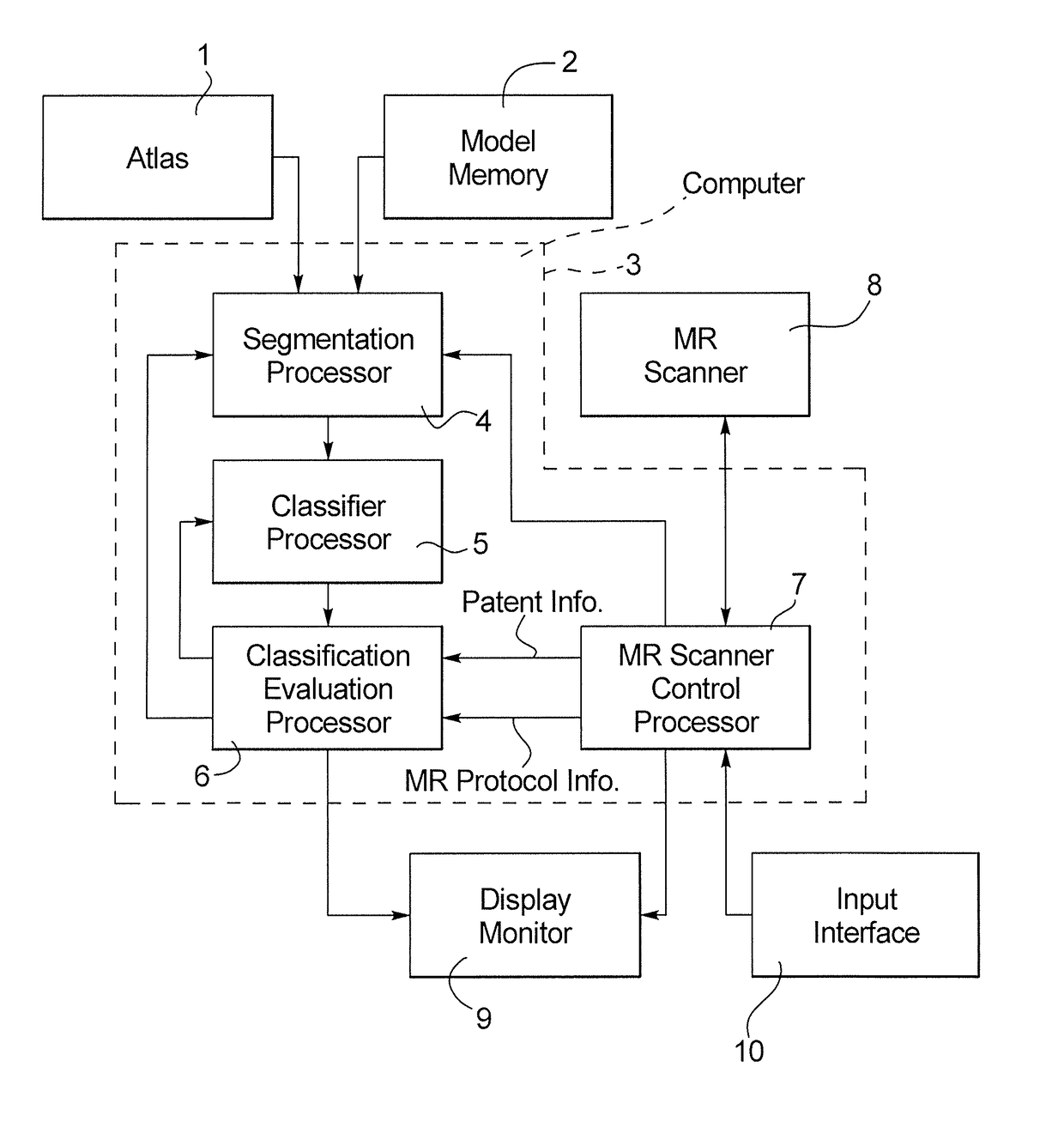
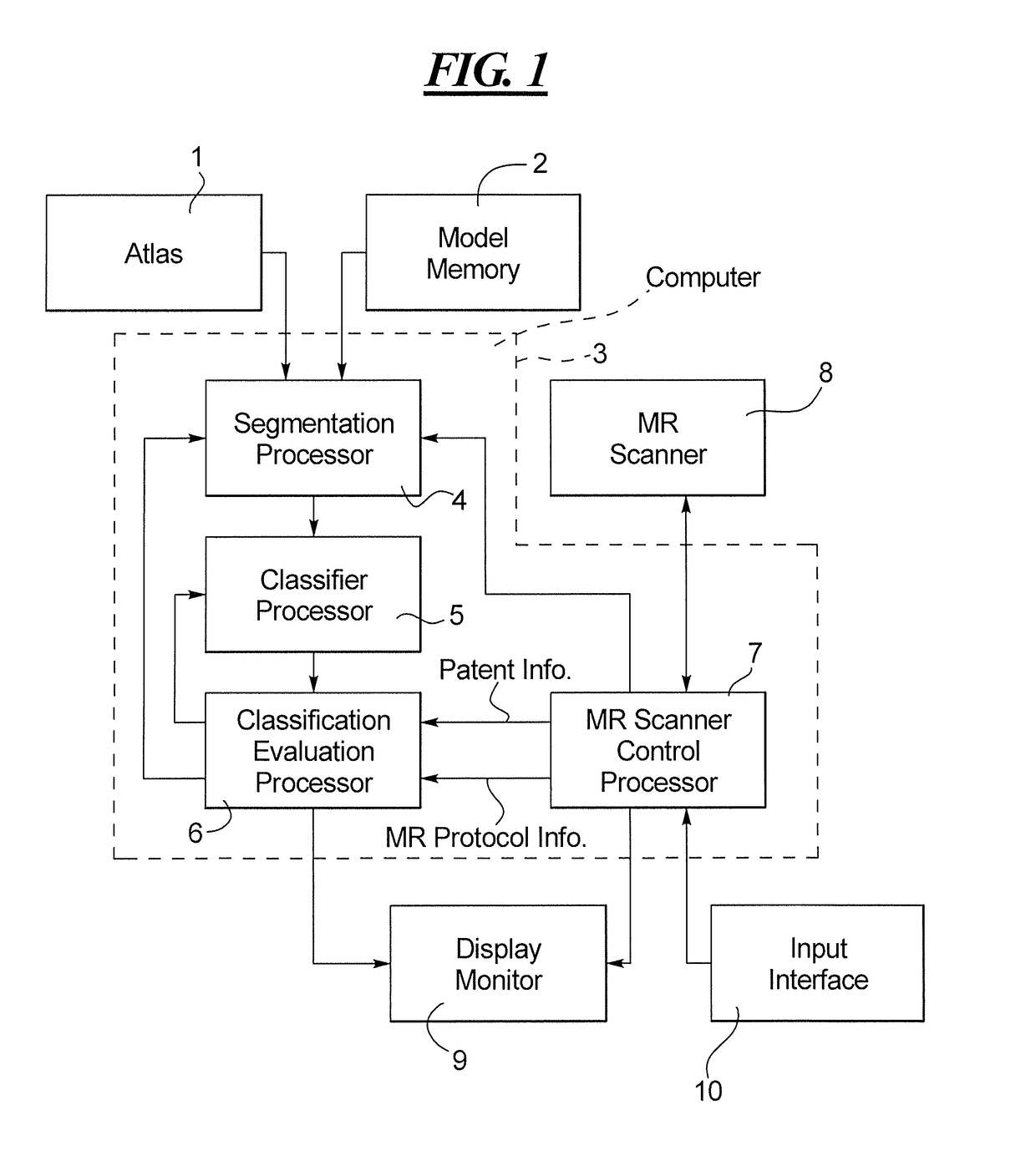
[0025]As shown in FIG. 1, the apparatus according to the invention makes use of an atlas 1 and / or a model memory 2 that provide a data file or model to a computer 3, specifically to a segmentation processor 4 of the computer 3. The segmentation processor 4 is also provided with a data file representing an MR image of a region of a patient, acquired by operation of an MR scanner 8. The MR image data file is provided from an MR scanner control processor 7 that operates the MR scanner 8 and generates image information from the acquired MR data in a known manner.

[0026]The acquired MR data represent image elements (pixels in the case of a 2D image, and voxels in the case of a 3D image), that each has different attributes or characteristics, such as intensity. The segmentation processor 4 operates in combination with a classifier processor 5 in order to classify the image elements of the provided MR image, in order to identify and extract image elements therefrom that have the image eleme...


PUM


Abstract

In a magnetic resonance (MR) apparatus and segmentation method, a region in an MR image, acquired from a scan of a patient with an MR scanner of the apparatus, is provided to a computer for segmentation of the region from the overall image dataset. The segmentation takes place based on a classification of image elements of the image dataset, and the classification is iteratively re-trained in a weakly supervised learning algorithm based on examination-specific information provided to the computer.

Description

BACKGROUND OF THE INVENTION[0001]Field of the Invention[0002]The present invention concerns the field of segmenting an organ or other region of interest in an image, or image data, acquired in a magnetic resonance data acquisition scan procedure, and in particular to implementing such segmentation using an atlas or a model that does not precisely cover or encompass the organ or region of interest that is to be segmented.[0003]Description of the Prior Art[0004]Magnetic resonance imaging is a widely used imaging modality for providing images of a patient to a physician in order to allow the physician to make a particular medical diagnosis. A general explanation of the fundamental concepts of magnetic resonance imaging is as follows.[0005]Magnetic resonance data are acquired from a patient by placing the patient on a movable bed or support within an opening of the magnetic resonance scanner that is designed to receive the patient therein. A basic field magnet of the magnetic resonance ...

Claims


Application Information

Patent Timeline
IPC IPC(8): G06K9/00G06T11/00G06T7/00G06K9/62G06V10/776G06V10/26G06V10/764
CPCG06T11/008G06K9/627G06K9/6262G06T7/0014G06T2207/10088G06T2207/20128G06T7/11G06V10/26G06V2201/033G06V2201/03G06V10/776G06V10/764G06F18/24G06F18/217
Inventor FENCHEL, MATTHIASZHAN, YIQIANG
Owner SIEMENS HEALTHCARE GMBH
Why Eureka
Try Eureka