
Method and system for 3D root canal treatment planning

a root canal treatment and 3d technology, applied in the field of 3d root canal treatment planning, can solve the problems of insufficient cleaning, inability to provide a method that is usable in the daily clinical practice of the patient, and insufficient cleaning of the available prior ar

Active Publication Date: 2017-10-31
DENTSPLY IMPLANTS NV +1
View PDF19 Cites 6 Cited by
  • Summary
  • Abstract
  • Description
  • Claims
  • Application Information

AI Technical Summary

Benefits of technology

The present invention provides a method and system for 3D root canal treatment planning. The system uses simulation to assess treatment risks and provide detailed information about the tooth anatomy. The dentist or dental specialist can then select the optimal tools for root canal treatment before the treatment itself. The invention allows for case-specific planning without the need for 3D imaging techniques that expose the patient to radiation doses higher than conventional X-ray. The technical effects of the invention include improved accuracy in root canal treatment planning and reduced risk of treatment-related risks.

Problems solved by technology

Root canal treatment can fail or lead to complications due to several reasons, e.g. insufficient cleaning of the root canals, incomplete filling of the root canals, untreated canals since these were missed by the practitioner, root perforations or file fracture during shaping of the canals, root fractures .
While the use of 3D computer images and models has been reported for the preparation of root canal treatment, the available prior art fails to provide a method that is usable in the daily clinical practice.
Current CBCT technology does not provide the required image resolution to allow for planning of the root canal treatment, since the root canals are often hardly if at all distinguishable from noise in the images.
Even in cases where the root canals can be discerned, the reliability of measurements performed on the images, e.g. with respect to the canal dimensions, is insufficient to provide added value when planning the clinical intervention.
In addition, radiation doses required for CBCT imaging can drastically exceed those of traditional X-rays, potentially adding risk to the patient, and making the technique currently unsuited for most endodontic indications. μCT imaging is even less suitable: currently, there is no commercially available equipment for acquiring μCT images on patients, since fields of view are too limited, required radiation doses too high and data acquisition times too long with this technology. μCT imaging currently only works on extracted teeth, in specific set-ups for research purposes.

Method used


Image

  • Method and system for 3D root canal treatment planning
  • Method and system for 3D root canal treatment planning
  • Method and system for 3D root canal treatment planning

Examples


Embodiment Construction

[0019]The present invention will be described with respect to particular embodiments and with reference to certain drawings but the invention is not limited thereto but only by the claims.

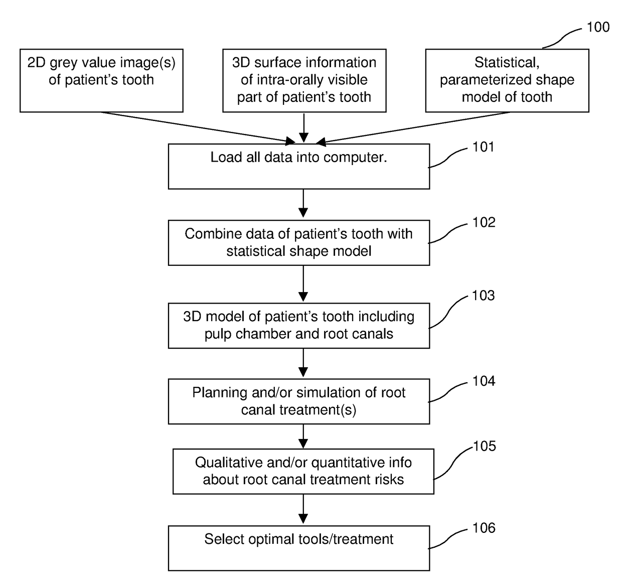
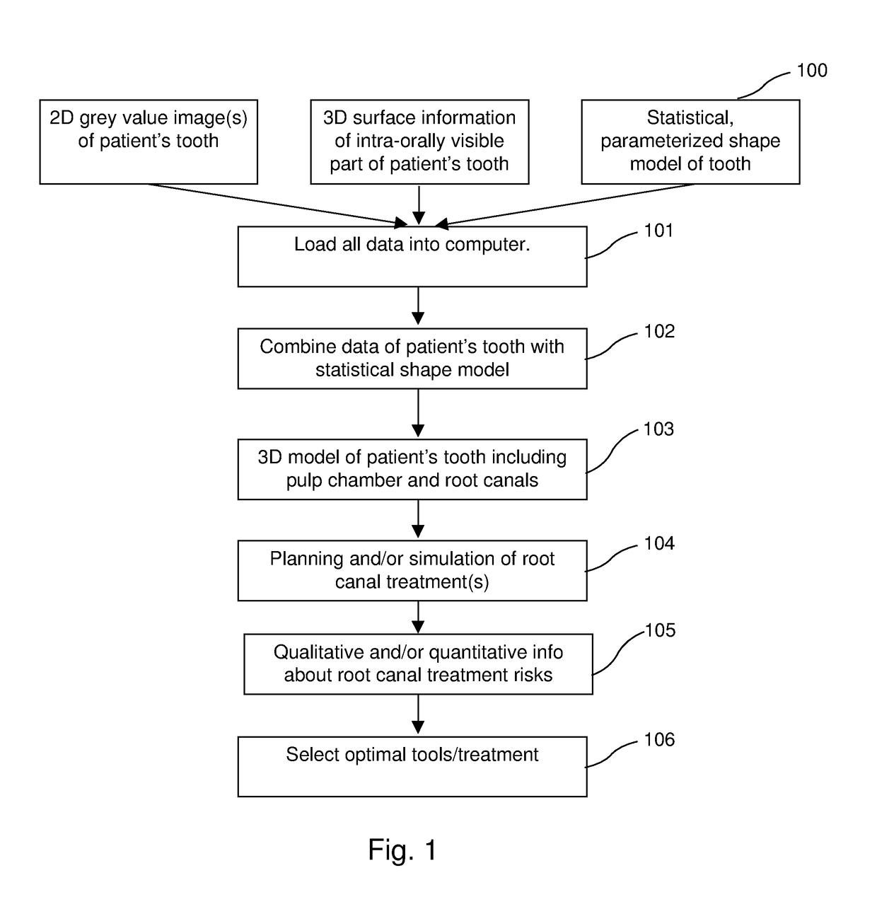
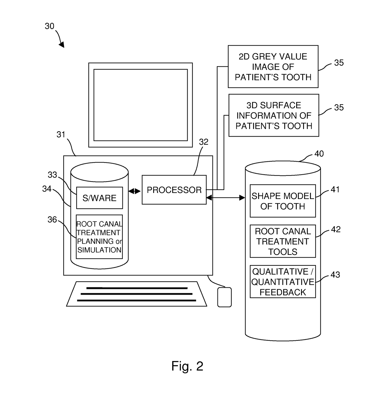
[0020]According to a preferred embodiment of the invention a first step according to a method for simulating root canal treatment simulation consists in creating and visualizing a three-dimensional model of a patient's tooth including the pulp chamber and the root canals. Therefore, the system consists at least of a computer including computer programs which can be utilized with the method for visualizing said three-dimensional model.

[0021]With reference to FIGS. 3 to 5, according to one embodiment the 3D model of the tooth with pulp chamber and root canals is generated based on the combination of 3D imaging data of the crown and one or more 2D radiographs of the tooth. Therefore the 3D crown information of the respective tooth is digitized. This can be done using different methods. A first method ...


PUM


Abstract

A three-dimensional computer model of the patient's tooth, including the pulp chamber and root canals, is created by combining at least one grey value image of the tooth and / or surface information about at least part of the intra-orally visible part of the tooth with a statistical, parameterized shape model of each tooth type (upper or lower incisors, canines, pre-molars, molars) as the patient's tooth to be treated. This allows for planning and / or simulation of one or more root canal treatments on the computer model of the patient's tooth and that the dentist or dental specialist is given qualitative and / or quantitative information by the system in order to aid in adequately analyzing the risks related to performing the root canal treatment with the proposed or user-selected endodontic tools.

Description

[0001]The present invention relates to a method and system for 3D root canal treatment planning as well as software for carrying out such methods.BACKGROUND[0002]Root canal treatment is a tooth-saving treatment that eliminates infections, protects the decontaminated tooth from future infections, and—if needed—restores the tooth at crown level either with or without use of a post. During this treatment the root canals, which mainly contain the nerve tissue and blood vessels are cleaned, shaped, decontaminated and subsequently filled with an inert filling such as gutta percha. In cases where the remaining tooth structure is insufficient to support a proper reconstruction of the dental crown, a metal or glass fiber post is positioned in one of the root canals and a core build-up is created in order to support and provide retention for the prosthetic restoration (i.e. crown).[0003]Root canal treatment can fail or lead to complications due to several reasons, e.g. insufficient cleaning o...

Claims


Application Information

Patent Timeline
Patent Type & Authority Patents(United States)
IPC IPC(8): G06F17/50G06F19/00A61C13/00G06T19/20A61C5/40
CPCG06T19/20A61C13/0004A61C5/40G06F19/3437G06T2210/41G16H50/50A61C13/00
Inventor VAN LIERDE, CARLPATTIJN, VEERLEVALLOTTON, PAUL-HENRI
Owner DENTSPLY IMPLANTS NV
Learn More