Ultrasonic diagnostic imaging system and method for detecting lesions of the liver

An imaging system and ultrasonic diagnosis technology, which is applied in the fields of ultrasonic diagnosis, ultrasonic/acoustic/infrasonic diagnosis, infrasonic diagnosis, etc., can solve the problems of liver damage detection and diagnosis, etc.

Active Publication Date: 2008-02-20
KONINKLIJKE PHILIPS ELECTRONICS NV
View PDF3 Cites 23 Cited by
  • Summary
  • Abstract
  • Description
  • Claims
  • Application Information

AI Technical Summary

Problems solved by technology

However, regardless of the complexity and versatility of these technologies, none of th

Method used


Image

  • Ultrasonic diagnostic imaging system and method for detecting lesions of the liver
  • Ultrasonic diagnostic imaging system and method for detecting lesions of the liver
  • Ultrasonic diagnostic imaging system and method for detecting lesions of the liver

Examples


Embodiment Construction

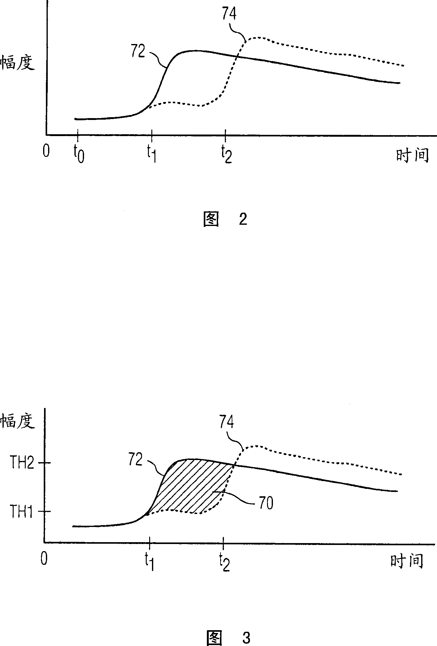
[0013] Blood flow to the liver comes from two sources: the hepatic artery and the portal vein. The hepatic artery provides blood flow from the aorta. Blood flow through the portal vein comes from the inferior vena cava. The inferior vena cava in turn receives blood from the kidneys, which purify the flow provided by the aorta. Blood flow from the liver travels through the hepatic vein to the superior vena cava. The liver receives 20% of its total blood supply from the aorta and hepatic artery, while 80% of its blood supply comes from the inferior vena cava and portal vein.

[0014] As a result of this vasculature, when a bolus of contrast agent is injected into a blood vessel, it will first emerge in the liver via the hepatic artery. After being pumped from the heart into the aorta, a portion of the contrast agent flows into the hepatic artery and into the liver. But since only 20 percent of the blood supplied to the liver is provided through the hepatic artery, this early...


PUM


Abstract

Significant liver growths such as HCC lesions are detected during a contrast agent ultrasound exam by their early reception of contrast and brightening following a bolus injection, as compared with surrounding normal tissue and benign growths. A pixel classifier (30) looks for and identifies pixel or voxel regions where this early wash-in of contrast occurs and denotes these pixel or voxel locations in a parametric image. The pixel classifier analyzes pixel or voxel values from a sequence of images and identifies suspicious regions in an image by uniquely coding the points in a parametric liver image where early wash-in occurs .

Description

technical field [0001] The present invention relates to an ultrasonic diagnostic imaging system, in particular to an ultrasonic system and method for diagnosing liver injury. Background technique [0002] It has been found that patients with hepatitis B and C have an increased risk of developing primary liver cancer, hepatocellular carcinoma (HCC). It was recently discovered that patients became infected with hepatitis C through blood transfusions in the early 1980s, so there are still a significant number of hepatitis C patients who need to be checked regularly to prevent the onset of HCC because the damage responds best when it is treated early. The usual disease progression is from hepatitis to cirrhosis to HCC. An easy-to-use technology for monitoring the progression of liver disease would have broad application in helping to detect this serious disease early. [0003] In addition to HCC, a number of lesions can develop in the liver. The article "Ultrasound Contrast I...

Claims


Application Information

Patent Timeline
IPC IPC(8): A61B8/08A61B8/00
CPCA61B8/08A61B8/481A61B5/7264A61B8/085G16H50/20A61B8/00
Inventor M·布鲁斯J·波沃斯R·加格D·斯基巴M·阿弗基奥
Owner KONINKLIJKE PHILIPS ELECTRONICS NV
Why Eureka
Try Eureka