Preparation method of human body stereo anatomy image as well as application

A production method and image technology, applied in application, 3D image processing, image data processing, etc., can solve problems such as insufficient understanding and difficulty in accurately describing the spatial structure of tissues and organs, and achieve the effect of improving the diagnostic level

Inactive Publication Date: 2008-05-14
宋岩峰 +1
View PDF0 Cites 8 Cited by
  • Summary
  • Abstract
  • Description
  • Claims
  • Application Information

AI Technical Summary

Problems solved by technology

In the past, in order to obtain the anatomical information of the patient, clinicians could only obtain it by reading two-dimensional images such as CT, MRI, MRA, PETCT, and ultrasound, and inferred its spatial anatomical structure, and could not directly read the three-dimensional anatomy Atlas, the understanding obtained in this way is not comprehensive and direct, and it is difficult to accurately describe the spatial structure, hierarchy and adjacent characteristics of tissues and organs

Method used


Image

  • Preparation method of human body stereo anatomy image as well as application
  • Preparation method of human body stereo anatomy image as well as application

Examples


Embodiment Construction

[0007] The content of the present invention will be described in detail below in conjunction with the accompanying drawings and specific embodiments:

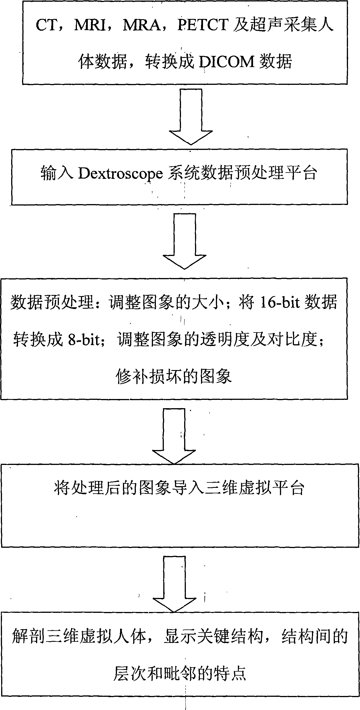
[0008] The present invention comprises the following manufacturing steps:

[0009] (1) Use CT, MRI, MRA, PETCT and ultrasound methods to collect human body data (the procedure is the same as taking CT, MRI, MRA, PETCT and ultrasound examination in the hospital), the data layer thickness is less than 2mm, and then converted into DICOM data ( An existing medical digital image and information language data format);

[0010] (2) Input the collected human body DICOM data into the data preprocessing platform of the Dextroscope system; the Dextroscope system is a software system developed by Singapore Volume interactions Pte Ltd, which can combine CT, MRI, MRA, PETCT and ultrasound The collected human body data is transformed into a virtual three-dimensional image;

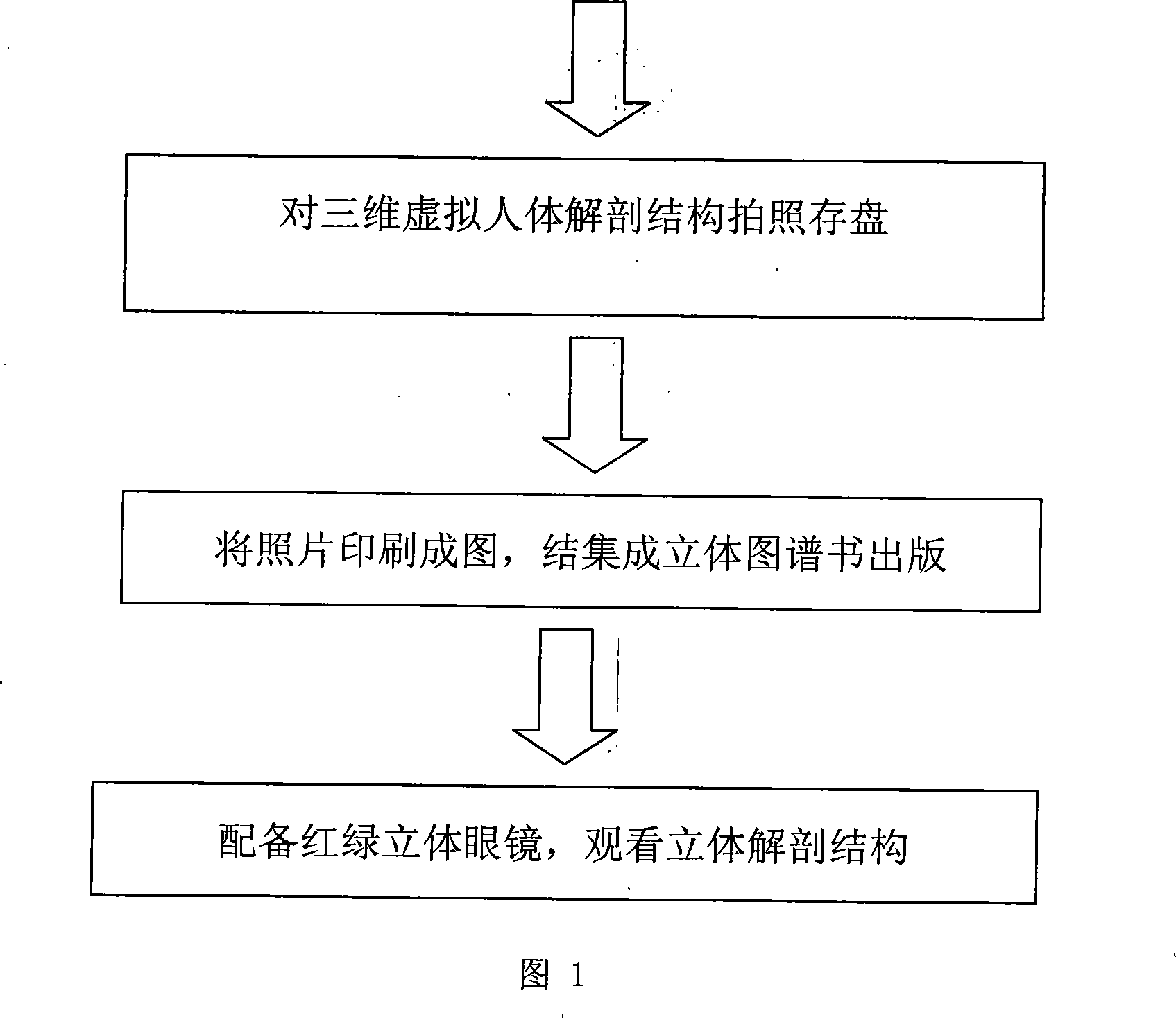
[0011] (3) Carry out the following steps of pre-analysis data proc...


PUM


Abstract

The utility model relates to a process and an application of a stereo dissection image of a human body. The technical proposal is that, gathering the somatic data by means of the X-ray computer tomography, the magnetic resonance imaging, the magnetic resonance angiography, the positron emission computed tomography and the ultrasonic; analyzing and processing the data by the Dextroscope system, to rebuild the three-dimensional virtual human, and taking pictures of each part, each angle of the virtual human, to form a solid image. The utility model allows observers to become conscious of the solid dissection image via a pair of red-and-green glasses or a pair of red-and-blue glasses, just like an elaborate specimen in front of us; the space structure, the inter-structural level and the adjoining feature are accurate and vivid, that is convenient for doctors to exactly master the complicated space dissection images of the patients, and is in favor of improving the diagnostic skills; the utility model is convenient for a clinician to draw up a plan before an operation, and simultaneously is a wonderful platform for the anthropotomy teaching.

Description

technical field [0001] The invention relates to a production method and application of a three-dimensional anatomical image of a human body. Background technique [0002] The anatomical atlas is used to describe the structure, hierarchy and adjacency of human organ tissues. In the past, in order to obtain the anatomical information of the patient, clinicians could only obtain it by reading two-dimensional images such as CT, MRI, MRA, PETCT, and ultrasound, and inferred its spatial anatomical structure, and could not directly read the three-dimensional anatomy Atlas, the understanding obtained in this way is not comprehensive and direct, and it is difficult to accurately describe the spatial structure, level and adjacent characteristics of tissues and organs. Contents of the invention [0003] The object of the present invention is to provide a production method and application of a human body stereoscopic anatomy image with good stereo effect and realistic image. [0004...

Claims


Application Information

Patent Timeline
Patent Type & Authority Applications(China)
IPC IPC(8): A61B19/00G06T1/00G06T15/00A61B34/10G06T15/08
Inventor 宋岩峰张文举
Owner 宋岩峰
Why Eureka
Try Eureka